• 검색 결과가 없습니다.

sp. nov., a new species identified from the gastrointestinal tract of striped field mice in Korea JVS

N/A
N/A
Protected

Academic year: 2022

Share "sp. nov., a new species identified from the gastrointestinal tract of striped field mice in Korea JVS"

Copied!
7
0
0

로드 중.... (전체 텍스트 보기)

전체 글

(1)

J Vet Sci 2015, 16(4), 475-481ㆍhttp://dx.doi.org/10.4142/jvs.2015.16.4.475

JVS

Received 8 Dec. 2014, Revised 2 Feb. 2015, Accepted 7 Mar. 2015

*Corresponding authors: Tel: +82-2-880-1259; Fax: +82-2-873-1213; E-mails: [email protected] (JK Seong), [email protected] (S Cho)

Journal of Veterinary Scienceㆍⓒ 2015 The Korean Society of Veterinary Science. All Rights Reserved.

This is an Open Access article distributed under the terms of the Creative Commons Attribution Non-Commercial License (http://creativecommons.org/licenses/

pISSN 1229-845X eISSN 1976-555X

Helicobacter apodemus sp. nov., a new Helicobacter species identified from the gastrointestinal tract of striped field

mice in Korea

Woo Jin Jeon

1,2

, Hee-Jin Dong

3

, Jae Hoon Shin

1

, Il Yong Kim

1

, Hungwui Ho

3

, Seung Hyun Oh

4

, Young Min Yoon

5

, Yang-Kyu Choi

6

, Jun Gyo Suh

7

, Ki-Hoan Nam

8

, Hyoung-Chin Kim

8

, Seongbeom Cho

3,

*, Je Kyung Seong

1,

*

Laboratories of

1

Developmental Biology and Genomics, and

3

Veterinary Public Health, College of Veterinary Medicine, BIO-MAX Institute, Program for Cancer Biology, and Interdisciplinary Program for Bioinformatics, BK21Plus Program for Creative Veterinary Science Research, Research Institute for Veterinary Science, Seoul National University, Seoul 08826, Korea

2

Incheon International Airport Imported Food Inspection Center, Gyeongin Regional Food and Drug Administration, Ministry of Food and Drug Safety, Incheon 22382, Korea

4

College of Pharmacy, Gachon University, Incheon 21936, Korea

5

College of Veterinary Medicine, Jeju National University, Jeju 63241, Korea

6

College of Veterinary Medicine, Konkuk University, Seoul 05029, Korea

7

Department of Medical Genetics, College of Medicine, Hallym University, Chuncheon 24252, Korea

8

Biomedical Mouse Resource Center, Korea Research Institute for Bioscience and Biotechnology, Ochang 34141, Korea

A novel Helicobacter species was identified from the gastrointestinal tract of the Korean striped field mouse (Apodemus agrarius).

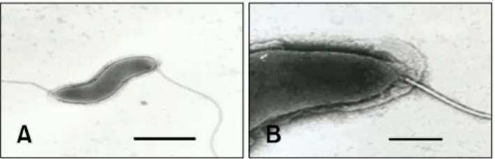
Biochemical testing, ultrastructure characterization, and 16S rRNA gene sequence analysis suggested that this bacterium represents a distinct taxon. The bacterium was positive for urease activity, susceptible to cephalothin and nalidixic acid, and weakly positive for oxidase and catalase activity. Electron microscopy revealed that the bacterium has spirally curved rod morphology with singular bipolar nonsheathed flagella.

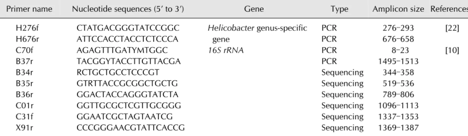
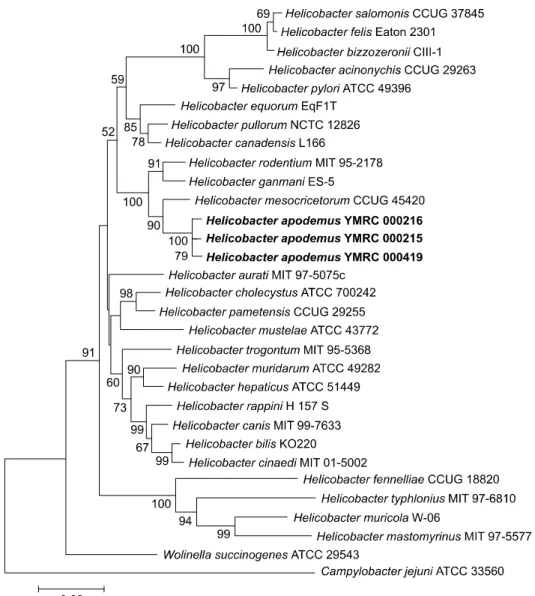
Genotypically, the isolated bacterial strains (YMRC 000215, YMRC 000216, and YMRC 000419) were most closely related to a reference strain of Helicobacter mesocricetorum (97.25%, 97.32%, and 97.03% 16S rRNA sequence similarities, respectively). The 16S rRNA sequences of these strains were deposited into GenBank under accession numbers AF284754, AY009129, and AY009130, respectively. We propose the name Helicobacter apodemus for this novel species.

Keywords: 16S rRNA, Apodemus agrarius, Helicobacter apodemus, striped field mouse

Introduction

Since the discovery of Helicobacter (H.) pylori in the human stomach, Helicobacter spp. have become the subject of intensive research and shown to be the causative agents of several gastric diseases, including gastritis, peptic ulcers, and stomach cancer [17,30]. In addition to humans, Helicobacter spp. can be found in various animal species, including horses, pigs, cattle, and birds [2,3,5,6]. These bacteria can colonize the liver, bile, gall bladder, and lining of the gastrointestinal tract [9,25] and can cause damage or reside in the host without causing any clinical signs. To date, 35 formally named

Helicobacter species have been identified. However, the majority of these bacteria were isolated from humans and domestic animals; thus, a potentially large number of Helicobacter spp. in wild animals are yet to be identified.

Over the last decade, the presence of Helicobacter spp. has

frequently been reported in laboratory facilities housing

rodents. To date, more than 10 rodent Helicobacter species have

been identified, several of which were shown to be related to

pathogenicity in the host. For instance, H. aurati was isolated

from adult Syrian hamsters with gastritis [19], and urease-

negative H. typhlonicus was recovered from the cecum and

fecal samples of interleukin-10-deficient laboratory mice with

(2)

Table 1. Oligonucleotide primer used in this study

Primer name Nucleotide sequences (5’ to 3’) Gene Type Amplicon size References H276f CTATGACGGGTATCCGGC Helicobacter genus-specific

gene

PCR 276–293 [22]

H676r ATTCCACCTACCTCTCCCA PCR 676–658

C70f AGAGTTTGATYMTGGC 16S rRNA PCR 8–23 [10]

B37r TACGGYTACCTTGTTACGA PCR 1495–1513

B34r RCTGCTGCCTCCCGT Sequencing 344–358

B35r GTRTTACCGCGGCTGCTG Sequencing 519–536

B36r GGACTACCAGGGTATCTA Sequencing 789–806

C01r GGTTGCGCTCGTTGCGGG Sequencing 1096–1113

C31f GGAATCGCTAGTAATCG Sequencing 1337–1353

X91r CCCGGGAACGTATTCACCG Sequencing 1369–1387

mastomyrinus was isolated and shown to possess a cytolethal distending toxin that can cause cell distention; however, the pathogenic potential of this species is unknown [28]. Some Helicobacter spp. may infect the host without causing any disease, such as H. muricola and H. ganmani, which were detected in asymptomatic Korean wild mice [36] and laboratory mice [23], respectively. However, the potential pathogenicity of these bacteria cannot be excluded, as immunocompromised hosts seem to be more susceptible to Helicobacter infections [34]. Moreover, some Helicobacter spp. in humans are believed to have originated from animals [18,30]. These include Helicobacter sp. flexispira, which belongs to the normal flora of the intestinal tract in animals and was found to be associated with bacteremia in an immunocompetent young man [14] and a child with pneumonia [33]. Therefore, the screening and characterization of Helicobacter spp. in rodents may have important benefits for rodent research and human health.

In Korea, the striped field mouse (Apodemus agrarius) is widespread in the wild [37] and is believed to be a natural reservoir of Hantaan virus [20] and intestinal fluke Echinostomas [4]. The presence of Helicobacter species in the striped field mouse is largely unknown; however, because this species has been developed as a new laboratory animal model in Korea, the transmission of these potential zoonotic pathogens, including Helicobacter, is of concern. Furthermore, the presence of Helicobacter may interfere with host immune responses and consequently affect the validity of data in immune-related studies, especially vaccine development [27]. Therefore, screening for the presence of Helicobacter in laboratory animals is important to ensure accuracy and reliability of data in future biomedical studies. Accordingly, this study was conducted to examine the presence of Helicobacter spp. in striped field mice in South Korea and conduct phenotypic and genotypic characterization of the newly identified Helicobacter isolates.

Materials and Methods

Animals

Overall, 24 male and 25 female striped field mice (Apodemus agrarius) aged 6 to 12 months were obtained from the Laboratory Animal Resources at the Korea Food and Drug Administration (KFDA) and screened for the presence of Helicobacter spp. The use of these animals and the experimental procedures were designed based on the Guide for Animal Experiments and approved by the Committee for Care and Use of Laboratory Animals of Yonsei University.

Bacterial isolation and culture

The 49 striped field mice were euthanized, and the gastrointestinal tract (stomach, liver, cecum, and colon) was aseptically removed. In addition, 83 freshly pooled fecal samples were collected from the mouse cages. Each tissue and fecal sample was homogenized in 1 mL of phosphate-buffered saline (PBS), after which the suspension was filtered through a 0.8 m syringe. The filtrates were then inoculated onto moist tryptic soy blood agar plates containing 5% sheep blood and incubated at 37

o

C under microaerophilic conditions (BBL GasPak System; BD Diagnostic Systems, USA) for up to 6 days.

PCR amplification

PCR was conducted to identify Helicobacter spp. using the previously designed primers H276f and H676r, after which the 16S rRNA genes of Helicobacter-positive isolates were amplified for further sequencing using primers C70f and B37r (Table 1) [10,22].

Biochemical testing

Biochemical tests commonly used to characterize Helicobacter

spp. were performed using an API Campy identification system

(VITEK; bioMérieux, USA) for colonies found to be

Helicobacter-positive by PCR. The optimum growth

temperature, tolerance to 1% glycine and 1.5% NaCl, Gram

(3)

Ta bl e 2. Bio chemical characteristics of nove l Helicobacter spp. i n com parison to closely related Helicobacters * Ta xo n C atalase pr oduc tion Ur ea se activ ity N itrate re duc tion A lk alin e phos pha ta se hy dr ol ys is

-G lu tam yl- tran sp ep tid ase activ ity Indoxy l- actetate hy dr ol ys is Gr ow th at 4 2

o

C Su scep tib ility to Per ipl as mi c fib ers Number of flag ella per c el l

D istrib utio n of flag ella N alid ix ic ac id ( 30 g) C ep ha lo th in (30 g) New isolates H. ap odemus (YM R C 000 215 ) ++ + + + − +S S − 2 B ipolar H. ap odemus (YM R C 000 216 ) ++ + + + − +S S − 2 B ipolar H. ap odemus (YM R C 000 419 ) ++ + + + − +S S − 2 B ipolar Reference organisms H. m esocricetorum + − ++ − ND ND R S − 2 B ipolar H. rodentium + − + −− − +R R − 2 B ipolar H. pu lloru m + − + −− − +R R − 1P ol ar H. pam entensis + − ++ −− +S S − 2 B ipolar H. chol ecystus + − ++ − ND ND R I − 1P ol ar H. cinaedi ++ + −− − − IS − 1 –2P ol ar , Bi polar H. hepaticus ++ + N D N D + − RR − 2 B ipolar H. mu rid arum ++ − ++ + − RR + 10 –14 Bipolar H. trogontu m ++ + − +N D + R R + 5 –7 B ipolar H. bili s ++ + N D N D − +R R + 3 –14 Bipolar H. pyl ori ++ − ++ −− RS − 4 –8 B ipolar

* Ref

erence organism descri ptions obtaine d f rom [12, 27,30]. + , posi tive react ion; − , negati ve reaction; R, resistant ; I , i nterm edi ate; S, suscept ible; N D , not determined.

(4)

Fig. 1. Transmission electron micrograph of a negatively stained Helicobacter sp. Nov. cell isolated from the cecum of Apodemus agrarius. (A) YMRC 000215. (B) YMRC 000215. Scale bars = 1

m (A), or 0.2 m (B).

Pharmaceutical, Korea), oxidase activity, catalase activity, and hydrolysis of indoxyl acetate were determined as previously described [7,8]. The isolates were tested for susceptibility to cephalothin (30 g/disc) and nalidixic acid (30 g/disc) using antibiotic-impregnated discs (Becton, Dickinson and Company, USA).

Electron microscopy

To prepare the isolates for observation by electron microscopy, bacterial colonies were harvested from blood agar plates, suspended in 10 mM Tris buffer (pH 7.4), and centrifuged at 12,000 g. The pellets were then gently washed three times with deionized water and spread on a carbon-coated grid, after which a small droplet of 4% phosphotungstic acid solution was added. The morphology of the isolates was examined using a Philips CM10 transmission electron microscope (Philips, The Netherlands) at 80 KeV and compared to the morphology of other known Helicobacter species (Table 2).

DNA extraction

Bacterial colonies on blood agar plates were propagated in Brucella broth supplemented with 5% fetal bovine serum and centrifuged at 10,000 g. The pellets were then washed with PBS, after which the bacterial DNA was extracted using a QIAamp Tissue Kit (Qiagen, USA) according to the manufacturer’s instructions. The concentration and purity of the DNA were then measured spectrophotometrically.

DNA sequencing

The products of PCR amplification of the 16S rRNA gene were purified using a JetSorb Kit (Genomed, USA), then sequenced using a Thermo Sequenase Cy 5 Dye Terminator Kit (Amersham Pharmacia Biotech, USA) according to the manufacturer’s instructions. The sequencing reactions were performed using the primers B34r, B35r, B36r, C01r, C31f, and X91r as previously described (Table 1) [10]. The sequences of Helicobacter, Wolinella, and Campylobacter spp. obtained from the GenBank database (The National Center for Biotechnology Information, USA) were compared with the 16S rRNA sequences (up to 1446 bp fragments) of our strains. The sequence data were assembled and aligned by ClustalW analysis using MEGA ver. 6.0 [32]. Phylogenetic trees were generated by the neighbor-joining method with bootstrapping based on 1,000 replications [26]. In addition, nucleotide changes at a single position were corrected using Jukes Cantor corrections [16], then analyzed to generate similarity matrices.

The 16S rRNA gene sequences for isolates YMRC 000215, YMRC 000216, and YMRC 000419 were deposited into GenBank under accession nos. AF284754, AY009129, and AY009130, respectively.

Results

Bacterial isolation

A Helicobacter-positive band was observed for two (8.3%), five (20.8%), and six (25%) samples of stomach, cecum, and colon, respectively, from 24 male striped field mice and three (12%), two (8%), seven (28%), and 12 (48%) samples of stomach, liver, cecum, and colon, respectively, from 25 female mice. Regardless of sex, 15 (18%) out of 83 fecal samples were positive for Helicobacter.

Biochemical testing and ultrastructure characterization The bacterial strains that were identified as Helicobacter by PCR were subjected to biochemical tests. Among these, three strains (YMRC 000419, YMRC 000215 and YMRC 000216, isolated from stomach, colon and cecum samples, respectively) that were phenotypically distinct from those of previously reported Helicobacter spp. were selected for further investigation.

The three strains isolated in this study differed from other Helicobacter spp. in 2 to 7 of the 12 phenotypic traits examined (Table 2). The strains grew well under microaerophilic conditions at 37

o

C and showed slight growth at 42

o

C. However, the isolates did not grow under similar conditions at 25

o

C or under aerobic conditions at 37

o

C. Brucella agar plates containing 1% glycine and 1.5% NaCl did not support their growth.

Morphological examination of the bacteria through Gram staining showed various short rods and coccal forms. The strains were positive for urease activity, nitrate reduction, alkaline phosphatase hydrolysis, and -glutamyl transpeptidase activity, weakly positive for oxidase and catalase activities, and negative for indoxyl acetate hydrolysis. Antibiotic susceptibility testing showed that the strains were susceptible to cephalothin and nalidixic acid. The bacterial cells were also observed by electron microscopy, which revealed spirally curved rods with singular bipolar nonsheathed flagella and no periplasmic fiber.

The cells were 2 to 5 m in length and 0.4 to 0.6 m in diameter (Fig. 1).

Phylogenetic analysis

Approximately 95% of the 1450 bp 16S rRNA gene fragment

(5)

Fig. 2. Phylogenetic tree of strains isolated in this study and members of other genera generated using the neighbor-joining method.

of the three Helicobacter isolates was sequenced. Two isolates, YMRC 000215 and YMRC 000216, differed by a single base C or U at position 1137 (Escherichia coli numbering), while YMRC 000216 differed by one base from YMRC 000215 and by two bases from YMRC 000419. Thus, the > 99% similarity of the 16S rRNA sequence of the three isolates indicates that they are the same species.

A similarity matrix table and phylogenetic tree were generated to investigate the genetic relatedness of our isolates to other Helicobacter species (Fig. 2). These analyses revealed that YMRC 000215, YMRC 000216, and YMRC 000419 are most closely related to the reference strain of H. mesocricetorum (97.25%, 97.32%, and 97.03% similarity, respectively).

Discussion

In the present study, the majority of presumptive Helicobacter

spp. were detected in samples of both male and female striped field mice. To the best of our knowledge, this is the first record of a novel Helicobacter sp. in the striped field mouse. In a previous study in Croatia, 22 wild rodents with gastritis were examined for the presence of Helicobacter using Warthin–

Starry staining. However, the results showed that one gastric sample from striped field mice was stained negative for Helicobacter [24]. Several murine Helicobacter species have been reported to commonly colonize in the extragastric region, particularly the large intestine, whereas gastric colonization is more rare [35]. Interestingly, our isolates were detected in stomach samples, as well as extragastric samples. These findings are similar to the reported distribution of H. aurati, which was present in the cecum and stomach samples of adult Syrian hamsters, and H. muridarum, a member of the intestinal flora that was shown to colonize the stomachs of mice [19,21].

The presence of flagella together with urease and catalase

(6)

well-known disease-causing species H. pylori, H. bilis, and H.

hepaticus [1]. The presence of flagella is believed to aid in the survival of Helicobacter in the stomach by allowing them to burrow into the gastric mucosa, while urease and catalase activities promote survival by neutralizing gastric acid and resisting the host’s antioxidant defense mechanism, respectively [13,31]. Thus, the biochemical and morphological traits of our isolates suggest that they are potentially pathogenic.

Our three isolates showed a similar morphology and size to H.

mesocricetorum under the electron microscope; however, they differed from H. mesocricetorum phenotypically as they exhibited urease activity, -glutamyl transpeptidase activity, and susceptibility to nalidixic acid. Together with H.

mesocricetorum, our strains were clustered with H. rodentium and H. ganmani in the phylogenetic tree (Fig. 2). The species in this cluster of Helicobacter were all isolated from the intestinal region and do not possess a sheathed flagella, which is believed to be an important factor for survival under the strongly acidic conditions of the stomach [29,35]. Interestingly, our isolates were able to colonize the stomach despite having non-sheathed flagella.

In recent years, there have been few reports of the presence of H. apodemus based on 16S rRNA sequences in GenBank. The presence of H. apodemus in wild rodents (Dipus sagitta, Euchoreutes naso, Meriones meridianus, and Nesokia indica) in different regions of China indicates the epidemic nature of H.

apodemus infection in the wild [12]. A study in Sweden also demonstrated the presence of H. apodemus with a 98.3%

sequence similarity to 16S rDNA in laboratory mice [15].

Taken together, these results indicate that H. apodemus may have multiple hosts.

In summary, a novel Helicobacter sp. isolated from the gastrointestinal tract of the striped field mouse (Apodemus agrarius) was described. The results of biochemical and phenotypic characterization and sequence analysis of the 16S rRNA gene strongly suggest that this isolate represents a distinct taxon; thus, we propose the name Helicobacter apodemus for this species. Further studies may be needed to investigate the pathogenic genes and possible pathogenesis of H. apodemus.

Acknowledgments

This research was supported by Korea Mouse Phenotyping Project (2013M3A9D5072550) of the Ministry of Science, ICT and Future Planning through the National Research Foundation to Je Kyung Seong.

Conflict of Interest

There is no conflict of interest.

References

1. Andersen LP, Wadström T, Basic bacteriology and culture.

In: Mobley HLT, Mendz GL, Hazell SL (eds.). Helicobacter pylori: Physiology and Genetics. Chapt. 4. ASM press, Washington, D.C., 2001.

2. Baele M, Decostere A, Vandamme P, Ceelen L, Hellemans A, Mast J, Chiers K, Ducatelle R, Haesebrouck F. Isolation and characterization of Helicobacter suis sp. nov. from pig stomachs. Int J Syst Evol Microbiol 2008, 58, 1350-1358.

3. Bezdekova B, Futas J. Helicobacter species and gastric ulceration in horses: a clinical study. Vet Med (Praha) 2009, 54, 577-582.

4. Chai JY, Park JH, Jung BK, Guk SM, Kim JL, Shin EH, Klein TA, Kim HC, Chong ST, Baek LJ, Song JW.

Echinostome infections in the striped-field mouse, Apodemus agrarius, and the Ussuri white-toothed shrew, Crocidura lasiura, caught near the demilitarized zone, Gyeonggi-do (Province), Republic of Korea. Korean J Parasitol 2009, 47, 311-314.

5. De Groote D, van Doorn LJ, Ducatelle R, Verschuuren A, Tilmant K, Quint WGV, Haesebrouck F, Vandamme P.

Phylogenetic characterization of ‘Candidatus Helicobacter bovis’, a new gastric Helicobacter in cattle. Int J Syst Bacteriol 1999, 49, 1707-1715.

6. Dewhirst FE, Seymour C, Fraser GJ, Paster BJ, Fox JG.

Phylogeny of Helicobacter isolates from bird and swine feces and description of Helicobacter pametensis sp. nov. Int J Syst Bacteriol 1994, 44, 553-560.

7. Finegold SM, Martin WJ, Scott EG. Baily and Scott’s Diagnostic Microbiology. 6th ed. pp. 663-673, Mosby, St.

Louis, 1982.

8. Fox JG, Dewhirst FE, Tully JG, Paster BJ, Yan L, Taylor NS, Collins MJ Jr, Gorelick PL, Ward JM. Helicobacter hepaticus sp. nov., a microaerophilic bacterium isolated from livers and intestinal mucosal scrapings from mice. J Clin Microbiol 1994, 32, 1238-1245.

9. Fox JG, Ge Z, Whary MT, Erdman SE, Horwitz BH.

Helicobacter hepaticus infection in mice: models for understanding lower bowel inflammation and cancer.

Mucosal Immunol 2011, 4, 22-30.

10. Fox JG, Yan LL, Dewhirst FE, Paster BJ, Shames B, Murphy JC, Hayward A, Belcher JC, Mendes EN. Helicobacter bilis sp. nov., a novel Helicobacter species isolated from bile, livers, and intestines of aged, inbred mice. J Clin Microbiol 1995, 33, 445-454.

11. Franklin CL, Gorelick PL, Riley LK, Dewhirst FE, Livingston RS, Ward JM, Beckwith CS, Fox JG. Helicobacter typhlonius sp. nov., a novel murine urease-negative Helicobacter species. J Clin Microbiol 2001, 39, 3920-3926.

12. Goto K, Jiang W, Zheng Q, Oku Y, Kamiya H, Itoh T, Ito M.

Epidemiology of Helicobacter infection in wild rodents in the Xinjiang-Uygur autonomous region of China. Curr Microbiol 2004, 49, 221-223.

13. Harris AG, Hinds FE, Beckhouse AG, Kolesnikow T, Hazell SL. Resistance to hydrogen peroxide in Helicobacter pylori:

role of catalase (KatA) and Fur, and functional analysis of a

novel gene product designated ‘KatA-associated protein’,

(7)

KapA (HP0874). Microbiology 2002, 148, 3813-3825.

14. Iten A, Graf S, Egger M, Täuber M, Graf J. Helicobacter sp.

flexispira bacteremia in an immunocompetent young adult. J Clin Microbiol 2001, 39, 1716-1720.

15. Johansson SK, Feinstein RE, Johansson KE, Lindberg AV.

Occurrence of Helicobacter species other than H. hepaticus in laboratory mice and rats in Sweden. Comp Med 2006, 56, 110-113.

16. Jukes TH, Cantor CR. Evolution of protein molecules. In:

Munro HN, Allison JB (eds.). Mammalian Protein Metabolism. pp. 21-132, Academic Press, New York, 1969.

17. Marshall B, Warren JR. Unidentified curved bacilli in the stomach of patients with gastritis and peptic ulceration.

Lancet 1984, 323, 1311-1315.

18. O’Rourke JL, Grehan M, Lee A. Non-pylori Helicobacter species in humans. Gut 2001, 49, 601-606.

19. Patterson MM, Schrenzel MD, Feng Y, Fox JG. Gastritis and intestinal metaplasia in Syrian hamsters infected with Helicobacter aurati and two other microaerobes. Vet Pathol 2000, 37, 589-596.

20. Peters CJ, Simpson GL, Levy H. Spectrum of hantavirus infection: hemorrhagic fever with renal syndrome and hantavirus pulmonary syndrome. Annu Rev Med 1999, 50, 531-545.

21. Queiroz DM, Contigli C, Coimbra RS, Nogueira AM, Mendes EN, Rocha GA, Moura SB. Spiral bacterium associated with gastric, ileal and caecal mucosa of mice. Lab Anim 1992, 26, 288-294.

22. Riley LK, Franklin CL, Hook RR Jr, Besch-Williford C.

Identification of murine helicobacters by PCR and restriction enzyme analyses. J Clin Microbiol 1996, 34, 942-946.

23. Robertson BR, O’Rourke JL, Vandamme P, On S, Lee A.

Helicobacter ganmani sp. nov., a urease-negative anaerobe isolated from the intestines of laboratory mice. Int J Syst Evol Microbiol 2001, 51, 1881-1889.

24. Robić M, Artuković B, Beck A, Turk R, Belić M, Svetina A, Grabarević Ž. Histopathological changes in the stomachs of wild rodents in Croatia and the first finding of the Helicobacter species. Vet Arh 2011, 81, 415-421.

25. Sabbaghian MS, Ranaudo J, Zeng L, Alongi AP, Perez-Perez G, Shamamian P. Identification of Helicobacter spp. in bile and gallbladder tissue of patients with symptomatic

gallbladder disease. HPB (Oxford) 2010, 12, 129-133.

26. Saitou N, Nei M. The neighbor-joining method: a new method for reconstructing phylogenetic trees. Mol Biol Evol 1987, 4, 406-425.

27. Shen Z, Fox JG, Dewhirst FE, Paster BJ, Foltz CJ, Yan L, Shames B, Perry L. Helicobacter rodentium sp. nov., a urease-negative Helicobacter species isolated from laboratory mice. Int J Syst Bacteriol 1997, 47, 627-634.

28. Shen Z, Xu S, Dewhirst FE, Paster BJ, Pena JA, Modlin IM, Kidd M, Fox JG. A novel enterohepatic Helicobacter species

‘Helicobacter mastomyrinus’ isolated from the liver and intestine of rodents. Helicobacter 2005, 10, 59-70.

29. Simmons JH, Riley LK, Besch-Williford CL, Franklin CL.

Helicobacter mesocricetorum sp. nov., a novel Helicobacter isolated from the feces of Syrian hamsters. J Clin Microbiol 2000, 38, 1811-1817.

30. Solnick JV. Clinical significance of Helicobacter species other than Helicobacter pylori. Clin Infect Dis 2003, 36, 349-354.

31. Strugatsky D, McNulty R, Munson K, Chen CK, Soltis SM, Sachs G, Luecke H. Structure of the proton-gated urea channel from the gastric pathogen Helicobacter pylori.

Nature 2013, 493, 255-258.

32. Tamura K, Stecher G, Peterson D, Filipski A, Kumar S.

MEGA6: molecular evolutionary genetics analysis version 6.0. Mol Biol Evol 2013, 30, 2725-2729.

33. Tee W, Leder K, Karroum E, Dyall-Smith M. “Flexispira rappini” bacteremia in a child with pneumonia. J Clin Microbiol 1998, 36, 1679-1682.

34. Ward JM, Anver MR, Haines DC, Melhorn JM, Gorelick P, Yan L, Fox JG. Inflammatory large bowel disease in immunodeficient mice naturally infected with Helicobacter hepaticus. Lab Anim Sci 1996, 46, 15-20.

35. Whary MT, Fox JG. Natural and experimental Helicobacter infections. Com Med 2004, 54, 128-158.

36. Won YS, Yoon JH, Lee CH, Kim BH, Hyun BH, Choi YK.

Helicobacter muricola sp. nov., a novel Helicobacter species isolated from the ceca and feces of Korean wild mouse (Mus musculus molossinus). FEMS Microbiol Lett 2002, 209, 45-51.

37. Yoon MH, Jung SJ, Oh HS. Population structure and

reproductive pattern of the Korean striped field mouse,

apodemus agrarius. Korean J Biol Sci 1997, 1, 53-61.

수치

Table 1. Oligonucleotide primer used in this study
Table 2. Biochemical characteristics of novel Helicobacter spp. in comparison to closely related Helicobacters* TaxonCatalase productionUreaseactivityNitratereductionAlkaline phosphatase hydrolysis
Fig. 1. Transmission electron micrograph of a negatively stained Helicobacter sp. Nov
Fig. 2. Phylogenetic tree of strains isolated in this study and members of other genera generated using the neighbor-joining method.

참조

관련 문서