© 2019 The Korean Ophthalmological Society
This is an Open Access article distributed under the terms of the Creative Commons Attribution Non-Commercial License (http://creativecommons.org/licenses /by-nc/3.0/) which permits unrestricted non-commercial use, distribution, and reproduction in any medium, provided the original work is properly cited.
Original Article
Long-term Outcome of Incision and Curettage Treatment in Patients with Lacrimal Gland Ductulitis
Jiseon An, Kyeongwook Lee
Department of Ophthalmology, Saevit Eye Hospital, Goyang, Korea
Purpose: To describe the effects and long-term outcomes of incision and curettage treatment in patients with lacrimal gland ductulitis.
Methods: Twenty-four patients (24 eyes) with lacrimal gland ductulitis who were treated at Saevit Eye Hospital from June 2010 to November 2016. All patients underwent incision and curettage through the lacrimal ductule, and granules or concretions were removed. After the procedure, oral and topical antibiotics, oral anti-inflam- matory agent were used for a week. Clinical presentations of the patients were analyzed. The resolution of symptoms and inflammatory signs and recurrence were evaluated more than 12 months after the procedure including telephone follow-up by a specialist nurse.
Results: Common symptoms were a painful, swelling mass with mucous discharge (17 eyes) and conjunctival injection (7 eyes) at the lateral canthal area. During the procedure, 22 patients (91.7%) had typical sulfur gran- ule of Actinomyces, and 10 patients (41.7%) had many cilia in the expressed debris from the ductule. Twen- ty-three of 24 patients had resolution of symptoms after the procedure and all but one patient (95.8%) showed no recurrence.
Conclusions: Incision and curettage is a simple and less invasive procedure that may be considered as a first treatment option for lacrimal gland ductulitis. Furthermore, incision and curettage of the affected lacrimal duct- ule has been shown to be effective at minimizing long-term recurrence of lacrimal ductulitis.
Key Words: Actinomyces, Curettage, Lacrimal apparatus, Lacrimal gland ductulitis
Lacrimal gland ductulitis is an inflammatory disorder characterized by infection of the lacrimal gland ductules [1,2]. Clinical manifestations of lacrimal gland ductulitis have been reported, but effective management approaches have not been well defined. Actinomyces—the probable
pathogen of lacrimal gland ductulitis—are anaerobic bac- teria first associated with the etiology of lacrimal canalicu- litis by Ellis et al. [3] in 1854. Since the chief symptoms of lacrimal gland ductulitis mimic other common ocular con- ditions (e.g., canthal mass, conjunctival injection and mu- cous discharge at the lateral canthal area), this condition is often misdiagnosed as chronic conjunctivitis, hordeolum or lacrimal gland ductal cyst (known as dacryops) infection.
Dacryops infection is a secondary infection of a pre-ex- isting sac which primarily occurs by bacterial infection followed by enlargement of the lacrimal gland ductule.
The same procedure used to treat dacryops infection and
Received: January 30, 2019 Final revision: February 15, 2019 Accepted: March 26, 2019
Corresponding Author: Kyeongwook Lee, MD. Department of Oph- thalmology, Saevit Eye Hospital, 1065 Jungang-ro, Ilsandong-gu, Goy- ang 10447, Korea. Tel: 82-31-900-7700, Fax: 82-31-900-7777, E-mail:
szera0306@saeviteye.com
lacrimal gland ductulitis is surgical excision of the affected cysts and ductule of the lacrimal gland [1,2]. However, de- spite different pathogenesis of these disorders, the thera- peutic approach for dacryops infection was applied to treat
lacrimal gland ductulitis. Complete excision of the ductule or cys ts for treatment of lacrimal gland ductulitis is a complicated and time-consuming procedure.
Under the supposition that both lacrimal gland ductulitis and lacrimal canaliculitis are caused by Actinomyces in- fection, the authors of this study attempted incision and curettage (I&C)—a simple and effective surgical interven- tion to manage lacrimal canaliculitis—to treat lacrimal gland ductulitis. Our study aimed to characterize the ef- fectiveness of I&C instead of complete excision of the lac- rimal gland ductule in patients diagnosed with lacrimal gland ductulitis, with a long-term follow-up of more than 12 months.
Materials and Methods
This study retrospectively reviewed patients who diag- nosed with lacrimal gland ductulitis at our hospital from June 2010 to November 2016. Chart reviews were conduct- ed to determine age, sex, clinical symptoms, onset site, ophthalmologic findings and histopathologic findings at di- agnosis. Furthermore, we reviewed treatment approaches and outcomes. This study was approved by the institution- al review board of Saevit Eye Hospital (SVEC 201908-003- 01). Informed consent was waived due to the retrospective nature of the study.
We thoroughly examined the lacrimal puncta, lacrimal canaliculi, eyelids, lateral canthus, medial canthus, lacri- mal glands, lacrimal gland ductule and conjunctiva in all patients at the time of the first visit. Patients with mucous discharge and conjunctival injection at the lateral canthus, redness and swelling of lacrimal gland ductule and dis- charge from the lacrimal ductule were diagnosed of having lacrimal gland ductulitis. No patients had a history of symptoms suggesting the presence of lacrimal gland duc- tal cysts and all underwent I&C of the lacrimal gland duct- ule under local anesthesia.
Subcutaneous and subconjunctival area in affected later- al canthus was anesthetized by injecting the combined solution of 2% lidocaine with 1 : 100,000 epinephrine. The eye was exposed using the eye speculum (K1-5675; Katena Products Inc., Denville, NJ, USA), and then the ductule wall of the enlarged lacrimal gland was partially excised and widened using Westcott stitch scissors (K4-4100, Kat- ena Products Inc.). Subsequently, curettage was applied to Fig. 1. Patient with lacrimal gland ductulitis underwent incision
and curettage. (A) An extended wall of the lacrimal gland ductile was incision and expanded using a Westcott stitch scissors (Katena K4-4100). (B) A Meyerhoefer chalazion curette 1.75 mm (Stephens S4-1005) was inserted into the lacrimal gland ductule and re- moved until no more contents. (C) Sulfur granules are suspected.
Informed consent was obtained from all study participants.
A
C B
completely remove infectious materials from the lacrimal ductule by inserting a Meyerhoefer chalazion curette,1.75 mm (S4-1005; Stephens Inc., Lexington, KY, USA) into the lacrimal gland ductule (Fig. 1A-1C). After surgery, antibi- otic eye drops (Cravit, levofloxacin 5 mg/mL; Santen Phar- maceutical, Osaka, Japan), steroid eye drops (Flumetholon 0.1, Santen Pharmaceutical), oral antibiotics (Augmentin 625 mg, amoxicillin 500 mg and clavulanate potassium 125 mg; Il Sung Pharmacia, Seoul, Korea), and an oral an- ti-inflammatory agent (Varidase, streptodornase 2500 IU and streptokinase 10000 IU; SK Chemical, Seoul, Korea) were given to all patients during the first postoperative week. Remission was defined as complete resolution of all clinical symptoms and signs after the surgery until the last follow-up. Patients follow up included outpatient visits and
telephone calls by a special nurse for a period of at least 12 months.
Results
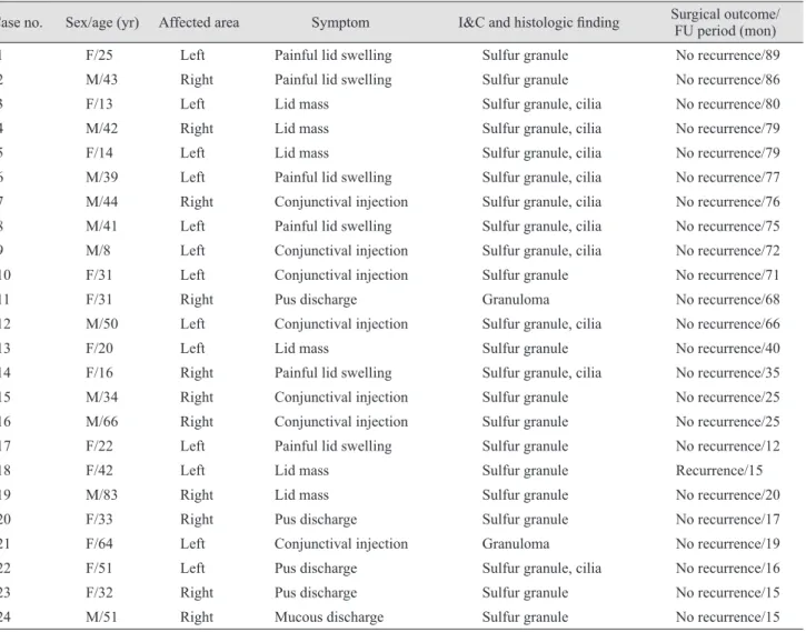
Twenty-four patients (24 eyes), including 11 males (11 eyes) and 13 females (13 eyes), with a mean age of 37.0 ± 18.0 years (range, 8 to 83 years) were included in the study (Table 1). Patients visited our hospital with a chief com- plaint of a painful mass with mucous discharge (17 eyes) and conjunctival injection at the lateral canthal area (7 eyes) (Fig. 2A). All patients were initially managed with local and oral antibiotic therapy due to a suspicion of chronic conjunctivitis, hordeolum, dacryops infection and others, however, were eventually referred to our hospital
Table 1. Demographics and clinical presentation and treatment of 24 cases of lacrimal gland ductulitis
Case no. Sex/age (yr) Affected area Symptom I&C and histologic finding Surgical outcome/
FU period (mon)
1 F/25 Left Painful lid swelling Sulfur granule No recurrence/89
2 M/43 Right Painful lid swelling Sulfur granule No recurrence/86
3 F/13 Left Lid mass Sulfur granule, cilia No recurrence/80
4 M/42 Right Lid mass Sulfur granule, cilia No recurrence/79
5 F/14 Left Lid mass Sulfur granule, cilia No recurrence/79
6 M/39 Left Painful lid swelling Sulfur granule, cilia No recurrence/77
7 M/44 Right Conjunctival injection Sulfur granule, cilia No recurrence/76
8 M/41 Left Painful lid swelling Sulfur granule, cilia No recurrence/75
9 M/8 Left Conjunctival injection Sulfur granule, cilia No recurrence/72
10 F/31 Left Conjunctival injection Sulfur granule No recurrence/71
11 F/31 Right Pus discharge Granuloma No recurrence/68
12 M/50 Left Conjunctival injection Sulfur granule, cilia No recurrence/66
13 F/20 Left Lid mass Sulfur granule No recurrence/40
14 F/16 Right Painful lid swelling Sulfur granule, cilia No recurrence/35
15 M/34 Right Conjunctival injection Sulfur granule No recurrence/25
16 M/66 Right Conjunctival injection Sulfur granule No recurrence/25
17 F/22 Left Painful lid swelling Sulfur granule No recurrence/12
18 F/42 Left Lid mass Sulfur granule Recurrence/15
19 M/83 Right Lid mass Sulfur granule No recurrence/20
20 F/33 Right Pus discharge Sulfur granule No recurrence/17
21 F/64 Left Conjunctival injection Granuloma No recurrence/19
22 F/51 Left Pus discharge Sulfur granule, cilia No recurrence/16
23 F/32 Right Pus discharge Sulfur granule No recurrence/15
24 M/51 Right Mucous discharge Sulfur granule No recurrence/15
I&C = incision and curettage; FU = follow-up.
because they were refractory to antibiotic therapy. All pa- tients underwent surgical intervention by I&C of the lacri- mal gland ductule under local anesthesia. Large granula- tion tissue and dacryolith visible to the naked eye were observed intraoperatively in 22 (91.7%) of 24 eyes and many cilia around dacryoliths in 10 (41.7%) of 24 eyes (Fig.
2B). Based on histopathologic examination, 22 eyes (91.7%) had dacryoliths formed by sulfur granules caused by Acti- nomyces infection, and 10 eyes (41.7%) had necrotic tissue consisting of ciliary follicles. After the procedure, 23 (95.8%) of 24 eyes showed resolution of the symptoms and signs of lacrimal gland ductulitis at the last follow-up.
There were no any postoperative complications. The aver- age length of follow-up was 48.3 ± 29.2 months (range, 12 to 89 months). All patients were followed up via outpatient visits and telephone calls and all but one patient (95.8%) showed no recurrence. The relapsing patient experienced the relapse on the first week after I&C, and then under- went excision of affected lacrimal glad ductule and biopsy revealing the formation of granulation tissue and dacryo- lith. Thereafter complete remission was achieved without relapse.
Discussion
Lacrimal gland ductulitis is an inflammatory disorder caused by infection of the lacrimal gland ductules; com- mon clinical manifestations are swelling of the lacrimal gland ductule, conjunctival injection and a painful mass
associated with mucous discharge from the lateral canthus.
This condition is characterized, in part by the formation of a yellowish stone or granulation tissue in the lacrimal gland ductule [1,2]. The subjects of this study had typical clinical signs of lacrimal gland ductulitis. However, some patients had mild symptoms and only conjunctival injec- tion and mucous or purulent discharge from the lacrimal ductules without clear swelling or redness of the lacrimal glnad ductule in slit-lamp biomicroscopy.
The etiology of lacrimal gland ductulitis still remains unclear but is presumed to be caused by Actinomyces in- fection of the lacrimal gland ductules [1,2]. The Actinomy- ces species—gram-positive, anaerobic bacteria that colo- nize the human mouth—may develop a chronic granulomatous infection through direct spread to the nor- mal flora of the mouth. The Actinomyces species have been identified as common pathogens causing lacrimal canalic- ulitis, conjunctivitis, dacryocystitis, keratitis and endoph- thalmitis [4-8]. Lacrimal gland ductulitis can be very diffi- cult to differentiate from dacryops infection. Dacryops is formed and enlarged by excessive production of IgA due to inflammation or trauma around the lacrimal ductules, contraction or destruction of the nerve root of the lacrimal ductules, and weakening of the ductal walls [9-12]. A lacri- mal ductal cyst presents as asymptomatic swelling in the lateral portion of the upper eyelid and appears as a bluish white, soft, mobile, translucent mass [13-15].
Dacryops infection is a very rare disease and there are only 5 cases reported in literatures listed on PubMed. Be- cause dacryops is a cystic form, there is no discharge. Da- Fig. 2. Patient with lacrimal gland ductulitis and dacryolith. (A) Patient with lacrimal gland ductulitis referred with painful lid swelling, persistent temporal conjunctival injection, and chronic mucopurulent discharge from the lacrimal gland ductule. (B) Many cilia in the expressed debris from the affected ductule. Informed consent was obtained from the study participant.
A B
cryops infection may be associated with sudden pain and/
or a gradual increase in mass size, and can be managed with complete excision of the cyst or marsupilization, but complete excision of the cyst is the preferred approach of treatment with no recurrence [12-17]. Differential diagno- sis between dacryops infection and lacrimal gland ductuli- tis is easily lead by the history of lacrimal ductal cyst, but challenging in those without this history. Also histological difference is able to distinguish, but only after surgery (e.g., local or complete excision of the cyst). Histopatholog- ical difference of dacryops infection and lacrimal gland ductulitis is often difficult since, in chronic cases, the cyst wall consists of pseudostratified nonkeratinized, basal co- lumnar, and superficial myoepithelial cell layers, dou- ble-layered lining of non-ciliated columnar cells and a sin- gle layer of cells [9-13,16,18,19]. Lee et al. [1] reported a case of pseudoepitheliomatous hyperplasia in the invaded ductule as one of the histologic features of lacrimal gland ductulitis. However, this new feature seems insufficient to distinguish between the two disorders. Because of difficul- ties in differential diagnosis, lacrimal gland ductulitis may have often be misdiagnosed as dacryops infection.
Lee et al. [1] and Hay-Smith and Rose [2] documented 7 and 12 patients with lacrimal gland ductulitis, respectively, and successfully treated them by performing complete ex- cision of the affected lacrimal gland ductule. In addition, anatomical pathological examination confirmed the pres- ence of sulfur granules and Actinomyces. The pathophysi- ology and histologic features of lacrimal gland ductulitis resemble those of lacrimal canaliculitis—an infection and inflammation of the lacrimal canaliculi. Lacrimal canalic- ulitis is treated with I&C of the canaliculus, a simple pro- cedure that is less invasive compared with complete exci- sion [20,21]. Based on these observations, the authors of this study performed I&C of the lacrimal gland ductules to manage lacrimal gland ductulitis of 24 eyes. During I&C, we detected sulfur granules visible to the naked eye in 22 of 24 eyes and a number of cilia in the expressed debris from the ductule in 10 eyes. Since infection by Actinomy- ces usually occurs because of direct extension, cilia may be the initial nidus for the development of lacrimal gland ductulitis. Although Actinomyces is suspected to be the causal organism for both lacrimal canaliculitis and lacri- mal gland ductulitis, cilia are frequently found in lacrimal gland ductulitis, unlike the rare presence of eyelashes in lacrimal canaliculitis. This might be because cilia at the
conjunctival fornix shift toward the horizontal lacrimal ductule in the lateral canthus by blinking because the punctum opens to a vertical orientation while the lacrimal ductule runs horizontally.
In this regard, Lee et al. [16] suggested that cilia may act as a nidus for lacrimal ductular inflammation or cyst for- mation as eyelashes shift toward the horizontal lacrimal ductule in the lateral canthus. Due to the retrospective na- ture of the analysis, this study was limited by the relatively small sample size and lack of a control group for compari- son. Our study was meaningful in that it successfully achieved complete remission in 23 of 24 eyes by perform- ing less invasive I&C compared with complete excision of the lacrimal gland ductule, and had no recurrence during follow-up (range, 12 to 89 months).
We concluded that, lacrimal gland ductulitis should be considered when swelling of the lacrimal gland ductule, chronic conjunctival injection and a painful mass associat- ed with mucous discharge from the lateral canthus are present. I&C is a simple and less invasive procedure that may be considered as a first treatment option for lacrimal gland ductulitis. Furthermore, I&C of the affected lacrimal ductule has been shown to be effective at minimizing long-term recurrence of lacrimal ductulitis.
Conflict of Interest
No potential conflict of interest relevant to this article was reported.
References
1. Lee MJ, Kim JE, Kim N, et al. Clinicopathological features of inflammatory lesions of the lateral canthal subconjuncti- val area. Ophthalmic Plast Reconstr Surg 2014;30:251-6.
2. Hay-Smith G, Rose GE. Lacrimal gland ductulitis caused by probable Actinomyces infection. Ophthalmology 2012;119:193-6.
3. Ellis PP, Bausor SC, Fulmer JM. Streptothrix canaliculitis.
Am J Ophthalmol 1961;52:36-43.
4. Kim MW, Son JO. 2 Cases of actinomycotic lacrimal cana- liculitis. J Korean Ophthalmol Soc 1975;16:225-8.
5. Hong JW, Lee TS. Two cases of chronic canaliculitis. J Ko- rean Ophthalmol Soc 1990;31:1096-102.
6. Lee YG, Kim HB. A case of canaliculitis caused by actino- myces odontolyticus. J Korean Ophthalmol Soc 1990;31:979- 82.
7. Freedman JR, Markert MS, Cohen AJ. Primary and sec- ondary lacrimal canaliculitis: a review of literature. Surv Ophthalmol 2011;56:336-47.
8. Hussain I, Bonshek RE, Loudon K, et al. Canalicular infec- tion caused by Actinomyces. Eye (Lond) 1993;7(Pt 4):542-4.
9. Bullock JD, Fleishman JA, Rosset JS. Lacrimal ductal cysts. Ophthalmology 1986;93:1355-60.
10. Remulla HD, Rubin PA. Giant dacryops in a patient with oc- ular cicatricial pemphigoid. Br J Ophthalmol 1995;79:1052-3.
11. Weatherhead RG. Wolfring dacryops. Ophthalmology 1992;99:1575-81.
12. Bay SW, Roh JH. Clinical characteristic of lacrimal ductal cyst. J Korean Ophthalmol Soc 2001;42;803-9.
13. Smith S, Rootman J. Lacrimal ductal cysts. Presentation and management. Surv Ophthalmol 1986;30:245-50.
14. Sen DK, Thomas A. Simple dacryops. Am J Ophthalmol 1967;63:161.
15. Nerad JA, Carter K, Folberg R. Simple dacryops. Arch Ophthalmol 1988;106:1129.
16. Lee JY, Woo KI, Suh YL, Kim YD. The role of entrapped cilia on the formation of lacrimal ductular cysts. Jpn J Ophthalmol 2015;59:81-5.
17. Salam A, Barrett AW, Malhotra R, Olver J. Marsupializa- tion for lacrimal ductular cysts (dacryops): a case series.
Ophthalmic Plast Reconstr Surg 2012;28:57-62.
18. Hornblass A, Herschorn BJ. Lacrimal gland duct cysts.
Ophthalmic Surg 1985;16:301-6.
19. Lee SH, Lew H, Yun YS. A case of lacrimal ductal cyst with dacryolith. J Korean Ophthalmol Soc 2004;45:131-4.
20. Kaliki S, Ali MJ, Honavar SG, et al. Primary canaliculitis:
clinical features, microbiological profile, and management outcome. Ophthalmic Plast Reconstr Surg 2012;28:355-60.
21. Lee MJ, Choung HK, Kim NJ, Khwarg SI. One-snip punc- toplasty and canalicular curettage through the punctum: a minimally invasive surgical procedure for primary canalic- ulitis. Ophthalmology 2009;116:2027-30.