305
Assessment of Left Atrial Function and Remodeling in Patients With Atrial Fibrillation by Performing Strain Echocardiography:
A Prospective Study to Assess the Influence of Renin-Angiotensin System Inhibitors on Atrial Fibrillation
Kyoung Im Cho, MD, Sang Hee Lee, RN, Sun Hee Jang, RN, Dong Won Lee, MD, Hyeon Gook Lee, MD and Tae Ik Kim, MD Division of Cardiology, Maryknoll Medical Center, Busan, Korea
ABSTRACT
Background and Objectives: Renin-angiotensin system (RAS) inhibitors are likely to reduce the development of atrial fibrillation (AF) by preventing atrial fibrosis. We attempted to assess the relevance of strain echocar- diography for quantitative assessment of the left atrial (LA) status in AF, its modification by RAS inhibitors and changes of biochemical markers during cardiac remodeling in AF. Subjects and Methods: Strain echocardiogra- phy is performed 2 times (baseline and 12 month) in 60 patients with AF (RAS inhibitors-used group: 30, non- used group: 30). In an apical 4-chamber view, the regional analysis consisted of placing the region of interest cursor at the basal segments of the septal and lateral wall of LA. Mean peak systolic and early diastolic strain/rate are measured with LA end-systolic antero-posterior, longitudinal and transverse dimensions. Results: Six pa- tients of RAS inhibitors-used group (group A, 20%) and three patients of non-used group (group B, 10%) were converted to normal sinus rhythm during the study. LA size, E wave velocity, E/E’, strain parameters showed no significant differences between groups at the baseline. There were no significant differences in LA size and E wave velocity between groups at the 12 months, however, peak systolic strain/rate were significantly higher in group A (36.71±13.63% and 2.98±0.59s-1, p<0.05, respectively) than group B (27.21±10.49% and 2.21±
0.47s-1). In addition, peak early diastolic strain/rate were significantly higher in group A (-1.89±3.30% and -2.32
±0.77s-1 p<0.05, respectively) than group B (-0.83±2.79% and -1.77±0.25s-1). There were no significant differences in C-reactive protein (CRP) and B-type natriuretic peptide (BNP) at the baseline, but BNP were sig- nificantly reduced in group A (822.9±798.3 pg/mL, p<0.05) than group B (1481.9±209.97 pg/mL) at the 12- month follow-up. Conclusion: The increased values of atrial peak systolic and diastolic strain/rate after treat- ment with RAS inhibitors revealed that passive stretching and shortening of LA wall might improve in some patients with AF even before LA size change possibly because of reduced atrial fibrosis and increased compliance.
Our results indicated that strain echocardiography provides clinically useful information of LA function and remodeling and treatment with RAS inhibitors appears to preserve LA reservoir function in AF patients without visible LA structural change. (Korean Circ J 2008;38:305-312)
KEY WORDS: Atrial fibrillation; Echocardiography; Strains.
Introduction
Atrial fibrillation (AF) is the most frequent arrhyth- mia found in clinical practice,1) and the management of AF remains a difficult clinical problem despite the
intense efforts that have been aimed at improving therapy. Several studies have focused on identifying the fundamental cellular processes that promote and main- tain AF. Emerging evidence has indicated that patho- logic remodeling within the atria plays a critical role in AF.2)3) Many of the recurrences are probably the clinical consequence of electrical remodeling,3)4) which is caused by changes in the refractory period of the atrial muscle and/or the occurrence of abnormal activity inside the pulmonary veins. An increasing body of evidence sug- gests that the atrial stretch induced by increased atrial pressure may precipitate atrial fibrillation through an
Received: January 25, 2008 Revision Received: March 5, 2008 Accepted: March 26, 2008
Correspondence: Kyoung Im Cho, MD,Division of Cardiology, Maryknoll Medical Center, 4-12 Daecheong-dong, Jung-gu, Busan 600-090, Korea
Tel: 82-51-461-2349, Fax: 82-51-465-7470 E-mail: [email protected]
effect on atrial refractoriness,5) so developing a therapy directed against remodeling could mark an important change in the management of atrial fibrillation.
Substantial evidence suggests that the renin-angio- tensin system (RAS) plays an important role in the remodeling that occurs with the development of the AF substrate. Stimulation of angiotensin II production promotes cardiac fibrosis, which contributes to left ven- tricular (LV) remodeling following myocardial infarc- tion,6)7) and rapid atrial stimulation can increase the angiotensin II plasma concentrations in animals.8) In- terestingly, it is known that angiotensin converting enzyme (ACE) inhibitors decrease the atrial pressure,9) and it is possible that treatment with RAS inhibitors minimizes the susceptibility to develop atrial fibrill- ation by lowering the atrial pressure and reducing left atrial enlargement. Elevated levels of C-reactive protein (CRP) may be related to the “burden” or type of AF,10) and the left atrium (LA) volume index, the LV mass index and the duration of AF were independent predic- tors of the plasma B-type natriuretic peptide (BNP) level in patients with chronic AF and preserved LV systolic function.11) So, we propose that the CRP and BNP levels would be decreased by treatment with RAS inhibitors.
The development of new echocardiographic techniques such as myocardial velocity, ultrasonic strain and strain rate imaging has enhanced our ability to noninvasively assess myocardial properties.12) In our previous study, we attempted to assess the relevance of the parameters of Tissue Doppler imaging (TDI) and 2 dimensional strain for quantitatively assessing the LA in control sub- jects and patients with AF. We concluded that strain echocardiography enabled qualitatively precise assess- ment of the LA contractile function and it provided clinically useful information on the LA function and remodeling.13)
We hypothesized that inhibition of endogenous an- giotensin II signaling by treatment with RAS inhibitors attenuates atrial remodeling in patients with AF and we objectively evaluated the changes of LA function by performing strain echocardiography and measuring the biochemical markers during cardiac remodeling in AF patients.
Subjects and Methods
Study population
This study is a randomized, single-center, open-label, placebo-controlled study to compare the efficacy of RAS inhibitors such as losartan for treating patients with AF. Institutional review board approval was obtained and all the subjects provided signed informed consent.
The subjects were enrolled in the study if they had per- sistent AF, which was defined as a duration of AF long-
er than 72 hours regardless of intervention. At the first visit to the arrhythmia unit, the patients were random- ized for treatment with losartan (group A; 30) or with- out losartan (group B: 30). Losartan was started soon after randomization and it was administered at 25-50 mg/d. The patients were examined at the first month, then at 12 months, and at any time the patient com- plained of palpitations or any other symptoms. A 24- hour Holter recording was performed at baseline and 12 months, and at any time the patient had any symp- toms such as palpitations, dizziness or syncope. Echo- cardiographic assessment and measurement of the bio- markers were performed 2 times (at baseline and 12 month). The exclusion criteria were a left atrium size
≥6 cm, myocardial infarction during the previous 6 months, uncontrolled hypertension, unstable angina, New York Heart Association (NYHA) heart failure class IV, the need to continue the use of digitalis, car- diac surgery during the previous 3 months, significant thyroid, pulmonary, kidney or hepatic disease, signifi- cant alterations of the atrioventricular conduction, the presence of a pacemaker, significant valve disease that indicated the need for operation, sick sinus syndrome and dilated or hypertrophied cardiomyopathy. Because antiarrhythmic therapy may have a negative inotropic effect on the atrium, we also excluded those patients who were under antiarrhythmic treatment. Informed consent for the physiologic assessment was obtained from all patients. The demographic data, including age, gender and the cardiovascular risk factors, were recorded.
Sample collection and analysis
Blood was collected before echocardiography in tubes containing ethylenediaminetetraacetic acid (Sarstedt, Nümbrecht, Germany) and the samples were promptly centrifuged for 10 minutes. After separation, plasma al- iquots were stored frozen at -70℃ until they were as- sayed within batches. Measurements of N-terminal-pro BNP (NT-pro-BNP) were performed on a Roche Elecsys 2010 automated analyzer (Roche Diagnostics, USA).
The plasma concentrations of high sensitivity CRP (hs CRP) were measured by performing fully automated latex-enhanced nephelometry. The laboratory person- nel involved in the laboratory measurements were un- aware of the clinical outcomes of the patients.
Echocardiographic evaluation
Standard 2D and strain echocardiographic exami- nations were performed on all the subjects with the subjects lying in the left lateral supine position and with using a 3.5-MHz transducer on Vivid 7 Dimension ultrasound equipment (General Electric, Horten, Nor- way). The LA diameter was measured during systole along the parasternal long-axis view from the 2D guided M-mode tracing. The LA volume was calculated from
the apical 4- and apical 2-chamber zoomed views of the LA with using the biplane method of disks, and the LA volume was divided by the body mass index to obtain the LA volume index. The LV global systolic function was evaluated by the LV ejection fraction. The mitral inflow velocity was obtained by pulsed wave Do- ppler sampling at the tips of the mitral leaflets from the apical four-chamber view at a sweep speed of 100 mm/s.
Color Doppler imaging and offline analysis
Images from the apical 4-chamber view at a high frame rate (150-180 frames per second) were obtained at end-expiratory apnea and these images were stored in a cineloop format for subsequent offline analysis.
Five heartbeats were collected from each view and they were analyzed off-line with an EchoPAC Dimension system (General Electric, Horten, Norway). The peak systolic and diastolic strain rates were obtained from 2 different areas of the basal segments of the LA free wall and the inter-atrial septum on the apical 4 chamber view by the tissue Doppler strain (Fig. 1A). An appro- priate velocity scale was chosen to avoid data aliasing.
The narrowest image sector angle possible (usually 30°
degrees) was used to achieve the maximum possible col- or Doppler frame rate. Careful attention was paid to keep the region of interest at the center of the ultra- sound sector to ensure an alignment as close to 0°as possible to the long-axis motion. For the longitudinal measurements, a computation area of 9×2 mm with an elliptical shape was chosen. Longitudinal direction changes (measured from the apical views) for the atria are better described by the terms “rate of lengthening”
in systole (the positive strain rate value) and the “rate of shortening” in diastole (the negative strain rate value).
The strain rate and strain curves were calculated for all the patients over 3 cardiac cycles and then the values
were averaged to obtain the mean strain rate and strain curves over 1 mean relative risk (RR) interval. End di- astole was defined as the electrocardiogram (ECG) R peak, and end systole was defined as the end of the ECG T wave. The peak positive systolic and early dia- stolic values were calculated from the extracted curve.
Statistical analysis
All the data is expressed as means±standard devia- tions (SDs). The data was analyzed using standard sta- tistical software {Statistical Package for Social Science (SPSS) package version 11.0, Chicago, IL} and compar- isons of all the measurements were done using paired Student’s t-test for the continuous variables and chi- square tests for the categorical variables. A p<0.05 was considered statistically significant.
Results
General characteristics of the patients
The major demographic and clinical characteristics are given in Table 1 with dividing the patients accord- ing to RAS inhibitors treatment. The total study dura- tion was 2.1 years and none of the patients were lost to follow-up. The mean age was 66.4±8.7 years (range:
42-75 years) from group A and 66.3±8.0 years (range:
43-78 years) from group B. The global LV systolic func- tion and, the LV chamber dimension and wall thick- ness were normal in all the patients with AF. The LA diameter, LA volume index, NT-pro-BNP and hs CRP showed no differences between the groups (Table 2).
Strain echocardiographic findings
There were no significant differences of the mitral inflow parameters at baseline and at the 12-month fol- low-up between the groups (Table 3). There were no
Fig. 1. The longitudinal peak strain/rates that obtained from 2 different areas of the basal segments of the LA free wall and the inter- atrial septum in the apical 4 chamber view. By tissue Doppler strain (A), and the peak systolic strain was measured (B). LA: left atrial.
A B
significant differences in the NT-pro-BNP and hsCRP levels at the baseline, but the NT-pro-BNP levels were significantly reduced more in group A (822.9±798.3 pg/mL, p<0.05) than in group B (1481.9±209.97 pg/
mL) at the 12-month follow-up (Table 4). There were no significant differences of the strain parameters at baseline between the groups; however, significant in- creases were noted in the values of the LA strain of group A versus group B at the 12-month follow-up, spe- cifically, there was higher mean peak systolic strain (36.71±13.63% vs. 27.21±10.49%, respectively, p=
0.019), higher mean peak systolic strain rate (2.98±
0.59 s-1 vs. 2.21±0.47s-1, respectively, p=0.003), higher mean peak early diastolic strain (-1.89±3.30% vs. -0.83
±2.79%, respectively, p=0.027) and higher mean peak early diastolic strain rate (-2.32±0.77 s-1 vs. -1.77
±0.25 s-1, respectively, p=0.035) in group A (Fig. 2) (Table 5). Six patients of group A (20%) and three pa- tients of group B (10%) were converted to normal sinus rhythm during the study (Fig. 3). Because there was a larger proportion of patients with sinus conversion in group A, and the values of LA strain in normal sinus rhythm would be higher than those in AF, we compared the inter-group differences of the strain parameters with excluding the patients with normal sinus rhythm conversion. There were also significant differences between the 2 subgroups for the mean peak systolic strain (32.61±2.38% vs. 25.18
±11.59%, respectively, p=0.025), the mean peak systolic strain rate (2.67±0.43 s-1 vs. 2.18±0.49 s-1, respectively,
Table 2. Parameters of 2-dimensional echocardiography and the biochemical markers between the groups at baseline
RAS inhibitor group (n=30)
No RAS inhibitor group (n=30) p
LVEDd (mm) 50.6±4.3 49.4±3.8 0.62
IVSd (mm) 11.9±1.9 11.4±0.9 0.75
EF (%) 60.8±5.0 59.6±4.6 0.45
RWT 0.44±0.06 0.43±0.05 0.68
LVMI (g/m2) 133.8±31.3 121.0±27.2 0.23 LA diameter
(mm) 48.7±5.7 47.6±3.3 0.73
LA volume index
(mL/m2) 43.8±17.3 46.5±10.6 0.66
HsCRP (mg/L) 3.64±2.79 7.85±6.25 0.21 NT-pro-BNP
(pg/mL) 1774.65±2555.01 1792.84±2724.21 0.82 Values are means±SDs. RAS: renin-angiotensin system, LVEDd:
left ventricular end diastolic volume, IVSTd: interventricular septal thickness, EF: ejection fraction, RWT: relative wall thickness, LVMI:
left ventricular mass index, LA: left atrial, hsCRP: high-sensitivity C- reactive protein, NT-pro-BNP: N-terminal plasma B-type natriuretic peptide
Table 1. Baseline demographic characteristics RAS inhibitor
group (n=30)
No RAS inhibitor group (n=30) p
Age (years) 66.4±8.7 66.3±8.0 0.65
Gender (M/F) 14/16 15/15 0.85
BMI (kg/m2) 23.4±3.5 24.5±2.8 0.78
SBP (mmHg) 134.4±12.9 138.3±14.0 0.75
DBP (mmHg) 83.1±5.6 87.1±9.7 0.68
HR (bpm) 71.6±9.2 74.2±11.6 0.48
Duration of AF
(years) 2.8±2.2 2.4±1.9 0.29
HTN (%) 13 (43) 10 (33) 0.18
DM (%) 11 (36) 12 (40) 0.14
Medication (%) Digoxin Beta blockers CCB Diuretics
14 (47) 21 (70) 17 (57) 10 (33)
16 (53) 20 (67) 18 (60) 08 (27)
0.18 0.32 0.24 0.17 Carotid IMT (mm) 1.26±0.23 0.99±0.13 0.10 Values are means±SDs. RAS: renin-angiotensin system, BMI: body mass index, SBP: systolic blood pressure, DBP: diastolic blood pres- sure, HR: heart rate, HTN: essential hypertension, DM: diabetes mel- litus, CCB: calcium channel blocker, IMT: intima-media thickness
Table 3. Baseline mitral inflow parameters and the values of TDI- based strain between the groups
RAS inhibitor group (n=30)
No RAS inhibitor group (n=30) p E velocity (cm/sec) 86.47±17.37 89.60±13.54 0.62 E/E’ 14.11±4.43 13.79±3.56 0.33 Peak, systolic
strain (%) 26.20±10.60 25.45±8.57 0.67 Peak, systolic strain
rate (s-1) 2.23±0.25 2.21±0.47 0.83 Peak early diastolic
strain (%) -0.79±0.40 -0.85±0.34 0.45 Peak early diastolic
strain rate (s-1) -1.85±0.40 -1.79±0.34 0.36 Values are means±SDs. RAS: renin-angiotensin system
Table 4. Parameters of 2-dimensional echocardiography and bio- chemical markers between the groups at 12-month follow-up
RAS inhibitor group (n=30)
No RAS inhibitor group (n=30) p SBP (mmHg) 132.7±13.8 135.3±12.7 0.46
DBP (mmHg) 82.5±6.7 81.8±10.3 0.52
HR (bpm) 70.9±10.3 72.5±12.6 0.29
hsCRP (mg/L) 3.35±3.06 3.07±2.37 0.75 NT-pro-BNP
(pg/mL) 822.9±798.3 1481.9±209.97 0.04
LVEDd (mm) 50.1±5.3 49.2±3.1 0.46
IVSd (mm) 11.5±0.9 11.2±0.6 0.13
EF (%) 60.9±4.6 60. 8±3.1 0.90
RWT 0.44±0.05 0.43±0.03 0.66 LVMI (g/m2) 127.3±24.4 120.7±17.4 0.24 LA diameter (mm) 47.5±5.4 48.3±3.8 0.52 LA volume index
(mL/m2) 44.6±16.2 47.0±10.2 0.50 Values are means±SDs. RAS: renin-angiotensin system, SBP: sys- tolic blood pressure, DBP: diastolic blood pressure, HR: heart rate, hsCRP: high-sensitivity C-reactive protein, NT-pro-BNP: N-ter- minal plasma B-type natriuretic peptide, LVEDd: left ventricular end diastolic volume, IVSTd: interventricular septal thickness, EF:
ejection fraction, RWT: relative wall thickness, LVMI: left ven- tricular mass index, LA: left atrial
p=0.008), the mean peak early diastolic strain (-1.59±
2.78% vs. -0.73±2.56%, resecively, p=0.035), and the mean peak early diastolic strain rate (-2.18±0.63 s-1 vs.
-1.65±0.34 s-1, respecively, p=0.028). During follow- up, the values of the peak systolic and diastolic strain were significantly increased and the NT-pro-BNP lev- els were significantly decreased in group A, but not in group B (Fig. 4). Interobserver and intraobserver var- iability were tested with performing independent ana- lysis by two independent observers and by repeated meas- urement of these segments at another occasion by the same observer. The interobserver variability was less than 20%, and the intraobserver variability was 15%
for strain and 18% for the strain rate. The main reason for the interobserver variability was the different loca-
tions of the sample volume. Once the sample volume was placed on a mutually agreed location within the my- ocardium, the measurements became virtually identical.
Discussion
In early diastole, the atria act as conduit that passively empty during ventricular relaxation when the blood is transferred from the systemic and pulmonary veins to the ventricles.14) Thus, the atrial function during early diastole is strongly influenced by LV compliance. Dur- ing ventricular systole, the atrial function as reservoirs to store blood when the AV valves are closed, and the reservoir function is influenced by atrial relaxation, ventricle contraction through the descent of the base
A A’
B B’
C C’
Fig. 2. An example of atrial deformation properties in the RAS inhibitors-used group. Peak systolic strain (A), peak early diastolic strain (B) and strain rate (C) at the baseline (37.5%, -5.8% and -2.16 s-1, respectively) and the increased peak systolic strain (A’), peak early diastolic strain (B’) and strain rate (C’) at the 12-month follow-up (50.6%, -11.4% and -3.78 s-1, respectively). RAS: renin-angiotensin system.
and the atrial chamber stiffness.15) Traditionally, studies on mitral inflow (the late diastolic A wave) and pul- monary vein flow (the A reversal wave) have been used to assess LA function. TDI has recently been employed as a sensitive, reproducible tool for assessing cardiac function.16) Analyses of clinical trials suggest that TDI would be a useful non-invasive method for assessing atrial function.17)18) In a recent study, strain/rate imag- ing has been proposed as strong index of the atrial re- servoir function, and the degree of the impairment in
atrial compliance, as assessed by strain and the strain rate, seems to be strongly predictive of maintaining sinus rhythm in patients with AF.19) In our previous study, the lower values of strain/rate in the AF patients suggest de- creased passive lengthening (stretching) and shortening of the atrial walls, and this is possibly because of atrial remodeling with fibrosis and the reduced compliance.13)
Electrical and structural remodeling AF is character- ized by atrial dilatation and shortening of the atrial effective refractory period20)21) and this may act as per- petuators, ensuring the persistence of AF for a longer period.22) Uneven distribution of stretch on myocyte groups comes from variations in the collagen net-work and non-uniform excitation-contraction coupling.23) El- ectrical remodeling plays a short-lasting role in the oc- currence and perpetuation of AF, and the structural atrial remodeling may be less reversible. So, the extent of structural changes is a predictor for failure of cardio- version. Acutely altered stress/strain patterns augment the synthesis of angiotensin II, which induces myocyte hypertrophy.24) Angiotensin II increases the intracellular calcium more in the atrial myocytes than in the ventric- ular myocytes in rats, and the density of angiotensin
Table 5. Follow up mitral inflow parameters and the values of TDI-based strain biochemical markers between the groups
RAS inhibitor group (n=30)
No RAS inhibitor group (n=30) p E velocity (cm/sec) 79.84±18.76 85.00±14.80 0.250 E/E’ 13.63±6.21 13.34±4.06 0.830 Peak, systolic strain (%) 36.71±13.63 27.21±10.49 0.019 Peak, systolic strain
rate (s-1) 2.98±0.59 2.21±0.47 0.003 Peak early diastolic
strain (%) -1.89±3.30 -0.83±2.79 0.027 Peak early diastolic
strain rate (%) -2.32±0.77 -1.77±0.25 0.035 Values are means±SDs. RAS: renin-angiotensin system
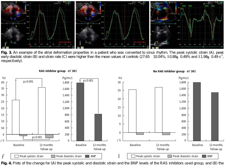
Fig. 3. An example of the atrial deformation properties in a patient who was converted to sinus rhythm. The peak systolic strain (A), peak early diastolic strain (B) and strain rate (C) were higher than the mean values of controls (27.65±10.04%, -0.88±0.49% and -1.98±0.49 s-1, respectively).
A B C
Fig. 4. Plots of the change for (A) the peak systolic and diastolic strain and the BNP levels of the RAS inhibitors used group, and (B) the peak systolic and diastolic strain and the BNP levels of the RAS inhibitors not used group. BNP:B-type natriuretic peptide, RAS: renin- angiotensin system.
30
25
20
15
10
5
0 -5
1,800 1,600 1,400 1,200 1,000 800 600 400 200 0 Baseline 12 months
follow up
Baseline 12 months follow up Peak systolic strain Peak diastolic strain BNP
No RAS inhibitor group (n=30)
B
40 35 30 25 20 15 10 50 0 -5
1,800 1,600 1,400 1,200 1,000 800 600 400 200 0 Baseline 12 months
follow up
Baseline 12 months follow up Peak systolic strain Peak diastolic strain BNP
RAS inhibitor group (n=30)
A
p<0.001
p<0.001
p<0.001 (%) (Pg/mL)
(Pg/mL) (%)
receptors in the atria is generally higher than that in the ventricles. Thus, angiotensin II-induced intracel- lular calcium overload may play a role not only in re- perfusion ventricular arrhythmias, but also in atrial el- ectrical remodeling.25)26) Therefore, RAS inhibitors would prevent or modify atrial remodeling by means of decreasing the atrial stretch, lowering the end-di- astolic left ventricular and atrial pressure, preventing atrial fibrosis and modifying the sympathetic tone, or modulating ion currents or the refractoriness. In a ca- nine rapid atrial pacing model, pharmacologic inhi- bition of endogenous angiotensin II prevented early electrical remodeling (this occurs within several hours),25) with a reduction of the AF duration and structural re- modeling during long-term stimulation.26) In the pre- sent study, the chronic atrial fibrillation patients who were treated with RAS inhibitors had significantly in- creased values of atrial peak systolic and diastolic strain/
rate, as compared with the patients without treatment, and this showed that treatment with RAS inhibitors appears to preserve the LA reservoir function in AF pa- tients without visible LA structural change. In addition, although there were too few to be statistically significant, the atrial fibrillation patients who were converted to normal sinus rhythm during the study showed more increased baseline atrial strain than did the permanent chronic atrial fibrillation patients, which would pos- sibly indicate that higher baseline atrial strain would be a predisposing factor for conversion to normal sinus rhythm in AF patients. The results of this investigation showed agreement with a previous study that reported patients with higher atrial strain and a higher strain rate appeared to have a greater likelihood of staying in sinus rhythm.19)
The atrial biopsy specimens of AF patients have dem- onstrated inflammatory changes and increased fibrosis, providing evidence of the possible role of inflammation in atrial structural remodeling.21) The relation of CRP and interleukin-6 to the left atrial size and AF duration before cardioversion indicates that inflammation could have a role in the atrial structural changes, which in turn may contribute to, or be a result of atrial remod- eling. An elevated BNP level has also been shown in some studies to be associated with atrial dilatation and the BNP levels have been shown by various studies to decrease after direct current cardioversion of AF and restoration of normal sinus rhythm.27) In the present study, there was no significant change in the hsCRP level, yet the NT-pro-BNP levels were significantly re- duced in the patients who were treated with RAS inhibitors at the 12 month follow-up without any visi- ble reduction of the LA size.
This study carries several limitations. Tracking of the region of interest is still suboptimal in some pa- tients with hyperdynamic LV function and poor echo-
cardiographic imaging. It still takes from 5 to 10 minutes to achieve satisfactory tracking, and the interobserver and intraobserver variability were relatively high because the atrial wall is too thin to be properly analyzed. Al- though analysis of the echocardiography parameters in AF patients would be more accurate if this was done over 5 cycles, the strain curves were calculated for all patients over 3 cardiac cycles because of the complexity.
Finally, we did not consider the AF duration, so there would be differences for a shorter duration and a long- er duration of AF, and the comparison between the AF patients may also have been influenced by other medications, but we tried to control the differences. To the best of our knowledge, no previous study has ever compared the change of atrial function between medical treatments by performing strain echocardiography. Our results indicate that strain echocardiography provides clinically useful information on the changes of LA func- tion and remodeling between RAS inhibitor treatments.
Acknowledgments
This study was supported by a grant from the Korean Society of Circulation (Industrial-educational cooperation 2005).
REFERENCES
1) Krahn AD, Manfreda J, Tate RB, Mathewson FA, Cuddy TE.
The natural history of atrial fibrillation: incidence, risk factors, and prognosis in the Manitoba follow-up study. Am J Med 1995;
98:476-84.
2) Allessie MA, Boyden PA, Camm AJ, et al. Pathophysiology and prevention of atrial fibrillation. Circulation 2001;103:769-77.
3) van Wagoner DR, Nerbonne JM. Molecular basis of electrical remodeling in atrial fibrillation. J Mol Cell Cardiol 2000;32:
1101-17.
4) Allessie MA. Atrial electrophysiological remodeling: another vicious circle? J Cardiovasc Electrophysiol 1998;9:1378-93.
5) Ravelli F, Allessie M. Effects of atrial dilatation on refractory period and vulnerability to atrial fibrillation in the isolated Lan- gendorff-perfused rabbit heart. Circulation 1997;96:1686-95.
6) Schnee JM, Hsueh WA. Angiotensin II, adhesion, and cardiac fibrosis. Cardiovasc Res 2000;46:264-8.
7) Ruiz-Ortega M, Lorenzo O, Ruperez M, et al. Role of the renin- angiotensin system in vascular diseases: expanding the field. Hy- pertension 2001;38:1382-7.
8) Willems R, Sipido KR, Holemans P, Ector H, van de Werf F, Heidbuchel H. Different patterns of angiotensin II and atrial na- triuretic peptide secretion in a sheep model of atrial fibrillation.
J Cardiovasc Electrophysiol 2001;12:1387-92.
9) Pedersen OD, Bagger H, Kober L, Torp-Pedersen C. Trandolapril reduces the incidence of atrial fibrillation after acute myocardial infarction in patients with left ventricular dysfunction. Circulation 1999;100:376-80.
10) Chung MK, Martin DO, Sprecher D, et al. C-reactive protein ele- vation in patients with atrial arrhythmias. Circulation 2001;104:
2886-91.
11) Hwang SJ, Kim BJ, Shin HS, et al. Assessment of factors in- fluencing plasma BNP level in patients with chronic atrial fibril- lation and preserved left ventricular systolic function. Korean Circ J 2005;35:605-12.
12) Hatle L, Sutherland GR. Regional myocardial function: a new
approach. Eur Heart J 2000;21:1337-57.
13) Cho KI, Lee HG, Kim TI, et al. Quantitative assessment of left atrial function changes in patients with atrial fibrillation by tissue Doppler strain and 2-dimensional strain imaging. Korean Circ J 2006;36:786-93.
14) Korinek J, Wang J, Sengupta PP, et al. Two-dimensional strain: a Doppler-independent ultrasound method for quantitation of re- gional deformation: validation in vitro and in vivo. J Am Soc Echo- cardiogr 2005;18:1247-53.
15) Barbier P, Alioto G, Guazzi MD. Left atrial function and ven- tricular filling in hypertensive patients with paroxysmal atrial fi- brillation. J Am Coll Cardiol 1994;24:165-70.
16) Sutherland GR, Hatle L, Rademakers FE, et al. Doppler Myocar- dial Imaging: A Textbook. Leuven, Belgium: Leuven University Press;2003. p.99-107.
17) Di Salvo G, Pacileo G, Del Giudice EM, et al. Atrial myocardial deformation properties in obese nonhypertensive children. J Am Soc Echocardiogr 2008;21:151-6.
18) Thomas L, Levett K, Boyd A, Leung DY, Schiller NB, Ross DL.
Changes in regional left atrial function with aging: evaluation by Doppler tissue imaging. Eur J Echocardiogr 2003;4:92-100.
19) Di Salvo G, Caso P, Lo Piccolo R, et al. Atrial myocardial de- formation properties predict maintenance of sinus rhythm after ex- ternal cardioversion of recent-onset lone atrial fibrillation: a color Doppler myocardial imaging and transthoracic and transesoph- ageal echocardiographic study. Circulation 2005;112:387-95.
20) Hobbs WJ, van Gelder IC, Fitzpatrick AP, Crijns HJ, Garratt CJ.
The role of atrial electrical remodeling in the progression of focal atrial ectopy to persistent atrial fibrillation. J Cardiovasc Electrophysiol 1999;10:866-70.
21) Hwang GS, Kim YH, Lee HS, et al. Electrophysiologic pro- perties of the atrium in patients with chronic and paroxysmal atrial fibrillation. Korean Circ J 2000;30:448-56.
22) Frustaci A, Chimenti C, Bellocci F, Morgante E, Russo MA, Ma- seri A. Histological substrate of atrial biopsies in patients with lone atrial fibrillation. Circulation 1997;96:1180-4.
23) Li D, Fareh S, Leung TK, Nattel S. Promotion of atrial fibrilla- tion by heart failure in dogs: atrial remodeling of a different sort.
Circulation 1999;100:87-95.
24) Sadoshima J, Izumo S. The cellular and molecular response of cardiac myocytes to mechanical stress. Annu Rev Physiol 1997;
59:551-71.
25) Nakashima H, Kumagai K, Urata H, Gondo N, Ideishi M, Ara- kawa K. Angiotensin II antagonist prevents electrical remodeling in atrial fibrillation. Circulation 2000;101:2612-7.
26) Kumagai K, Nakashima H, Urata H, Gondo N, Arakawa K, Saku K. Effects of angiotensin II type 1 receptor antagonist on electri- cal and structural remodeling in atrial fibrillation. J Am Coll Cardiol 2003;41:2197-204.
27) Wazni OM, Martin DO, Marrouche NF, et al. Plasma B-type natriuretic peptide levels predict postoperative atrial fibrillation in patients undergoing cardiac surgery. Circulation 2004;110:
124-7.