INTRODUCTION
Renal ischemia and reperfusion (I/R) injury is a common prob- lem in clinical surgeries, such as renal transplantation, and pro- longed ischemia results in irreversible damage to renal function.
1Studies have reported that apoptosis is essential to the patho- genesis of renal injury secondary to I/R.
2,3Proximal tubular ep- ithelial cells (PTCs) are particularly vulnerable to hypoxia-in- duced injury. More importantly, the vital pathogenesis of renal
I/R has been widely considered as renal tubular epithelial cells apoptosis.
4The close connection between renal tubular epithe- lial cell apoptosis and renal I/R injury has drawn substantial at- tention in developing anti-apoptosis therapies.
Propofol (2,6-disopropylphenol), an intravenous sedative- hypnotic agent well-known for its utilization in anesthesia, has also been demonstrated to alleviate I/R injury due to its anti-in- flammatory and anti-oxidant activities.
5However, the precise mechanism thereof remains largely unresolved. Meanwhile, au- tophagy is an evolutionarily conserved multi-step process that maintains normal function and the structure of the cell and is reported to be involved in renal I/R injury.
6Recent research re- ported that propofol attenuated hypoxia/reoxygenation-trig- gered neuronal injury via inhibition of autophagy.
7However, little is known about the underlying mechanisms by which pro- pofol regulates autophagy.
c-Jun N-terminal kinase (JNK, including JNK 1, 2, 3), also known as stress-activated protein kinase, is another subclass of the mi- togen-activated protein kinase signaling pathway in mamma-
Propofol Attenuates Hypoxia/Reoxygenation-Induced Apoptosis and Autophagy in HK-2 Cells
by Inhibiting JNK Activation
Huaxin Wang, Xuan Peng, Yayi Huang, Yeda Xiao, Zhuo Wang, and Liying Zhan
Department of Anesthesiology, Wuhan University Renmin Hospital, Wuhan, China.
Purpose: The aim of this study was to investigate whether propofol could attenuate hypoxia/reoxygenation-induced apoptosis and autophagy in human renal proximal tubular cells (HK-2) by inhibiting JNK activation.
Materials and Methods: HK-2 cells were treated with or without propofol or JNK inhibitor SP600125 for 1 hour and then subject- ed to 15 hours of hypoxia and 2 hours of reoxygenation (H/R). Cell viability and LDH release were measured with commercial kits.
Cell apoptosis was evaluated by flow cytometry. The expressions of p-JNK, cleaved-caspase-3, Bcl-2, and autophagy markers LC3 and p62 were measured by Western blot or immunofluorescence.
Results: HK-2 cells exposed to H/R insult showed higher cell injury (detected by increased LDH release and decreased cell viabil- ity), increased cell apoptosis index and expression of cleaved-caspase-3, a decrease in the expression of Bcl-2 accompanied by increased expression of p-JNK and LC3II, and a decrease in expression of p62. All of these alterations were attenuated by propofol treatment. Similar effects were provoked upon treatment with the JNK inhibitor SP600125. Moreover, the protective effects were more obvious with the combination of propofol and SP600125.
Conclusion: These results suggest that propofol could attenuate hypoxia/reoxygenation induced apoptosis and autophagy in HK-2 cells, probably through inhibiting JNK activation.
Key Words: Propofol, HK-2 cells, apoptosis, autophagy
pISSN: 0513-5796 · eISSN: 1976-2437
Received: July 23, 2019 Revised: September 13, 2019 Accepted: September 23, 2019
Corresponding author: Liying Zhan, MD, Department of Anesthesiology, Wuhan University Renmin Hospital, No. 238, Jiefang Road, Wuhan 430060, Hubei, China.
Tel: 86-027-88041911-81026, Fax: 86-027-88042292, E-mail: [email protected]
•The authors have no potential conflicts of interest to disclose.
© Copyright: Yonsei University College of Medicine 2019
This is an Open Access article distributed under the terms of the Creative Com- mons Attribution Non-Commercial License (https://creativecommons.org/licenses/
by-nc/4.0) which permits unrestricted non-commercial use, distribution, and repro- duction in any medium, provided the original work is properly cited.
Yonsei Med J 2019 Dec;60(12):1195-1202
https://doi.org/10.3349/ymj.2019.60.12.1195
lian cells. JNK is closely related to inflammation, cell apoptosis, and autophagy,
8and excessive activation of JNK has been im- plicated in nephropathy and renal I/R injury.
9,10Accordingly, we hypothesized that propofol could attenuate hypoxia/reoxygen- ation-induced apoptosis and autophagy in human renal proxi- mal tubular cells (HK-2) by inhibiting JNK activation.
MATERIALS AND METHODS
Cell culture and treatments
HK-2 cells were purchased from Procell Life Science & Technol- ogy Co., Ltd. (Wuhan, China). The cells were cultured in mini- mum essential medium supplemented with 10% fetal bovine serum both from Gibco/Thermo Fisher Scientific (Shanghai, Chi- na) and 1% penicillin and streptomycin under a humidified at- mosphere consisting of 5% CO
2and 95% air at 37°C (control group). Hypoxia and reoxygenation (H/R) injury was introduced by exposing the cells to hypoxic conditions (1% O
2, 5% CO
2and 94% N
2) for 15 h, followed by reoxygenation under normoxic conditions (reoxygenation) for 2 h in fresh normal medium (H/R group).
Propofol (Aladdin Reagent Int., Shanghai, China) was dissolved in dimethyl sulfoxide (DMSO) and subsequently diluted in min- imum essential medium to final concentrations of 10, 25, 50, and 100 μM prior to H/R injury (propofol groups); the final con- centrations of DMSO were below 0.01%, thereby minimizing its effects. A positive control was set up using cells pretreated with 10 μM SP600125 (MedChemExpress, Shanghai, China), which is a specific JNK inhibitor. Finally, a combination of 50 μM pro- pofol and 10 μM SP600125 was added to the cell cultures at 1 h before H/R injury to ascertain synergistic effects.
Cell viability analysis
Cell viability was assessed with Cell Counting Kit-8 from Biosharp (Guangzhou, China). Briefly, after reoxygenation for 2 h, each well was supplemented with 10 μL of CCK-8 solution, followed by 4 h of incubation at 37°C. Subsequently, optical density of each well was measured with a microplate reader at a wavelength of 450 nm (MULTISKAN MK3, Thermo, USA). Higher cell via- bility leads to higher optical density reading.
Measurement of lactate dehydrogenase by ELISA A lactate dehydrogenase (LDH) release assay (Changchun Huili Biotech Co., Ltd, Changchun, China) was used to determine the extent of cell injury by measuring the amount of LDH released into the medium by the cells following the manufacturer’s guid- ance.
Flow cytometry analysis
Flow cytometry analysis was carried out to assess apoptotic in- dex values in HK-2 cells. Following treatment, HK-2 cells were detached from wells by addition of 0.25% trypsin, resuspended
in phosphate-buffered saline (PBS), and spun down at 1500 rpm for 5 mins. Subsequently, the cells were resuspended in 500 μL of binding buffer, followed by incubation with Annexin V-FITC (5 μL) and propidium iodide (PI, 5 μL) for 10–15 mins at room temperature (20–25°C) in the dark. Apoptotic cell ratio was de- tected using a flow cytometer (CytoFLEX, Beckman Coulter Life Sciences, Indianapolis, IN, USA). Early apoptotic cells were dif- ferentiated by unique characteristics: positive for Annexin V- FITC and negative for PI.
Western blotting
Cell cultures were harvested and homogenized in ice-cold ra- dioimmunoprecipitation assay buffer with protease inhibitors (Beyotime, Shanghai, China) and incubated for 30 mins at 4°C.
After centrifugation, the supernatants, which contained our tar- get proteins, were transferred to a new vial for storage at -70°C or immediate use. A BCA protein assay kit was then used to quan- tify the concentrations of proteins (Beyotime, Shanghai, China).
The proteins (40 μg) were run on 12% SDS-PAGE gel and then transferred electrophoretically to a polyvinylidene fluoride mem- brane (Millipore, Shanghai, China). The blots were blocked for 2 h at 25°C with 5% skim milk in Tris-buffered saline containing 0.1% Tween 20 (TBST), followed by incubation with primary an- tibodies, including anti-JNK, anti-p-JNK, anti-caspase-3, anti- Bcl-2, anti-LC3, anti-p62, and anti-β-actin antibodies (Protein- tech Group, Inc., Wuhan, China) at 4°C overnight. Subsequently, membranes were washed with TBST five times for 5 mins each.
Then, respective horseradish peroxidase-linked secondary an- tibodies (Beyotime, Shanghai, China) were added and incubat- ed for 2 h at 37°C, followed by washing with TBST. Lastly, ECL solution was applied to the membrane evenly prior to detection of chemiluminescence (Thermo Scientific, Shanghai, China).
Immunofluorescence assay
The HK-2 cells were fixed in 4% paraformaldehyde for 15 mins and washed three times with PBS for 3 mins each time. Then, the samples were treated with 0.5% Triton X-100 for 20 mins, washed, and blocked for 30 mins at 37˚C. Next, the samples were incubated in a wet box at 4°C overnight with primary antibod- ies against LC3 (mouse anti-LC3 antibody or p62 (rabbit anti- p62 antibody) diluted 100 times in blocking solution. After wash- ing with PBS containing 0.1% Tween 20, the cells were incubated with secondary antibodies diluted 100 times (LC3, Cy3-labeled goat anti-mouse IgG; p62, Cy3-labeled goat anti-rabbit IgG) for 1 h in the dark at 37˚C. Lastly, 4,6-diamidino-2-phenylindole (Beyotime, C1002) was used as a counterstain. After staining, cells were visualized with a fluorescence microscope (BX53, Olym- pus, Tokyo, Japan).
Statistics analysis
Statistical analyses were conducted with GraphPad Prism ver-
sion 6 (GraphPad Software Inc., San Diego, CA, USA). Each ex-
periment was repeated six times. Results are presented as mean±
standard deviation (SD). Statistical significance was analyzed first with one-way analysis of variance and then with Tukey’s multiple comparisons test. p values less than 0.05 were consid- ered to represent statistically significant differences.
RESULTS
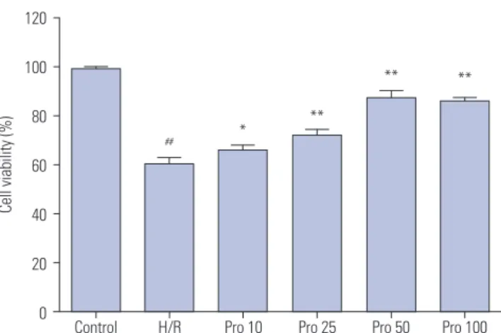
Propofol alleviates the reduced cell viability induced by H/R injury
In the present study, we investigated the effect of H/R insult on HK-2 cells. As shown in Fig. 1, H/R decreased cell viability by 39%, compared with the no-insult control group (p<0.001). Propofol significantly alleviated the decrease in cell viability induced by H/R insult in a dose-dependent manner (H/R vs. Pro 10, p=0.045;
H/R vs. Pro 25, p<0.001; H/R vs. Pro 50, p<0.001; H/R vs. Pro 100, p<0.001). However, a peak increase in cell viability was observed with pretreatment of propofol at a dose of 50 μM, probably due to the saturation of ligand binding (Fig. 1).
Propofol attenuates LDH release induced by H/R injury in HK-2 cells
We then evaluated the effects of propofol on LDH release in HK-2 cells. As shown in Fig. 2, H/R increased LDH release 3.8-fold over that in the control group, which was significantly attenuat- ed by propofol in a dose-dependent manner. Maximal attenu- ation of LDH release (1.5-fold that of the control group) was ob- served at a dose of 50 μM propofol. Interestingly, 100 μM propofol seemed to increase LDH leakage, compared to 50 μM propofol, although there was no statistical difference (p>0.05). The pos- sible cause was related to cytotoxicity from the high concentra- tion of propofol (Fig. 2).
Propofol pretreatment attenuates H/R induced cell apoptosis in HK-2 cells
The effects of propofol pretreatment were also determined by detecting cell apoptosis using flow cytometry. As shown in Fig. 3, H/R injury lead to a 3.5-fold increase in cell apoptosis, compared to the control group (p<0.001), and this alteration was reduced by propofol treatment (H/R vs. Pro 10, p=0.038; H/R vs. Pro 25, p=0.003; H/R vs. Pro 50, p<0.001; H/R vs. Pro 100, p<0.001). Con- sistently, peak reduction of cell apoptosis was obtained with treatment of 50 μM propofol. Based on the above results, we chose 50 μM propofol for subsequent experiments.
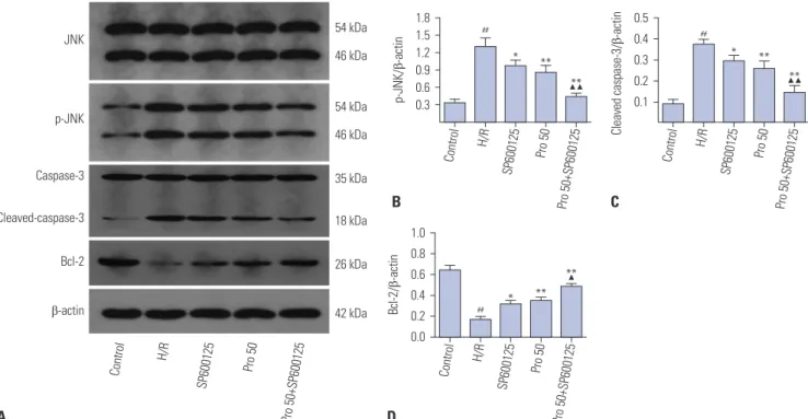
Propofol pretreatment attenuates H/R-triggered cell apoptosis of HK-2 cells by inhibiting JNK activation Given the essential role of JNK signal transduction pathway in cell apoptosis, we hypothesized that propofol would alleviate H/R-induced cell apoptosis in HK-2 cells by inhibiting JNK ac- tivation. To test this, we first ascertained the percentages of apop- totic cells in total cell cultures by measuring Annexin V-FITC and PI with flow cytometry (Fig. 4). As expected, we found that H/R
injury induced cell apoptosis, while pretreatment with 50 μM propofol alleviated the induced cell apoptosis (p<0.01). Similar effects were also observed with pretreatment of 10 μM SP600125 (p<0.01). Interestingly, a combination of 50 μM propofol and 10 μM SP600125 offered stronger protection against cell apop- tosis (p<0.01), indicating potentially synergistic effects (com- bination of 50 μM propofol and 10 μM SP600125 versus 50 μM propofol, p<0.05) We also examined the protein expressions lev- els of JNK, p-JNK, cleaved caspase-3, and Bcl-2 by Western blot- ting. As shown in Fig. 5, H/R injury resulted in elevated protein levels of p-JNK and cleaved caspase-3 and decreased protein levels of Bcl-2 (all p<0.01). All these changes were attenuated by pretreatment with 50 μM propofol or 10 μM SP600125 (all p<0.05), and more obvious attenuation was observed with a Fig. 1. Effects of propofol on hypoxia and reoxygenation (H/R)-induced cytotoxicity to HK-2 cells. Cell viability of the control group with neither H/
R injury nor propofol pretreatment was used as a 100% benchmark. H/R injury led to reduced cell viability (
##p<0.01 against the control group), and propofol pretreatment alleviated decreases in cell viability induced by H/
R injury (*p<0.05 and **p<0.01 against the H/R injury group). No signifi- cant difference was observed between pretreatments with 50 µM and 100 µM propofol (Pro 10, 10 µM; Pro 25, 25 µM; Pro 50, 50 µM; Pro 100, 100 µM).
120
100
80
60
40
20
0 Control H/R Pro 10 Pro 25 Pro 50 Pro 100
##
* **
** **
Cell viability (%)
Fig. 2. Effects of propofol on LDH leakage in HK-2 cells. LDH levels were measured for the same control and experimental groups as in Fig. 1. Con- sistently, hypoxia and reoxygenation (H/R) injury led to significantly higher levels of LDH (
##p<0.01 against the control group), and propofol pretreat- ment alleviated increases in LDH caused by H/R injury (**p<0.01 against H/R group).
1200
1000
800
600
400
200
0 Control H/R Pro 10 Pro 25 Pro 50 Pro 100
##
**
**
**
LDH (IU/L)
combination thereof (all p<0.01).
Propofol alleviates H/R-induced autophagy
To investigate whether propofol pretreatment attenuates the activation of H/R-induced autophagy, we also tested the levels of autophagy markers (LC3 and p62) via immunofluorescence and Western blot. As shown in Fig. 6A–C, the fluorescence in- tensity of anti-LC3II antibody was higher in the H/R group than that in the no-H/R control group (p<0.01), suggesting elevated
levels of autophagy. This alteration was partially reversed by 50 μM propofol or 10 μM SP600125 (50 μM propofol versus H/R, p<0.01; 10 μM SP600125 vs. H/R, p<0.01). Like our findings with apoptosis, the combination of propofol and SP600125 provided greater alleviation against H/R-induced autophagy (p<0.01).
In a similar vein, changes in the fluorescence intensity of anti- p62 antibody also suggested that propofol alleviated H/R in- duced autophagy (all p<0.05). Western blots further supported our hypothesis. Propofol administration significantly inhibited Fig. 3. Effects of propofol on apoptosis of HK-2 cells. Flow cytometry was conducted on the same control and experimental groups as in Fig. 1 (A: Control group; B: Hypoxia and reoxygenation (H/R) group; C: Pro 10 group; D: Pro 25 group; E: Pro 50 group; F: Pro 100 group). (G) Flow cytometric analysis for (A–F) was carried out as described in the Materials and Methods section. H/R injury increased the number of apoptotic cells (
##p<0.01 against the control group), and propofol pretreatment alleviated the observed increases (*p<0.05 and **p<0.01 against H/R group).
20
15
10
5
0 Control H/R Pro 10 Pro 25 Pro 50 Pro 100
##
* **
** **
% of cells Anenexin V+/PI-
G
PI-A 10²
10²
10
310
310
410
4FITC-A
10
510
510
610
610
7A
PI-A 10²
10²
10
310
310
410
4FITC-A
10
510
510
610
610
7D
PI-A 10²
10²
10
310
310
410
4FITC-A
10
510
510
610
610
7B
PI-A 10²
10²
10
310
310
410
4FITC-A
10
510
510
610
610
7E
PI-A 10²
10²
10
310
310
410
4FITC-A
10
510
510
610
610
7C
PI-A 10²
10²
10
310
310
410
4FITC-A
10
510
510
610
610
7F
autophagy as demonstrated by decreased LC3II and increased P62 expression. Moreover, SP600125 strengthened the benefi- cial effect of propofol in HK2 cells exposed to hypoxia/reoxy- genation injury (Fig. 6D–F; p<0.05).
DISCUSSION
To examine the effect of propofol on H/R injury and its underly- ing mechanism, an in vitro model was established by subjecting HK-2 cells to H/R insult in this study. Decades of experimental results have shown that PTCs play a vital role in the histopathol- ogy of renal injury, especially in I/R impairment.
11,12After an acute ischemic insult, PTCs are the most sensitive cells to I/R injury, which is mainly due to physiological hypoxia in the med- ullary region and the high metabolic activity of PTCs.
12In recent years, numerous studies have used PTC models of H/R to in- vestigate mechanisms of renal I/R injury or to search for drugs that lower the risk of I/R injury.
13,14Several studies have shown that propofol is an excellent anti-inflammation and anti-oxi-
dation agent, and may help to prevent and treat I/R injury,
15,16although the exact mechanisms remain unclear. We investigat- ed the protective effects and underlying mechanisms of propo- fol against H/R-induced injury in HK-2 cells in the absence or presence of the JNK inhibitor SP600125. H/R injury was eval- uated by measuring cell viability, LDH leakage, and apoptotic index. Studies have indicated that the clinically relevant con- centrations of propofol were approximately 11–56 μM.
17,18Thus, the concentration range of 10–100 μM was used in this study, which made our results more clinically relevant. Our present study found a dose-dependent increase in cell viability, as well as dose-dependent decreases in LDH release and cell apopto- sis, upon propofol treatment. All these indicate that propofol has a definite protective effect against H/R injury in HK-2 cells. Li, et al.
16reported that propofol prevented H/R injury in rat renal tubular epithelial cell line (NRK-52E cells), similar to our re- sults. Compared with 50 μM of propofol, 100 μM offered no ex- tra protection against H/R injury.
JNK is involved in cell apoptosis; however, it also acts syner- gistically with JAK/STAT, NF-κB, and other signaling molecules
PI-A 10²
10²
10
310
310
410
4FITC-A
10
510
510
610
610
7D
PI-A 10²
10²
10
310
310
410
4FITC-A
10
510
510
610
610
7E
Fig. 4. Potential synergistic effect of propofol and SP600125 on alleviating cell apoptosis induced by hypoxia and reoxygenation (H/R) injury. (A–E) Repre- sentative figures of flow cytometry results for (A) the control group with neither H/R injury nor propofol/SP600125 pretreatment, (B) the H/R injury group with no pretreatment, (C) the SP600125 group pretreated with 10 µM SP600125 prior to H/R injury, (D) the Pro 50 group pretreated with 50 µM propofol prior to H/R injury, and (E) the Pro 50+SP600125 group pretreated with a combination of 50 µM propofol and 10 µM SP600125 prior to H/R injury. (F) Flow cyto- metric analysis for (A–E) was carried out as described in the Materials and Methods section. H/R injury increased cell apoptosis significantly (
##p<0.01 against the control group), while a combination of propofol and SP600125 attenuated the increase in a potentially synergistic manner (*p<0.05 against H/R group; **p<0.01 against H/R group;
▲p<0.05 against Pro 50 group).
PI-A 10²
10²
10
310
310
410
4FITC-A
10
510
510
610
610
7A
PI-A 10²
10²
10
310
310
410
4FITC-A
10
510
510
610
610
7B
PI-A 10²
10²
10
310
310
410
4FITC-A
10
510
510
610
6C
% of cells Anenexin V+/PI-
F 20 15 10 5
Control H/R
SP600125 Pro 50
Pro 50+SP600125
##
*
**
▲*
to promote cancer cell survival.
19Studies have reported that JNK activation after H/R is excessively increased in human brain vas- cular smooth muscle cells,
20H9c2 cardiomyocytes,
21and HK-2 cells.
22In the present study, we selected the specific JNK inhib- itor SP600125 to ascertain the effect of propofol against H/R in- jury. Our results showed that propofol significantly attenuates H/R-induced cell apoptosis and JNK activation. Similar effects were shown by treatment with JNK inhibitor SP600125. These results indicated that the protective effects of propofol against H/R injury may be related to JNK inhibition. An interesting find- ing in our study was that the beneficial effects of the combined use of propofol and SP600125 were more obvious than use of only one them. These indicate their combination has better su- perimposed protective effects.
Autophagy is a critical mechanism for cellular homeostasis and survival in response to renal I/R injury. However, it is be- lieved to be a double-edged sword in cell injury. On the one hand, physiologic autophagy is involved in degradation of harmful pro- teins and damaged organelles to prevent the accumulation of harmful substances, which limits the transmission of harmful signaling. On the other hand, excessive autophagy will cause ir- reversible injury and transform cells to autophagic cell death.
To ascertain the role of autophagy in propofol-induced protec- tion, HK-2 cells were pretreated with propofol in the absence or presence of SP600125 prior to H/R injury. We showed that au- tophagy was significantly increased after H/R as indicated by a significant increase in LC3II protein levels and a decrease in p62
protein levels, and propofol attenuated these changes, which were further alleviated by a combination of propofol and SP600125.
Likewise, Xie, et al.
23reported that autophagosomes and autol- ysosomes were significant increased at 2 h after reoxygenation in HK-2 cells. According to previous reports, inhibition of the JNK pathway by SP600125 could block autophagy activation in can- cer cells.
24We showed here that SP600125 attenuated H/R-trig- gered autophagy activation. Although the exact mechanism by which propofol-inhibited autophagy protects from H/R injury is unclear, our results showed that it is likely that propofol in- hibits excessive autophagy during the injury partly through JNK pathway. It was confirmed that some situations, including over- digestion of cytoplasmic contents due to excessive autophagy, might result in autophagic cell death.
25Recent studies have re- ported that propofol antagonizes H/R-triggered neuronal inju- ry via inhibition of autophagy.
7Other studies have stated that propofol postconditioning protects H9c2 cells from H/R inju- ry by inducing autophagy via JNK pathway.
26This discrepancy could result from differences in the degree of autophagy acti- vation or test subjects.
More should be done in future studies to verify the protective effect of propofol on H/R injury particularly with in vivo assay.
Also, reoxygenation time may affect the expression levels of re- lated proteins after H/R injury,
14and in this study, we harvested the cells only at 2 h after reoxygenation due to limited resourc- es. Therefore, it would be informative to measure the effects of propofol against H/R injury at different reoxygenation times.
Fig. 5. Effects of propofol and SP600125 on the protein expression levels of JNK, p-JNK, caspase-3, cleaved-caspase-3, and Bcl-2 in HK-2 cells. Western blots were performed for the same control and experimental groups as in Fig. 4 to measure expression levels of the above-mentioned proteins. (A) Raw data. (B–D) Hypoxia and reoxygenation (H/R) injury increased the protein expression levels of p-JNK and cleaved-caspase-3, and decreased those of Bcl-2. Both SP600125 and propofol attenuated the changes in expression levels, and a combination of both alleviated the changes further (
##p<0.01 against the control group; *p<0.05 and **p<0.01 against the H/R group;
▲p<0.05 and
▲▲p<0.01 against the Pro 50 group).
p-JNK/ β-actin
B 1.8 1.5 1.2 0.9 0.6 0.3
Control H/R
SP600125 Pro 50
Pro 50+SP600125
##
*
**
▲▲
**
Cleaved caspase-3/ β-actin
C 0.5 0.4 0.3 0.2 0.1
Control H/R
SP600125 Pro 50
Pro 50+SP600125
##
*
**
▲▲
**
Bcl-2/ β-actin
D 1.0 0.8 0.6 0.4 0.2 0.0
Control H/R
SP600125 Pro 50
Pro 50+SP600125
##
*
**
▲**
Control H/R
SP600125 Pro 50
Pro 50+SP600125
54 kDa
54 kDa
35 kDa 46 kDa
46 kDa
18 kDa
26 kDa
42 kDa JNK
p-JNK
Caspase-3
Cleaved-caspase-3
Bcl-2
β-actin
A
Despite the limitations, our study has demonstrated unambig- uously that propofol helps to protect HK-2 cells against H/R in- jury by alleviating apoptosis and autophagy, and suggests that
the protective effects are derived from its inhibition of JNK ac- tivation.
Fig. 6. Effects of propofol and SP600125 on autophagy induced by hypoxia and reoxygenation (H/R) injury. The same control and experimental groups as in Fig. 4 were subjected to fluorescence microscopy (A–C) and Western blots (D–F) to measure the protein expression levels of LC3II and p62 (autophagy markers). Both techniques showed that H/R injury increased the protein expression levels of LC3II and decreased those of p62. Both SP600125 and pro- pofol attenuated the changes, and their alleviating effects were stronger when applied together (
##p<0.01 against the control group; *p<0.05 and **p<0.01 against the H/R group;
▲p<0.05 against the Pro 50 group).
LC3II/LC3I ratio
E 4 3 2 1 0
Control H/R
SP600125 Pro 50
Pro 50+SP600125
##
*
**
▲*
p62/ β-actin
F 0.7 0.6 0.5 0.4 0.3 0.2 0.1
Control H/R
SP600125 Pro 50
Pro 50+SP600125
##
*
**
▲*
Punctate p62 cells
C 0.18 0.15 0.12 0.09 0.06 0.03
Control H/R
SP600125 Pro 50
Pro 50+SP600125
##
*
**
▲*
Punctate LC3II cells
B 0.10 0.08 0.06 0.04 0.02
Control H/R
SP600125 Pro 50
Pro 50+SP600125
##
**
**
▲**
D
Control H/R
SP600125 Pro 50
Pro 50+SP600125 18 kDa
60 kDa 16 kDa
42 kDa LC3 I
LC3 II p62
β-actin
Control
H/R
SP600125
LC3II DAPI Merge p62 DAPI Merge
Pro 50
Pro 50+SP600125 A
20 µm 20 µm
20 µm
20 µm
20 µm
20 µm 20 µm
20 µm
20 µm
20 µm
20 µm 20 µm
20 µm
20 µm
20 µm
20 µm 20 µm
20 µm
20 µm
20 µm
20 µm 20 µm
20 µm
20 µm
20 µm
20 µm 20 µm
20 µm
20 µm
20 µm