INTRODUCTION
Retained foreign body (FB) is a complication of
Ultrasound-Guided Percutaneous Removal of Wooden Foreign Bodies in the Extremities with Hydro-Dissection Technique
Hee Jin Park, MD
1, Sung Moon Lee, MD
2, So Yeon Lee, MD
1, Eun Seok Son, MD
3, Eun Chul Chung, MD
1, Myung Ho Rho, MD
1, Sun Joo Lee, MD
41Department of Radiology, Sungkyunkwan University School of Medicine, Kangbuk Samsung Hospital, Seoul 03181, Korea; Departments of
2Radiology and 3Orthopaedic Surgery, Dongsan Medical Center, Keimyung University School of Medicine, Daegu 41931, Korea; 4Department of Radiology, Inje University College of Medicine, Busan Paik Hospital, Busan 47392, Korea
Objective: We described the technique of ultrasound (US)-guided percutaneous removal of the foreign bodies (FB) with hydro-dissection in the radiologic department and presented video files of several cases.
Materials and Methods: Four patients referred to the radiology department for US evaluation and US-guided percutaneous removal of the FBs in the upper and lower extremities between November, 2006 and November, 2013 were included in this study. The procedures started with US evaluation for the exact location and shape of the FB. A 5 mm-sized skin incision was made at the site of the nearest point from the FB where no passing arteries or tendons were present. We adopted a hydro- dissection technique to separate the FB from adjacent tissue using a 2% lidocaine solution. Injected anesthetics detached the FBs from surrounding tissue and thereby facilitated removal. After the tip of the mosquito forceps reached the FB, the wooden FBs were removed.
Results: The mean time required for the entire procedure was approximately 20 minutes. There were no significant complications during the US-guided removal or long-term complications after the procedure. All 4 FBs were successfully removed from the soft tissue under US guidance.
Conclusion: Ultrasound-guided percutaneous removal of the FBs with hydro-dissection in the radiology department is a less invasive and safe method over surgical removal in the operating room. Additionally, the use of a guide wire and serial dilator may help minimize soft tissue injury and facilitate the introduction of forceps.
Index terms: Ultrasound; Intervention; Foreign bodies; Surgery Korean J Radiol 2015;16(6):1326-1331
traumatic soft tissue injuries, including laceration and penetration injuries (1). FBs may be composed of wood, glass, metal, plastic, or gravel (1, 2). Because many FBs are asymptomatic and some are radiolucent, FB detection is occasionally difficult (1). According to a previous study, about 38% of the FBs are initially overlooked and may result in inflammation and infection (3).
It is well known that high-resolution and real-time ultrasound (US) is useful in the detection of the radiolucent FBs (4, 5). Additionally, US is an effective imaging
technique for localization of various types of embedded superficial FBs (5). Surgical removal of FBs in the operating room is an invasive and expensive procedure and sometimes
pISSN 1229-6929 · eISSN 2005-8330
Received May 27, 2015; accepted after revision August 19, 2015.
Corresponding author: Sung Moon Lee, MD, Department of Radiology, Dongsan Medical Center, Keimyung University School of Medicine, 56 Dalseong-ro, Jung-gu, Daegu 41931, Korea.
• Tel: (8253) 250-7767 • Fax: (8253) 250-7766
• E-mail: [email protected]
This is an Open Access article distributed under the terms of the Creative Commons Attribution Non-Commercial License (http://creativecommons.org/licenses/by-nc/3.0) which permits unrestricted non-commercial use, distribution, and reproduction in any medium, provided the original work is properly cited.
carries the risk of surgical complications (2). US-guided percutaneous removal of FBs in the radiology department, which can be concurrently performed with diagnostic US, has many advantages over a conventional surgical procedure owing to the fact that US-guided procedures permit
excellent visualization of major surrounding anatomic structures including arteries, veins, and nerves (1). The hydro-dissection technique, which can be used in carpal tunnel release or nerve release in meralgia paresthetica, is a very useful method for FBs that have been retained for an extended period of time and can be easily used during FB removal (4, 6).
In this article, we described the technique of US-guided percutaneous removal of the FBs with hydro-dissection in the radiologic department and presented video files of cases.
MATERIALS AND METHODS
Patients
Inclusion criteria were as follows: cases that did not show a FB on plain radiography, the shape of the FB was elongated and did not cause damage to adjacent normal tissue, and the FB had a safe removal tract. Exclusion criteria were glass or metallic FBs. Because glass FB could be broken into pieces, which makes it difficult to be removed and irregular shaped FBs such as metal or glass might damage adjacent neural or vascular tissue.
Four patients referred to the radiology department for US evaluation and US-guided percutaneous removal of the FBs in the upper and lower extremities between November, 2006 and November, 2013 were included in this study. The patients were referred by orthopedic surgeons. Two males and 2 females aged 10 to 74 years (mean 50.5 ± 28 years) were included. Radiographic examinations of the lesion were performed in all 4 patients. The histories of the patients and body location of the lesions were briefly described in
Table 1. The mean interval between trauma onset and US- guided FB removal was 55 days (range, 10–120 days). This research was approved by the Institutional Ethics Review Board of our medical center, and the requirement for informed consent was waived due to the retrospective study design.
US Evaluation and Removal of FBs
A musculoskeletal radiologist performed all diagnostic US and US-guided removal of the FBs using a Logiq E9 (GE Medical Systems, Milwaukee, WI, USA) imaging device equipped with linear 6–15 MHz probes. The diagnosis was confirmed by US by identifying FBs ranging from 10 mm to 40 mm. US-guided removal of the FBs in the radiology department was initiated with US evaluation for the exact location and shape of the FB. Adjacent major vascular structures and tendons were also evaluated. After disinfecting the overlying skin, local anesthetics (lidocaine chlorhydrate 2%, Huons, Jecheon, Korea) were injected around the targeted skin area. A 5 mm-sized skin incision was made at the site of the nearest point from the FB where no passing arteries or tendons were present. The incision was made large enough for mosquito forceps (Halsted- Mosquito Forceps, Solco Medical, Seoul, Korea, 12 cm) to be inserted and wide enough to accommodate the passage of the FB. We adopted a hydro-dissection technique to separate the FB from adjacent tissue using a 2% lidocaine solution (Figs. 1-3, Videos 1, 2 in the online-only Data Supplement). Injected anesthetics detached the FBs from surrounding tissue and thereby facilitated removal.
If the FBs were located in a deeper portion of the soft tissue, such as the intramuscular layer, an 18-gauge spinal needle was introduced, through which a guide wire (0.03 inch, Terumo, Tokyo, Japan) was inserted. After removal of the spinal needle, we created a tract for introducing mosquito forceps with serial dilators (7–12 Fr, Cook, Bloomington, IN, USA) along the guide wire. When the tip Table 1. Patient’s Data with Wooden Foreign Bodies Removed with Ultrasonography-Guidance and Hydro-Dissection Technique
Patient No. Sex Age (Year) Location
Size of Foreign Bodies (mm)
Material Kind Trauma History
Time Interval between Trauma and Removal (Day)
1 M 10 Knee 30 Wood Tree Sports injury 10
2 F 64 Wrist 20 Wood Toothpick Cleaning activity 60
3 M 54 Ankle 10 Wood Tree Construction site 120
4 F 74 Wrist 40 Wood Tree During seed harvest 30
of the mosquito forceps reached the FB, the wooden FBs were removed. All procedures were performed under real- time US guidance. The mean time required for the entire procedure was approximately 20 minutes. Prophylactic oral antibiotics (penicillin and amoxicillin) were administered to all patients for 5 days to prevent infectious complications after the procedure. There were no significant complications during the US-guided removal or long-term complications after the procedure.
RESULTS
Foreign bodies of all 4 patients that were undetected on plain radiography, were detected with US. The sizes of each lesion were described in Table 1. All 4 FBs were successfully removed from the soft tissue under US guidance. All FBs were pure wooden materials. There was 1 case of a FB around the knee joint referred from a local medical center (LMC) after incision and drainage of the subcutaneous fat
A
C
B
D
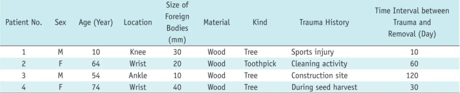
Fig. 1. Case of retained foreign body in 10-year-old male with complaint of knee pain after soccer.
A. Plain radiography reveals diffuse soft tissue swelling in pre-patellar area but discrete foreign body is not seen. B, C. Ultrasound shows linear echogenic foreign body (arrows) with posterior shadowing superficial to patellar tendon. There is skin defect (arrowheads) with sinus tract from distal end of foreign body. Foreign body was removed with ultrasound guidance and hydro-dissection techniques. See also Video 1 in the online- only Data Supplement. D. Removed wooden foreign body.
layer with the impression of focal abscess because the patient self-treated the lesion with local ointment himself for 3 days after a sports injury. The 30 mm-sized FB made of tree wood was located in the subcutaneous fat layer around the anteromedial portion of the patellar tendon (Fig.
1, Video 1 in the online-only Data Supplement). A case of FB in the wrist was referred from a LMC after intravenous antibiotic therapy for 2 months due to wrist infection, which developed after an injury during dish washing. A 20 mm-sized, toothpick-like wooden material was removed (Fig.
A
C
B
D
Fig. 2. Case of retained foreign body in 64-year-old female who complained of painful wrist swelling for 1 week. Patient had history of stab injury with toothpick.
A. Plain radiography reveals no discrete foreign body. B, C. Longitudinal (B) and transverse (C) ultrasonography show foreign body (arrows) surrounded by hypoechoic granulation tissue (asterisk). Foreign body was removed with ultrasound guidance and hydro-dissection techniques.
Open arrow: median nerve, arrowhead: palmaris longus tendon. D. Removed wooden foreign body.
2). A 54-year-old man was referred to our department with clinical impression of cellulitis. He was injured with a rusty nail at a construction site 4 months prior to presentation.
We removed a 10 mm-sized wooden FB from the left ankle and the patient was discharged from the hospital after intravenous antibiotics therapy for 1 month following the US-guided removal in the radiology department (Fig. 3, Video 2 in the online-only Data Supplement). The fourth case was a 74-year-old woman whose wrist was injured during the harvest of perilla seeds, which is a source of perilla oil rich in α-linolenic acid omega-3 fatty acids.
She was treated with intravenous antibiotics for 1 month because of purulent discharge. We removed a 4 cm-sized wooden FB from the wrist.
DISCUSSION
Failure to find FBs in the soft tissue can result in many complications such as allergies and infections (2, 7).
Retained FBs can cause peritendinitis, tenosynovitis and in some cases neuroma or neuropathies (2, 8). US is the first choice for detection of FBs in the soft tissue and has a sensitivity and specificity of more than 90% (9, 10). On US, FBs appear as echogenic lesions with posterior shadowing
(2). Chronic FBs are surrounded by a hypoechogenic halo, which results from granulomatous inflammation (11).
US also aids in the evaluation of surrounding ligaments, tendons, and neurovascular structures that could hinder the safe removal of FBs (11). Since surgical removal of FBs can result in severe bleeding and surgical infection, it is recommended that small FBs be removed with narrow entry hole under US-guidance (1, 2). US-guided FB removal from soft tissue is inexpensive, repeatable, and carries less risk.
Moreover, if US-guided FB removal fails, surgical removal can be performed subsequently. Skin incision of the US- guided removal is less than a centimeter and results in minimal scar formation, which can be a serious cosmetic problem when FBs are surgically removed (2). Our successful results are encouraging and suggest a good model of practice in the radiology department. The hydro-dissection technique originated from blind steroid injection in the carpal tunnel to release compression of the median nerve (12). This technique can separate FBs from compressive surrounding structures throughout the entire length of the FBs to facilitate removal. For safe removal, the creation of a tract using a guide-wire and serial dilators was necessary because relatively large mosquito forceps were needed to contact the shallow end of the FBs to minimize tissue injury
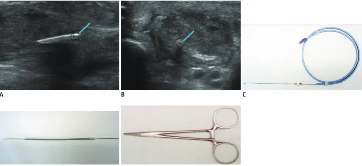
Fig. 3. Case of retained foreign body in 54-year-old male who complained of painful foot swelling for duration of 4 months. He received stab injury at construction site and removed rusted nail at time of original injury.
A, B. Longitudinal (A) and transverse (B) ultrasounds show echogenic foreign body (between markers, arrows) in abductor halluces longus muscle. As foreign body was located in deep area and adhesion with surrounding soft tissue was suspected due to long term presence of FB, serial dilators with guide wire were employed. C. Guide wire was inserted through 18-G spinal needle. D. After removal of 18-G spinal needle, tract was dilated with 7 to 12 Fr serial dilators introduced along guide wire. E. Mosquito forceps (Halsted-Mosquito Forceps, 12 cm) were used to remove FB. FB = foreign body
A B
D E
C
during removal of the FBs. No serious complication such as massive bleeding occurred during the creation of a tract.
One limitation to this study was the small number of cases. Another limitation was a lack of long-term follow- up results. We could not evaluate whether unexpected complications developed after discharge from the hospital more than 6 month after the procedure because all patients were transferred back to the original referring LMCs. The third limitation was that our cases were limited to subacute or chronic stage of FB. However, Budhram and Schmunk (1) reported that they could safely remove acute stage (less than 2 hours) cutaneous FBs with US-guided bed side procedure in the emergency department. It is likely that the time interval from the injury to removal of the FB is not critical.
In conclusion, the US-guided percutaneous removal of the FBs with hydro-dissection in the radiology department is a less invasive and safe method over surgical removal in the operating room. Additionally, the use of a guide wire and serial dilator may help minimize soft tissue injury and facilitate the introduction of forceps.
Acknowledgments
We declare that one of the patients (patient number 3) was reported previously in J Korean Orthop Assoc 2012;
47:150-155, however, this article mainly deals with video file of FB removal in the radiologic department with multiple cases. This article contains none of the same image files or paragraphs used in the previous article.
Supplementary Materials
The online-only Data Supplement is available with this article at http://dx.doi.org/10.3348/kjr.2015.16.6.1326.
REFERENCES
1. Budhram GR, Schmunk JC. Bedside ultrasound AIDS identification and removal of cutaneous foreign bodies: a case series. J Emerg Med 2014;47:e43-e48
2. Callegari L, Leonardi A, Bini A, Sabato C, Nicotera P, Spano’ E, et al. Ultrasound-guided removal of foreign bodies: personal experience. Eur Radiol 2009;19:1273-1279
3. Anderson MA, Newmeyer WL 3rd, Kilgore ES Jr. Diagnosis and treatment of retained foreign bodies in the hand. Am J Surg 1982;144:63-67
4. Lee SM, Cho CH. Ultrasound-guided percutaneous removal of foreign body using hydrodissection and serial dilators. J Korean Orthop Assoc 2012;47:150-155
5. Nwawka OK, Kabutey NK, Locke CM, Castro-Aragon I, Kim D.
Ultrasound-guided needle localization to aid foreign body removal in pediatric patients. J Foot Ankle Surg 2014;53:67-70 6. Mulvaney SW. Ultrasound-guided percutaneous neuroplasty of
the lateral femoral cutaneous nerve for the treatment of meralgia paresthetica: a case report and description of a new ultrasound- guided technique. Curr Sports Med Rep 2011;10:99-104 7. Peterson JJ, Bancroft LW, Kransdorf MJ. Wooden foreign
bodies: imaging appearance. AJR Am J Roentgenol 2002;178:557-562
8. Choudhari KA, Muthu T, Tan MH. Progressive ulnar neuropathy caused by delayed migration of a foreign body. Br J Neurosurg 2001;15:263-265
9. Jacobson JA, Powell A, Craig JG, Bouffard JA, van Holsbeeck MT. Wooden foreign bodies in soft tissue: detection at US.
Radiology 1998;206:45-48
10. Bray PW, Mahoney JL, Campbell JP. Sensitivity and specificity of ultrasound in the diagnosis of foreign bodies in the hand.
J Hand Surg Am 1995;20:661-666
11. Boyse TD, Fessell DP, Jacobson JA, Lin J, van Holsbeeck MT, Hayes CW. US of soft-tissue foreign bodies and associated complications with surgical correlation. Radiographics 2001;21:1251-1256
12. Malone DG, Clark TB, Wei N. Ultrasound-guided percutaneous injection, hydrodissection, and fenestration for carpal tunnel syndrome: description of a new technique. J Appl Res 2010;10:116-123