334
MATERIALS AND METHODS Population
A total of 168 premature neonates whose birth weight ≤1500 g or gestational age ≤34 weeks were examined by cranial ultra- sound (CUS) for detection of GM-IVH among the babies admit- ted between January 2011 and December 2012 in our medical center neonatal intensive care unit. Antenatal steroids were inject- ed to all premature neonates. Patient’s data were collected retro- spectively such as sex, gestational age, birth weight, delivery meth- od, disease diagnosis and its grade, initial blood glucose, sodium levels and presence of comorbidities such as respiratory distress syndrome (RDS), patent ductus arteriosus (PDA) and anemia.
And maternal data were collected retrospectively such as mater- nal age, gravidity, presence of preeclampsia and premature rupture of membrane (PROM).
INTRODUCTION
Germinal matrix-intraventricular hemorrhage (GM-IVH) is one of the most common complications in the premature in- fant. The incidence of this disease has been decreased in recent years. But it is still important because of two reasons. First, the outbreak is directly related with the degree of prematurity. Sec- ond, survival rate for smaller premature infants increases con- sistently. Therefore, there are many previous reports in order to identify risk factors associated with development of GM-IVH and to establish effective strategies. Perinatal risk factors such as a low birth weight and low gestational age, vaginal delivery, in- trauterine infection, low Apgar score, sepsis have been proposed as associated with the pathogenesis of GM-IVH16). Therefore, the purpose of this study is to identify the risk factors associated with the development of GM-IVH and the relationship of the severity of disease and prematurity.
Risk Factors Associated with Germinal Matrix-Intraventricular Hemorrhage in Preterm Neonates
Kwang-Ryeol Kim, M.D., Sang-Won Jung, M.D., Dong-Won Kim, M.D., Ph.D.
Department of Neurosurgery, Dongsan Medical Center, Keimyung University School of Medicine, Daegu, Korea
Objective : The purpose of this study is to identify the risk factors associated with the development of germinal matrix-intraventricular hemorrhage (GM-IVH) and the relationship of the severity of disease and prematurity.
Methods : A total of 168 premature neonates whose birth weight ≤1500 g or gestational age ≤34 weeks were examined by cranial ultrasound (CUS) for detection of GM-IVH among the babies admitted between January 2011 and December 2012 in our medical center neonatal intensive care unit. The babies were divided into two groups : GM-IVH and non-IVH. Clinical presentations, precipitating factors of the patients and maternal factors were analyzed.
Results : In univariate analysis, gestational age, birth weight, delivery method, presence of premature rupture of membrane (PROM) and level of so- dium and glucose were statistically meaningful factors (p<0.05). But only two factors, gestational age and presence of patent ductus arteriosus (PDA) were statistically meaningful in multivariate logistic regression (p<0.05). Delivery method [normal vaginal delivery (NVD) to Caeserean section]
was borderline significant (p<0.10).
Conclusion : Presence of PDA and gestational age were the important risk factors associated with development of GM-IVH.
Key Words : Germinal matrix-intraventricular hemorrhage · Gestational age · Ductus arteriosus, patent.
Clinical Article
•Received : June 26, 2014 •Revised : August 31, 2014 •Accepted : September 18, 2014
•Address for reprints : Dong-Won Kim, M.D., Ph.D.
Department of Neurosurgery, Dongsan Medical Center, Keimyung University School of Medicine, 194 Dalseong-ro, Jung-gu, Daegu 700-712, Korea Tel : +82-53-250-7333, Fax : +82-53-250-7356, E-mail : [email protected]
•This is an Open Access article distributed under the terms of the Creative Commons Attribution Non-Commercial License (http://creativecommons.org/licenses/by-nc/3.0) which permits unrestricted non-commercial use, distribution, and reproduction in any medium, provided the original work is properly cited.
J Korean Neurosurg Soc 56 (4) : 334-337, 2014
http://dx.doi.org/10.3340/jkns.2014.56.4.334
Copyright © 2014 The Korean Neurosurgical Society Print ISSN 2005-3711 On-line ISSN 1598-7876
www.jkns.or.kr
online © ML Comm
335
Risk Factors Associated with GM-IVH in Preterm Neonates | KR Kim, et al.
Detection of IVH and grading system
CUS was performed for all neonates in this study in the first week of life for detection of IVH and its grading were done. If a neonate had GM-IVH, then serial CUS would be done weekly.
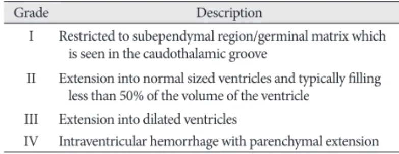
If not, follow up CUS was taken after 2 weeks. All subjects were divided into two groups–GM-IVH group and Non-IVH groups according to presence of GM-IVH. GM-IVH group was subdi- vided by grading system proposed by Papile et al.19) (Table 1).
Grade I and II group were considered as mild GM-IVH group, Grade III and IV group were as severe GM-IVH group.
Statistical analysis
Statistical analysis was performed by using SPSS version 21.0 (SPSS, Chicago, IL, USA). To compare the continuous variables, the Student’s t-test was used. Pearson’s chi-square test was used for categorical variables. Logistic regression analysis was used to find the independent association between development of GM- IVH and explanatory variables. Variables that were p≤0.20 as de- termined by univariate analysis were entered into multivariate logistic regression. The odds ratios (ORs) with a 95% confidence intervals (CIs) were calculated. p value<0.05 was considered sta- tistically significant.
RESULTS
General characteristics
Total 168 patients were included. 93 patients (55.36%) were male.
Infants born from mother with multigravida were 44 (26.19%).
Mean maternal age was 32 years. Mean gestational age and birth weight are 221 days and 1668 g, respectively. 35 babies (20.83%) were born by normal vaginal delivery (NVD). The mothers who had past history were 31 cases (18.45%) for preeclampsia and 44 cases (26.19%) for PROM. 34 babies (20.24%) had PDA, 95 babies (56.55%) with RDS, 67 babies (39.88%) with anemia. Among 168 subjects, GM-IVH was diagnosed in 31 cases (18.45%); 15 cases (48.39%) with grade 1, 9 cases (29.03%) with grade 2, 2 cases (6.45%) with grade 3, 5 cases (16.13%) with grade 4.
Univariate analysis
In univariate analysis, gestational age, birth weight, delivery method, presence of PROM and level of sodium and glucose were statistically meaningful factors (p<0.05). To operate multivariate analysis, gestational age, birth weight, delivery method, presence of PROM, PDA, RDS, anemia and level of sodium and glucose were selected. All these results were demonstrated on Table 2.
Multivariate analysis
Only two factors–gestational age and presence of PDA were statistically meaningful in multivariate logistic regression (p<
0.05). Delivery method (NVD to Caeserean section) was border- line significant (p<0.10). All these results were demonstrated in Table 3.
Comparison of mild and severe IVH group
Unpaired t-test was done between two groups by variables. Mild group was 24 cases, severe groups was 7 cases. Meaningful differ- ence in gestational age between two groups was revealed (p<0.05).
No other variable was statistically meaningful factor.
DISCUSSION
GM-IVH is rare in full-term babies11), but more common in preterm babies and it leaves significant sequelae such as cerebral Table 1. Papile’s classification of preterm intraventricular hemorrhage19)
Grade Description
I Restricted to subependymal region/germinal matrix which is seen in the caudothalamic groove
II Extension into normal sized ventricles and typically filling less than 50% of the volume of the ventricle
III Extension into dilated ventricles
IV Intraventricular hemorrhage with parenchymal extension
Table 2. Comparison of neonatal and maternal characteristics between two groups
Variables Total (n=168) IVH (n=31) Non-IVH (n=137) p value
Sex (M : F) 93 : 75 18 : 13 75 : 62 0.737
Multigravida 44 08 36 0.957
Maternal age (year) 32.63±3.93 31.87±3.41 32.80±4.03 0.237
Gestational age (weeks) 221.66±19.91 199.32±22.77 226.72±15.27 0.000
Weight (g) 1668.93±540.83 1222.58±516.27 1769.93±494.66 0.000
Delivery method (normal delivary vs. caesarean section) 35 : 133 12 : 19 23 : 114 0.009
Preeclampsia 31 06 25 0.886
PROM 44 13 31 0.030
PDA 34 13 21 0.001
RDS 95 22 73 0.077
Anemia 67 16 51 0.143
Sodium (mEq/L) 140.71±3.920 138.03±5.830 141.32±3.060 0.000
Glucose (mg/dL) 0069.78±44.860 099.90±54.53 062.96±39.51 0.000
Data presented as number or mean±standard deviation. *p value was calculated by logistic regression. IVH : intraventricular hemorrhage, PROM : premature rupture of membrane, PDA : patent ductus arteriosus, RDS : respiratory distress syndrome
336 J Korean Neurosurg Soc 56 | October 2014
palsy and mental retardation. Therefore, it is very important to find the risk factors associated with development of GM-IVH and prepare for them. Many risk factors for the development of GM-IVH have been reported. These contain gender, lack of an- tenatal steroids, low Apgar score, PROM, intrauterine infection, vaginal delivery, in vitro fertilization, mechanical ventilation, a large PDA, RDS and transfusion of blood products7,12,14,16,18,25,26).
However, in our study, there was no significant difference in gender, birth weight, delivery method, preeclampsia, PROM, anemia. The only two factors, gestational age and presence of PDA, were statistically meaningful risk factors for development of GM- IVH. This result could be understood easily because gestational age reflects fetal maturity more accurately than birth weight. As several reports15,21,23) and our results has shown, it is certain that younger gestational age is related with higher risk for severe GM- IVH. It can be described in terms of the anatomy and pathophys- iology of it. It occurs in germinal matrix, an intensively vascular- ized glioblast tissue13). If children were born before the 32 weeks of gestation, the subependymal region is equipped with a tight net of capillaries that are mainly supplied by the Heubner’s ar- tery. After the 32 weeks into pregnancy, the germinal matrix in- volutes and the vessels will be differentiated10). Preterm infants are quite helpless against cerebrovascular injury due to a unique constellation of pathophysiological factors. Preterm newborns have difficulty maintaining an adequate cerebral perfusion pres- sure. Maintain cerebral perfusion pressure is more difficult due to normally reduced hypotension and low cardiac output for the new borns to adjst extrauterine life, especially in the first day of life. In addition, intrinsic cerebral vasoreactivity and autoregula- tory mechanisms are poorly developed in the immature brain. As decreasing gestational age, the autoregulation pressure range is narrower and lower5).
Normal vaginal delivery was borderline significant risk factor (p<0.1). Although there is a objection20,21,24), some retrospective studies reported that Caesarean delivery does not improve neo- natal survival of very low birth weight infants but decreases GM- IVH occurrence2,4,17,27). In addition, following hypoperfusion- reperfusion hypothesis seemed to support this5). The germinal matrix is a transient neural cell proliferative zone with poor de-
veloped vasculature and located on the head of caudate nucleus.
This involutes gradually in third trimester. Fragile vessels and the lack of cerebrovascular resistance make GM-IVH1,22). The re- sistance to ischemic injury is weaker and autoregulation range of blood pressure is lower as decreasing gestational age. Certain situ- ation like presence of PDA or normal vaginal delivery could re- sult in hypoperfusion-reperfusion state. Several possible factors are suggested cardiorespiratory system (hypotension, PDA, hy- poperfusion-reperfusion pattern, hypercarpnia, hypoglycemia, hypernatremia), hematologic (anemia, thrombocytopenia), im- munologic response, impared cerebral reactivity, immature anat- omy, eventhough they are not proven yet1,5,7,10,12-14,16,18,22,25,26). An- other reason why the hemorrhage occurs in this region, beside the massive vascularization, is the fact that the area around the caudate nucleus represents a border zone between ventriculop- etal and fugal blood supply3). Furthermore, the endothelium of the vessels located in the brain of premature newborns is more sensitive to hypoxemia. Hydrostatic and osmotic changes lead more often to the ruptures of the vessels9).
An important pathophysiological parameter is the missing or disruptured autoregulation mechanism of premature newborns.
This means that the cerebral blood flow is directly influenced by changes of the systemic blood pressure. Hence hypertension, as well as hypotension, will lead directly to changes in the cerebral blood flow4,6,8). Other triggers that can cause an ICH are rupture of the alveolars, RDS, pneumothorax and artificial ventilation of the new born. Endotracheal intubation with positive pressure ventilation increases central venous pressure, which can lead to episodic poor cerebral perfusion. But, in the present study, high risk pregnancy situations such as preeclampsia, PROM and RDS were not related to the development of GM-IVH.
There are several limitations in our study. First, the population of our study was small in number. So we couldn’t sort all the cas- es by gestational age and estimate odds ratio by decreasing ges- tational age. Second is a retrospective study from a single institu- tion, although this also might be an advantage due to consistency of practice and expertise. Last, this study did not evaluate the neu- rodevelopmental outcome of patients.
CONCLUSION
Despite of several limitations, our study revealed that presence of PDA and gestational age were the important risk factors asso- ciated with development of GM-IVH. How the severity of disese relates to gestational age will be necessary to study with larger population in multicenter.
References
1. Ballabh P : Intraventricular hemorrhage in premature infants : mecha- nism of disease. Pediatr Res 67 : 1-8, 2010
2. Dani C, Poggi C, Bertini G, Pratesi S, Di Tommaso M, Scarselli G, et al. : Method of delivery and intraventricular haemorrhage in extremely pre- term infants. J Matern Fetal Neonatal Med 23 : 1419-1423, 2010 Table 3. Multivariate logistic regression analysis of several factors
Variables p value Odds ratio 95% CI
Gestational age 0.035 1.056 1.004–1.111
Weight 0.575 1.001 0.999–1.003
Delivery 0.065 2.952 0.934–9.334
PROM 0.600 1.348 0.442–4.109
PDA 0.019 4.273 1.267–14.404
RDS 0.862 1.105 0.361–3.383
Anemia 0.554 1.377 0.476–3.984
Sodium 0.484 1.049 0.918–1.199
Glucose 0.696 1.002 0.991–1.014
CI : confidence interval, PROM : premature rupture of membrane, PDA : patent ductus arteriosus; RDS : respiratory distress syndrome
337
Risk Factors Associated with GM-IVH in Preterm Neonates | KR Kim, et al.
3. De Reuck JL : Cerebral angioarchitecture and perinatal brain lesions in premature and full-term infants. Acta Neurol Scand 70 : 391-395, 1984 4. Deulofeut R, Sola A, Lee B, Buchter S, Rahman M, Rogido M : The im-
pact of vaginal delivery in premature infants weighing less than 1,251 grams. Obstet Gynecol 105 : 525-531, 2005
5. du Plessis AJ : Cerebrovascular injury in premature infants : current un- derstanding and challenges for future prevention. Clin Perinatol 35 : 609- 641, 2008
6. Dykes FD, Lazzara A, Ahmann P, Blumenstein B, Schwartz J, Brann AW : Intraventricular hemorrhage : a prospective evaluation of etiopathogen- esis. Pediatrics 66 : 42-49, 1980
7. Ertan AK, Tanriverdi HA, Meier M, Schmidt W : Perinatal risk factors for neonatal intracerebral hemorrhage in preterm infants. Eur J Obstet Gynecol Reprod Biol 127 : 29-34 2006
8. Goldberg RN, Chung D, Goldman SL, Bancalari E : The association of rapid volume expansion and intraventricular hemorrhage in the pre- term infant. J Pediatr 96 : 1060-1063, 1980
9. Goldstein GW : Pathogenesis of brain edema and hemorrhage : role of the brain capillary. Pediatrics 64 : 357-360, 1979
10. Hambleton G, Wigglesworth JS : Origin of intraventricular haemorrhage in the preterm infant. Arch Dis Child 51 : 651-659, 1976
11. Hayden CK Jr, Shattuck KE, Richardson CJ, Ahrendt DK, House R, Swischuk LE : Subependymal germinal matrix hemorrhage in full-term neonates. Pediatrics 75 : 714-718, 1985
12. Heuchan AM, Evans N, Henderson Smart DJ, Simpson JM : Perinatal risk factors for major intraventricular haemorrhage in the Australian and New Zealand Neonatal Network, 1995-97. Arch Dis Child Fetal Neonatal Ed 86 : F86-F90, 2002
13. Jensen A : Das hirnblutungsrisiko des neugeborenen. Gynakologe 25 : 176-186, 1992
14. Lee JY, Kim HS, Jung E, Kim ES, Shim GH, Lee HJ, et al. : Risk factors for periventricular-intraventricular hemorrhage in premature infants. J Ko- rean Med Sci 25 : 418-424, 2010
15. Levene MI, Fawer CL, Lamont RF : Risk factors in the development of in- traventricular haemorrhage in the preterm neonate. Arch Dis Child 57 : 410-417, 1982
16. Linder N, Haskin O, Levit O, Klinger G, Prince T, Naor N, et al. : Risk
factors for intraventricular hemorrhage in very low birth weight prema- ture infants : a retrospective case-control study. Pediatrics 111 (5 Pt 1) : e590-e595, 2003
17. Ment LR, Oh W, Ehrenkranz RA, Philip AG, Duncan CC, Makuch RW : Antenatal steroids, delivery mode, and intraventricular hemorrhage in preterm infants. Am J Obstet Gynecol 172 : 795-800, 1995
18. Osborn DA, Evans N, Kluckow M : Hemodynamic and antecedent risk factors of early and late periventricular/intraventricular hemorrhage in premature infants. Pediatrics 112 (1 Pt 1) : 33-39, 2003
19. Papile LA, Burstein J, Burstein R, Koffler H : Incidence and evolution of subependymal and intraventricular hemorrhage a study of infants with birth weights less than 1500 gm. J Pediatr 92 : 529-534, 1978
20. Paul DA, Sciscione A, Leef KH, Stefano JL : Caesarean delivery and out- come in very low birthweight infants. Aust N Z J Obstet Gynaecol 42 : 41-45, 2002
21. Riskin A, Riskin-Mashiah S, Bader D, Kugelman A, Lerner-Geva L, Boyko V, et al. : Delivery mode and severe intraventricular hemorrhage in sin- gle, very low birth weight, vertex infants. Obstet Gynecol 112 : 21-28, 2008 22. Robinson S : Neonatal posthemorrhagic hydrocephalus from prematu- rity : pathophysiology and current treatment concepts. J Neurosurg Pe- diatr 9 : 242-258, 2012
23. Spinillo A, Ometto A, Bottino R, Piazzi G, Iasci A, Rondini G : Antenatal risk factors for germinal matrix hemorrhage and intraventricular hem- orrhage in preterm infants. Eur J Obstet Gynecol Reprod Biol 60 : 13-19, 1995
24. Vergani P, Locatelli A, Doria V, Assi F, Paterlini G, Pezzullo JC, et al. : In- traventricular hemorrhage and periventricular leukomalacia in preterm infants. Obstet Gynecol 104 : 225-231, 2004
25. Vergani P, Patanè L, Doria P, Borroni C, Cappellini A, Pezzullo JC, et al. : Risk factors for neonatal intraventricular haemorrhage in spontaneous prematurity at 32 weeks gestation or less. Placenta 21 : 402-407, 2000 26. Vermeulen GM, Bruinse HW, de Vries LS : Perinatal risk factors for ad-
verse neurodevelopmental outcome after spontaneous preterm birth.
Eur J Obstet Gynecol Reprod Biol 99 : 207-212, 2001
27. Wylie BJ, Davidson LL, Batra M, Reed SD : Method of delivery and neo- natal outcome in very low-birthweight vertex-presenting fetuses. Am J Obstet Gynecol 198 : 640.e1-e7; discussion e1-e4, 2008