Effect of Obesity and Diabetes on Alzheimer’s APP Gene Expression in Mouse Adipose Tissues
Jin Woo Kim and Yong-Ho Lee*
Department of Biomedical Science, Catholic University of Daegu, Gyeongsan 712-702, Korea
Received May 6, 2010 /Accepted May 24, 2010The aim of this study was to determine whether Alzheimer’s amyloid precursor protein (APP) is dys- regulated in adipose tissues of C57BL/6 male mice by high-fat diet (HFD) induced obesity, aging, or streptozotocin (STZ)-induced diabetes. APP mRNA expression was examined by quantitative real-time PCR (QPCR) in subcutaneous (SAT) and epididymal adipose tissues (EAT) from mice in 8 different condition groups. By combining conditions of age (16 weeks/26 weeks of age), diet (normal diet (ND)/high-fat diet), and induction of diabetes (non-diabetic/diabetic), 88 mice were divided into 8 dif- ferent groups. QPCR demonstrated that APP expression in SAT was significantly increased by about two-fold in HFD-induced obese mice compared to both 16 week-old and 26 week-old mice in the ND group (16 weeks p=0.001; 26 weeks p<0.0001), but no changes in EAT was found. Particular effects of aging on APP gene expression were not observed in either adipose tissue depots. Significantly de- creased APP expression was found in SAT in STZ-induced diabetic mice fed on ND or HFD at 16 weeks of age (ND p<0.05; HFD p<0.01). Linear regression analysis demonstrated that APP expression levels correlated with body weight in both the non-diabetic group (R=0.657, p<0.0001, n=39) and the diabetic group (R=0.508, p=<0.0001, n=49), but did not correlate with plasma glucose levels, which suggests that decreased APP expression in STZ-induced diabetic mice is most likely due to weight loss rather than hyperglycemia. These data confirm APP dysregulation by weight changes in humans and suggest a possible role linking midlife obesity with the later development of amyloidogenesis in the brain of older patients with Alzheimer’s disease.
Key words : Amyloid precursor protein, adipose tissue, obesity, diabetes, Alzheimer’s disease
*Corresponding author
*Tel:+82-53-850-3773, Fax:+82-53-850-3727
*E-mail : [email protected]
Introduction
Alzheimer’s disease (AD) is the most common cause of dementia in the elderly [6,29]. The deposition of amyloid beta (Aβ) peptides, including Aβ40 and Aβ42, is an early and consistent finding in AD [8,11]. Both over-expression of amyloid precursor protein (APP) and abnormal APP en- zymatic processing play key roles, resulting in the pro- duction of Aβ fragments, that are neurotoxic and pro- inflammatory. These peptides aggregate to form an insoluble extracellular deposit constituting the core of the neuritic pla- ques pathognomonic of AD [2,3,22,30].
APP is a type 1 transmembrane protein normally present in wide variety of cell types, including neurons, adrenal, liv- er, spleen, heart, fibroblasts, intestine, and adipose cells [7,14,16,28]. The human APP gene, located on the long arm of chromosome 21 (21q21), spans approximately 240 kb [34], while the mouse App gene has been mapped to a syntenic
region of mouse chromosome 16 spanning 220 kb [19]. Three major mRNA isoforms of APP are generated by alternative spicing. In non-neuronal tissues, the APP751 and APP770 variants predominate in human and mouse [24,32,33]. These two variants contain a Kunitz proteinase inhibitor domain in the extracellular portion of the protein. In neuronal tissue, the APP695 variant, which lacks a Kunitz domain, predom- inates [24].
While the link between obesity and insulin resistance, metabolic syndrome, and T2DM is widely appreciated, emerging data indicate that mid-life obesity also increases risk for late-life dementia and AD [9,10,12,20,26,27,31].
Abdominal obesity, indexed as the waist-hip ratio, has also
been associated with an increased risk for AD [26]. In addi-
tion, recent studies demonstrated that plasma Aβ levels
were directly related to BMI in a small group of young,
non-demented individuals [1] and adipocyte APP expression
levels [14]. It is possible that up-regulated expression of APP
and production of Aβ peptides in adipose cells of obese in-
dividuals during adulthood could alter circulating levels and
CNS clearance of Aβ peptides and thereby directly enhance
risk for AD in later life [14].
Patients with AD, like patients with T2DM, have been re- ported to be insulin resistant and hyperinsulinemic com- pared to non-demented controls [5]. Several studies have al- so demonstrated that hyperinsulinemia and T2DM appear to be risk factors for developing AD [4,13,21,25]. Thus, in- sulin resistance in obesity and T2DM may predispose to AD.
In previous studies [15-17,23], we have demonstrated that APP mRNA was highly expressed in adipocytes, up-regu- lated in obesity and correlated to insulin resistance, hyper- insulinemia and a pro-inflammatory gene expression profile in adipocytes.
In a recent study, we have shown that APP gene ex- pression levels in subcutaneous adipocytes in obese in- dividuals are correlated with plasma Aβ40 levels and sig- nificantly decreased with weight loss [14]. These novel ob- servations suggest that plasma concentrations of Aβ40 may be derived, at least in part, from adipose tissue and that APP expression in adipocytes may be related to insulin re- sistance [14].
In this study using male C57BL/6 mice, we aimed to con- firm the overexpression of APP with obesity as we pre- viously demonstrated in human subjects and further inves- tigate APP regulation by a couple of treatments which can not be easily applied to human subjects, such as HFD-in- duced obesity, STZ-induced diabetes, and weight loss by diabetes.
Materials and Methods Animals
The protocols used in this study were approved by the Animal Experimentation and Ethics Committee of Catholic University of Daegu. Male C57BL/6J mice were obtained from Haeehan Biolink (Eumseong, Korea) at 4 weeks of age.
They were housed four per cage in a temperature-controlled room with a 12 hr light/12 hr darkness cycle. Six-week-old male C57BL/6J mice were divided into a normal diet group or a high-fat diet group. Mice in normal diet group were given chow containing 4.0% (wt/wt) total fat (Rodent NIH-31 Open Formula Auto, Zeigler Bros., Inc., Gardners, PA, USA). Mice on the high-fat diet were given chow con- taining 45% fat on a caloric basis (Feedlab Korea Co., Ltd, Korea). Mice were given ad libitum access to food and water and weighed on an electronic balance once weekly. After 10 and 20 weeks of feeding (at 16 and 26 weeks of age),
mice were anesthetized with sodium pentobarbital and exsanguinated. The adipose tissues (subcutaneous abdomi- nal and epididymal depots) were isolated, flushed with phosphate-buffered saline and then quickly frozen and stor- ed in -80°C.
Induction of diabetes mellitus
Diabetic mice were generated according to the multiple low-dose streptozotocin (STZ) induction protocol of Animal Models of Diabetic Complications Consortium. STZ (Sigma- Aldrich, St. Louis, MO) was dissolved in 0.1M sodium citrate buffer (pH 4.5) at a concentration of 0.75%. C57BL/6J male mice of 11 and 21 weeks of age were given intraperitoneal injections of STZ at a dose of 50 mg/kg for five consecutive days. Diabetes induction was confirmed by the presence of hyperglycemia at four weeks after STZ injection. Blood glu- cose levels were measured by a glucose meters. The mini- mum blood glucose level accepted for a diabetic mouse was higher than 250 mg/dl. Mice that did not exhibit hyper- glycemia at this time were given an additional one injection of STZ, as necessary for hyperglycemia to develop. Adipose tissue depots were isolated from injected mice week 5 after the first injection (mice at 16 or 26 weeks of age).
Total RNA extraction and cDNA synthesis
Total RNA was extracted from isolated adipose tissues using an RNeasy Lipid Tissue Mini Kit from Qiagen (Valencia, CA). During the extraction, RNA was treated with DNase I using the RNase free DNase Set (Qiagen) according to the manufacturer’s instructions to minimize potentially contaminating genomic DNA. The analyzed total RNA sam- ples were reverse-transcribed using the High Capacity cDNA Reverse Transcription Kit (Applied Biosystems, Foster City, CA).
Quantitative real-time PCR
Quantitative real-time PCR was performed using cDNA samples from adipose tissues to assess APP gene expression levels. Real-time PCR was carried out as previously de- scribed [18], using the TaqMan Gene Expression Assay (assay ID: APP Mm00431827_m1, Applied Biosystems), which can detect all 3 major APP mRNA variants, on a 7500 Real-Time PCR System (Applied Biosystems). The transcript level of APP was normalized to that of GAPDH (TaqMan Mouse Endogenous Control, Applied Biosystems).
The 20 μl reaction mixture included a cDNA template cor-
responding to 10 ng of the original total RNA. The PCR con- ditions were as follows: 1 cycle at 50°C for 2 min, 1 cycle at 95°C for 10 min, and 40 cycles at 95°C for 15 sec and 60°C for 1 min. Standard curves for each gene were gen- erated by serial dilution of a cDNA mixture, which was made using equal amounts of cDNA from subcutaneous and epididymal adipose tissue samples.
All results are expressed as means±SD unless otherwise specified. Real-time PCR expression data for APP mRNA were analyzed using a student’s t-test. General linear re- gression models were used to assess the relationships be- tween APP gene expression levels and plasma glucose levels and body weight. P values less than 0.05 were considered significant.
Results
Weight change by high-fat diet and induction of diabetes
Six-week-old male C57BL/6J mice were divided into a normal diet (ND) group or a high-fat diet (HFD) group. Mice eating the HFD exhibited a significant increase in body weight gain one week after dividing (p<0.05), and ~1.5 fold body weight at 26 weeks of age at the end of the ex- perimental trial as compared with normal-diet control ani- mals (HFD 47.7±1.5 g, n=10 vs. ND 31.9±2.0 g, n=10, p<0.0001).
Induction of diabetes with STZ was associated with the characteristic development of hyperglycemia and loss of body weight. All STZ-induced diabetic mice in groups of 16-week-old mice and 26-week-old mice fed on normal or high-fat diet had significantly lower body weight compared to non-diabetic controls (all 4 groups, p<0.0001).
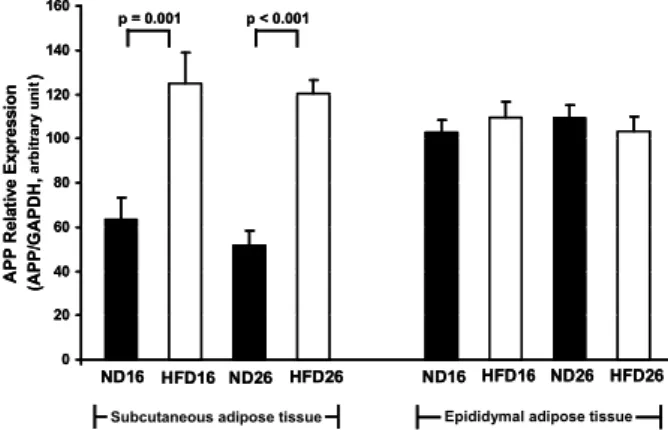
Effect of HFD-induced obesity on APP gene expression in adipose tissue
The expression levels of APP mRNA transcripts from adi- pose tissue depots were compared between high-fat diet group and normal diet group. APP gene expression in sub- cutaneous abdominal adipose tissue was significantly in- creased by about 2-fold in both 16 and 26-week-old HFD-in- duced obese mice compared to mice in normal diet group (16 weeks 125.0±13.9 vs. 63.5±9.9, arbitrary number, p=0.001;
26 weeks 120.2±6.0 vs. 51.8±6.3, p<0.0001, Fig. 1). Linear re- gression analysis demonstrated that APP expression in sub- cutaneous abdominal adipose tissue from euglycemic mice
ND16 HFD16 ND26 HFD26 ND16 HFD16 ND26 HFD26 0
20 40 60 80 100 120 140 160
APP Relative Expression (APP/GAPDH, arbitrary unit)
p = 0.001 p < 0.001
Subcutaneous adipose tissue Epididymal adipose tissue ND16 HFD16 ND26 HFD26 ND16 HFD16 ND26 HFD26 0
20 40 60 80 100 120 140 160
APP Relative Expression (APP/GAPDH, arbitrary unit)
p = 0.001 p < 0.001
Subcutaneous adipose tissue Epididymal adipose tissue
Fig. 1. Effect of diet induced obesity on APP gene expression.
APP gene expression levels were significantly increased in high-fat diet induced obese mice in subcutaneous adi- pose tissue, but not in epididymal adipose tissue. Data are means±SEM of 10 mice in each group of 16-week-old mice fed on normal diet (ND16) and high-fat diet (HFD16) and 26-week-old mice fed on normal (ND26) and high-fat diet (HFD26).
without STZ treatment used in this study significantly corre- lated with body weight (R=0.657, p<0.0001, n=39, Fig. 2).
However, APP gene expression in epididymal adipose tissue was not changed by high-fat diet feeding (16 weeks p=0.46;
26 weeks p=0.52, Fig. 1).
In addition, APP gene expression levels in subcutaneous and epididymal adipose tissues were not significantly changed by aging between 16- and 26-week-old mice in both HFD group and ND group (p>0.4, Fig. 1)
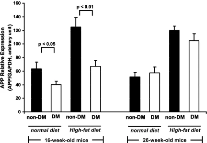
Effect of hyperglycemia on APP gene expression in adipose tissue
Induction of diabetes by STZ led to significantly de-
R = 0.657 p < 0.0001
0 40 80 120 160 200
0 10 20 30 40 50 60
Body Weight (g)
APP/GAPDH
Fig. 2. Relation of APP gene expression to body weight. APP gene expression levels in subcutaneous adipose tissue of non-diabetic mice (n=39) without STZ treatment were directly correlated with body weight (R=0.657,
p
<0.0001).creased APP gene expression in subcutaneous adipose tissue from ND or HFD fed mice at 16 weeks of age (ND16 diabetic 63.5±9.9 vs. non-diabetic 40.2±5.0, p<0.05; HFD16 125.0±13.9 vs. 67.0±9.0, p<0.01, Fig. 3). However, APP gene expression in subcutaneous adipose tissue of 26-week-old mice was not significantly changed by STZ-induced diabetes (ND26 51.8±6.3 vs. 57.4±8.8, p=0.6; HFD26 120.2±6.0 vs. 104.8±10.3, p=0.3, Fig. 3).
Linear regression analysis also demonstrated APP in sub- cutaneous adipose tissue from diabetic mice correlated with body weight (R=0.508, p<0.0001, n=49, Fig. 4), but not with plasma glucose levels (R=0.095, p=0.52, data not shown).
non-DM DM non-DM DM non-DM DM non-DM DM 0
20 40 60 80 100 120 140 160
APP Relative Expression (APP/GAPDH, arbitrary unit)
p < 0.05
p < 0.01
16-week-old mice 26-week-old mice normal diet High-fat diet normal diet High-fat diet non-DM DM non-DM DM non-DM DM non-DM DM 0
20 40 60 80 100 120 140 160
APP Relative Expression (APP/GAPDH, arbitrary unit)
p < 0.05
p < 0.01
16-week-old mice 26-week-old mice normal diet High-fat diet normal diet High-fat diet
Fig. 3. Effect of streptozotocin (STZ)-induced diabetes on APP gene expression in subcutaneous adipose tissue. APP gene expression levels were significantly decreased in STZ-induced diabetic mice at 16 weeks of age, but not at 26 weeks of age. Data are means±SEM of at least 10 mice in each group of diabetic (DM) and non-diabetic (non-DM) mice fed on normal or high-fat diet.
R = 0.508 p < 0.0001
0 40 80 120 160 200
0 10 20 30 40 50
Body Weight (g)
APP/GAPDH
Fig. 4. Relation of APP gene expression under hyperglycemic condition to body weight. APP gene expression levels in subcutaneous adipose tissue of STZ-induced diabetic mice (n=49) were directly correlated with body weight (R=0.508,
p
<0.0001).APP gene expression in epididymal adipose tissue did not tend to be affected by induction of hyperglycemia in most conditions investigated (HFD16 p=0.34; ND26 p=0.36;
HFD26 p=0.33), except 16-week-old mice fed on normal diet (102.6±5.5 vs. 85.8±6.6, p<0.05, data not shown).
Discussion
This study was aimed to elucidate the effects of HFD-in- duced obesity, aging, hyperglycemia on APP gene regu- lation in subcutaneous and epididymal adipose tissue in mice. This study demonstrates, for the first time, that APP mRNA expression is up-regulated by HFD-induced obesity and down-regulated by diabetes related weight loss in mouse subcutaneous abdominal adipose tissues. These find- ings were consistent with the previously published data de- scribing the APP regulation associated with obesity or weight loss in human subjects.
We have previously demonstrated that APP mRNA was highly expressed in subcutaneous abdominal and visceral adipose tissues and levels in subcutaneous adipocytes were upregulated in obesity and related to in vivo measures of insulin resistance [16]. In a gene-chip microarray expression profiling study, APP was overexpressed approximately 2.5 fold (p<0.0001) in subcutaneous adipocytes from 19 obese compared to 20 nonobese human subjects. We also found that the APP gene-chip signal was highly correlated to in- sulin concentration and HOMA-IR [15,16]. In a recent study, we have shown that APP gene expression levels in subcuta- neous adipocytes in obese individuals are correlated with plasma Aβ40 levels and significantly decreased with weight loss and changes in APP expression levels of subcutaneous adipocyte with weight loss were related to changes in plas- ma Aβ40 levels and changes in the 2-hour insulin levels [14].
These novel observations suggest that plasma concentrations of Aβ40 may be derived, at least in part, from adipose tissue and that APP expression in adipocytes may be related to insulin resistance [14].
Increased APP mRNA expression in subcutaneous adipo- cytes in obesity demonstrated in our previous studies [16]
was corroborated by the current study in mice which con-
firmed that APP mRNA expression increased by about
2-fold in subcutaneous abdominal adipose tissue from
HFD-induced obese mice compared to control mice fed on
normal chow (Fig. 1). These data suggest that APP ex-
pression in mice may be regulated in a similar way to hu-
man beings. However, APP expression in epididymal adi- pose tissue was not affected by HFD-induced obesity, which suggests a depot specific pattern of APP expression.
In the present study, we demonstrated that induction of diabetes by STZ led to significantly decreased APP gene ex- pression in subcutaneous adipose tissue (Fig. 3). With re- gression analysis, we also found correlation between APP expression levels in subcutaneous adipose tissue and body weight of mice in both euglycemic (without STZ treatment, Fig. 2) and diabetic (with STZ treatment, Fig. 4) groups, which is similar to our previous observation that APP ex- pression in human subcutaneous adipocytes was sig- nificantly decreased after 6 months weight loss by diet and exercise intervention [14]. However, a relation between APP expression and plasma glucose levels was not observed (p=0.52). STZ-induced diabetes is an animal model for type I diabetes mellitus without accompanying insulin resistance or hyperinsulinemia. In this study, diabetes by STZ treat- ment was induced to investigate the effects of hyperglycemia and consequent weight loss without insulin resistance. These data imply that APP down-regulation in STZ-induced dia- betic mice is due to weight loss or attenuated weight gain rather than hyperglycemia.
Aging between the weeks 16 to weeks 26 did not appear to change APP mRNA expression in mouse adipose tissues (Fig. 1). AD is usually associated with older age and some studies have indicated that global APP gene expression is increased with aging. No relation between age and the level of APP gene expression in adipose tissue was found in this study. This is most likely due to the fact that animals were mainly young adults and the range of ages was not broad enough to detect significant changes in APP expression levels.
Previous studies, together with present results, support the potential importance of peripheral tissues including adi- pose tissues in the regulation of APP and Aβ in individuals at risk for AD, which will have to be ascertained in future studies of the physiological significance of adipose tissue APP.
In summary, this study indicates that APP expression is significantly up-regulated in adipose tissue from diet in- duced obese compared with non-obese control mice. In addi- tion, APP gene expression in adipose tissue was significantly down-regulated by induction of diabetes by STZ treatment.
This down-regulation is most likely due to weight loss. The mechanism by which adipose tissue from obese animals in-
creases APP gene expression needs further investigation. In addition, it will be important to determine whether up-regu- lated adipose tissue APP expression contributes to higher circulating Aβ peptides in obesity. Finally, the relationship between adipose tissue expression of APP and Aβ peptides and amyloidogenesis in brain risk for AD are not known and will require long-term studies to address.
Acknowledgement
“This research was supported by Basic Science Research Program through the National Research Foundation of Korea (NRF) funded by the Ministry of Education, Science and Technology (No. 2009-0069167).”
References
1. Balakrishnan, K., G. Verdile, P. D. Mehta, J. Beilby, D.
Nolan, D. A. Galvao, R. Newton, S. E. Gandy, and R. N.
Martins. 2005. Plasma Abeta42 correlates positively with in- creased body fat in healthy individuals.
J. Alzheimers Dis.
8, 269-282.
2. Butterfield, D. A., S. Griffin, G. Munch, and G. M. Pasinetti.
2002. Amyloid beta-peptide and amyloid pathology are cen- tral to the oxidative stress and inflammatory cascades under which Alzheimer's disease brain exists.
J. Alzheimers Dis.
4, 193-201.3. Combs, C. K., D. E. Johnson, J. C. Karlo, S. B. Cannady, and G. E. Landreth. 2000. Inflammatory mechanisms in Alzheimer's disease: inhibition of beta-amyloid-stimulated proinflammatory responses and neurotoxicity by PPARgamma agonists.
J. Neurosci.
20, 558-567.4. Craft, S. 2007. Insulin resistance and Alzheimer's disease pathogenesis: potential mechanisms and implications for treatment.
Curr. Alzheimer Res.
4, 147-152.5. Craft, S., E. Peskind, M. W. Schwartz, G. D. Schellenberg, M. Raskind, and D. Jr. Porte. 1998. Cerebrospinal fluid and plasma insulin levels in Alzheimer's disease: relationship to severity of dementia and apolipoprotein E genotype.
Neurology
50, 164-168.6. Desai, A. K. and G. T. Grossberg. 2005. Diagnosis and treat- ment of Alzheimer's disease.
Neurology
64, S34-S39.7. Galloway, S., L. Jian, R. Johnsen, S. Chew, and J. C. Mamo.
2007. beta-amyloid or its precursor protein is found in epi- thelial cells of the small intestine and is stimulated by high-fat feeding.
J. Nutr. Biochem.
18, 279-284.8. Gandy, S. 2005. The role of cerebral amyloid beta accumu- lation in common forms of Alzheimer disease.
J. Clin. Invest
. 115, 1121-1129.9. Gorospe, E. C. and J. K. Dave. 2007. The risk of dementia with increased body mass index.
Age Ageing
36, 23-29.10. Gustafson, D., E. Rothenberg, K. Blennow, B. Steen, and I.
Skoog. 2003. An 18-year follow-up of overweight and risk
of Alzheimer disease.
Arch. Intern. Med.
163, 1524-1528.11. Hardy, J. and D. J. Selkoe. 2002. The amyloid hypothesis of Alzheimer's disease: progress and problems on the road to therapeutics.
Science
297, 353-356.12. Kivipelto, M., T. Ngandu, L. Fratiglioni, M. Viitanen, I.
Kareholt, B. Winblad, E. L. Helkala, J. Tuomilehto, H.
Soininen, and A. Nissinen. 2005. Obesity and vascular risk factors at midlife and the risk of dementia and Alzheimer disease.
Arch. Neurol.
62, 1556-1560.13. Kuusisto, J., K. Koivisto, L. Mykkanen, E. L. Helkala, M.
Vanhanen, T. Hanninen, K. Kervinen, Y. A. Kesaniemi, P.
J. Riekkinen, and M. Laakso. 1997. Association between fea- tures of the insulin resistance syndrome and Alzheimer's disease independently of apolipoprotein E4 phenotype:
cross sectional population based study.
BMJ
315, 1045-1049.14. Lee, Y. H., J. M. Martin, R. L. Maple, W. G. Tharp, and R. E. Pratley. 2009. Plasma amyloid-beta peptide levels cor- relate with adipocyte amyloid precursor protein gene ex- pression in obese individuals.
Neuroendocrinology
90, 383-390.15. Lee, Y. H., S. Nair, E. Rousseau, D. B. Allison, G. P. Page, P. A. Tataranni, C. Bogardus, and P. A. Permana. 2005.
Microarray profiling of isolated abdominal subcutaneous adipocytes from obese vs non-obese Pima Indians: increased expression of inflammation-related genes.
Diabetologia
48, 1776-1783.16. Lee, Y. H., W. G. Tharp, R. L. Maple, S. Nair, P. A. Permana, and R. E. Pratley. 2008. Amyloid precursor protein ex- pression is upregulated in adipocytes in obesity.
Obesity
16, 1493-1500.17. Lee, Y. H., W. G. Tharp, M. Peshavaria, S. Nair, P. A.
Permana, and R. E. Pratley. 2006. Overexpression of amy- loid precursor protein (APP) in adipose tissue of obese individuals.
Obesity
, 13, A214.18. Lee, Y. H., S. Tokraks, R. E. Pratley, C. Bogardus, and P.
A. Permana. 2003. Identification of differentially expressed genes in skeletal muscle of non-diabetic insulin-resistant and insulin-sensitive Pima Indians by differential display PCR.
Diabetologia
46, 1567-1575.19. Lovett, M., D. Goldgaber, P. Ashley, D. R. Cox, D. C.
Gajdusek, and C. J. Epstein. 1987. The mouse homolog of the human amyloid beta protein (AD-AP) gene is located on the distal end of mouse chromosome 16: further ex- tension of the homology between human chromosome 21 and mouse chromosome 16.
Biochem. Biophys. Res. Commun.
144, 1069-1075.
20. Luchsinger, J. A. and R. Mayeux. 2007. Adiposity and Alzheimer's disease.
Curr. Alzheimer Res.
4, 127-134.21. Luchsinger, J. A., M. X. Tang, S. Shea, and R. Mayeux. 2004.
Hyperinsulinemia and risk of Alzheimer disease.
Neurology
63, 1187-1192.22. Lue, L. F., R. Rydel, E. F. Brigham, L. B. Yang, H. Hampel, G. M., Jr. Murphy, L. Brachova, S. D. Yan, D. G. Walker, Y. Shen, and J. Rogers. 2001. Inflammatory repertoire of Alzheimer's disease and nondemented elderly microglia
in
vitro
.Glia
35, 72-79.23. Nair, S., Y. H. Lee, E. Rousseau, M. Cam, P. A. Tataranni, L. J. Baier, C. Bogardus, and P. A. Permana. 2005. Increased expression of inflammation-related genes in cultured pre- adipocytes/stromal vascular cells from obese compared with non-obese Pima Indians.
Diabetologia
48, 1784-1788.24. Palmert, M. R., T. E. Golde, M. L. Cohen, D. M. Kovacs, R. E. Tanzi, J. F. Gusella, M. F. Usiak, L. H. Younkin, and S. G. Younkin. 1988. Amyloid protein precursor messenger RNAs: differential expression in Alzheimer's disease.
Science
241, 1080-1084.25. Peila, R., B. L. Rodriguez, L. R. White, and L. J. Launer.
2004. Fasting insulin and incident dementia in an elderly population of Japanese-American men.
Neurology
63, 228-233.26. Razay, G., A. Vreugdenhil, and G. Wilcock. 2006. Obesity, abdominal obesity and Alzheimer disease.
Dement. Geriatr.
Cogn. Disord.
22, 173-176.27. Rosengren, A., I. Skoog, D. Gustafson, and L. Wilhelmsen.
2005. Body mass index, other cardiovascular risk factors, and hospitalization for dementia.
Arch. Intern. Med.
165, 321-326.28. Selkoe, D. J., M. B. Podlisny, C. L. Joachim, E. A. Vickers, G. Lee, L. C. Fritz, and T. Oltersdorf. 1988. Beta-amyloid precursor protein of Alzheimer disease occurs as 110- to 135-kilodalton membrane-associated proteins in neural and nonneural tissues.
Proc. Natl. Acad. Sci. USA
85, 7341-7345.29. Small, G. W., P. V. Rabins, P. P. Barry, N. S. Buckholtz, S. T. DeKosky, S. H. Ferris, S. I. Finkel, L. P. Gwyther, Z.
S .Khachaturian, B. D. Lebowitz, T. D. McRae, J. C. Morris, F. Oakley, L. S. Schneider, J. E. Streim, T. Sunderland, L.
A. Teri, and L. E. Tune. 1997. Diagnosis and treatment of Alzheimer disease and related disorders. Consensus state- ment of the American Association for Geriatric Psychiatry, the Alzheimer's Association, and the American Geriatrics Society.
Jama
278, 1363-1371.30. White, J. A., A. M. Manelli, K. H. Holmberg, L. J. Van Eldik, and M. J. Ladu. 2005. Differential effects of oligomeric and fibrillar amyloid-beta 1-42 on astrocyte-mediated inflammation.
Neurobiol Dis.
18, 459-465.31. Whitmer, R. A., E. P. Gunderson, E. Barrett-Connor, C. P., Jr. Quesenberry, and K. Yaffe. 2005. Obesity in middle age and future risk of dementia: a 27 year longitudinal pop- ulation based study.
BMJ
330, 1360.32. Yamada, T., H. Sasaki, K. Dohura, I. Goto, and Y. Sakaki.
1989. Structure and expression of the alternatively-spliced forms of mRNA for the mouse homolog of Alzheimer's dis- ease amyloid beta protein precursor.
Biochem. Biophys. Res.
Commun.
158, 906-912.33. Yamada, T., H. Sasaki, H. Furuya, T. Miyata, I. Goto, and Y. Sakaki. 1987. Complementary DNA for the mouse homo- log of the human amyloid beta protein precursor.
Biochem.
Biophys. Res. Commun.
149, 665-671.34. Zheng, H. and E. H. Koo. 2006. The amyloid precursor pro- tein: beyond amyloid.