Types of Extended-Spectrum β-Lactamase Produced in Enteric Bacteria Isolated from Sewage Plant Drain Water
Gun-Do Kim and Hun-Ku Lee*
Department of Microbiology, College of Natural Sciences, Pukyong National University, 599-1 Daeyeon3-Dong, Nam-Gu, Busan 608-737, Korea
Received April 6, 2010 /Accepted May 25, 2010This study focused on typing of the extended-spectrum β-lactamase (ESBL) produced in organisms isolated from a natural environment, rather than a clinical setting. Samples were collected from drain water issuing from a sewage plant in Kwanganri (Busan, Korea). Following double disk synergy test- ing, 29 strains were selected as potential ESBL positive strains. Of these, 15 strains were trans- conjugants of the sodium azide resistant recipient strain Escherichia coli J53 and analyzed biochemically including indole, methyl-red, Voges-Proskauer, Simmon's citrate, decarboxylase-dihydrolase and sug- ar-fermentation tests. The tests classified the 15 strains as Klebsiella pneumoniae (n=13) and Escherichia coli (n=2). The type of ESBL from each strain was deduced by isoelectric focusing point analysis and DNA sequencing. The results indicated that the types of ESBL were SHV-12 (n=4) and SHV-12/TEM-1 (n=9) from K. pneumoniae and TEM-1 (n=2) from E. coli strains.
Key words : Extended-spectrum β-lactamase (ESBL), SHV-12, TEM-1, Klebsiella pneumoniae, Escherichia coli
*Corresponding author
*Tel:+82-51-629-5613, Fax:+82-51-629-5619
*E-mail : hunku@pknu.ac.kr
Introduction
β -lactamase (EC 3.5.2.6) is an enzyme that hydrolyses amide, amidine, and other C-N bonds, especially ring-asso- ciated amides [3,5]. The hydrolysis of amide connection in penicillin, cephalosporin or other related β-lactam antibiotics hydrolyzing the β-lactam ring, inactivating the antibiotic [3].
β -lactamases are classified based on either the difference in amino acid sequence [1] or, more commonly, DNA molec- ular structure [3]. DNA-based classification further divides β -lactamases into TEM and SHV types. These types are mu- tants made by a point mutation involving transposition of 1–4 amino acids [4]. In terms of substrates utilized and anti- biotic specificity, β-lactamases can be classified in four groups [4,5,8,9,13].
Since plasmid-mediated extended-spectrum β-lactamase (ESBL) producing strains were isolated first in 1983, they have spread globally and increasingly become the source of clinical infections [16,17,21]. Bacteria capable of producing several types of β-lactamase have been identified clinically and in non-clinical environments including slaughter hous- es, drain waters and rivers [11,12]. The Enterobacteriaceae gen- era Klebsiella and Escherichia constitute the most commonly isolated ESBL producers from the clinical and more natural
environment [11,12]. Their antibiotic resistance is both plas- mid- and chromosome- mediated [21].
In Korea, the strains possessing a plasmid-mediated ceph- alosporinase type of β-lactamase (group 1) are suspected to be widely spread type [14,15]. Our studies have focused on slaughter houses, waste water, and plant drain water in Busan. Presently, we report the isolation and character- ization of plasmid-mediated ESBL-producing strains from the overflow water draining from a sewage treatment plant at Kwanganri in Busan.
Materials and Methods Isolation of strains
Samples were collected once a month immediately follow- ing a day of precipitation from the overflow drain of a sew- age plant at Kwanganri in Busan from June–August, 2007.
Aliquots (0.1 ml) of drain water were spread onto MacConkey agar (Difco, Detroit, MI, USA) containing 4 μg/
ml of the third generation antibiotic ceftazidime (Young-Jin Pharmaceuticals, Seoul, Korea) [20]. After 17 hr of incubation at 37
oC, representative colonies were isolated and used for two cycles of pure culture on Brain Heart Infusion (BHI) agar (Difco).
Tests of biochemical and antibiotic susceptibility
The isolated pure colonies were analyzed by biochemical
tests and antimicrobial susceptibility. Biochemical tests in- cluded indole, methyl-red, Simmon's citrate, decarboxylase- dihydrolase activity, and fermentation of 10 selected sugars.
Preparation of culture media and strain verification followed previously described protocols [6,7]. Antimicrobial suscepti- bility tests were performed against mother strains which formed conjugants in a transconjugation test [14,15].
Antibiotic disk diffusion testing utilized commercially avail- able antibiotic disks (BBL, Cokeysville, MD, USA). Results for the eight tested antibiotics (ampicillin, amikacin, cefalo- thin, chloramphenicol, kanamycin, tetracyclin, gentamycin and nalidixic acid) were determined using National Committee for Clinical Laboratory Standards protocol [18].
Double disk synergy test
Bacteria obtained from a representative pure colony were resuspended in a McFarland turbidity No. 0.5, and aliquots (0.1 ml) were spread evenly on Mueller-Hinton agar (MHA) (Difco). Double disk synergy test was performed by position- ing antibiotic-containing disks 25 mm apart at the center of each plate as previously described [14,15]. The third-gen- eration of cephalosporins [cefotaxime (30 μg), ceftazidime (30 μg) and ceftriaxone (30 μg)] and ticarcillin/clavulanate (75/10 μg) were applied for the examination. After 18 hr of incubation at 37
oC, the inhibitory zone formed between each cephalosporin containing disk and the ticarcillin/clav- ulanate disk.
Analysis of isoelectric focusing (IEF)
Bacteria were inoculated into 30 ml of BHI broth and in- cubated for 24 hr at 37
oC. The bacteria were recovered by centrifugation for 10 min at 5,000× g and the cell pellet was resuspended in 1 ml of triple distilled water. The bacteria were ultrasonically homogenized using a model 4710 appa- ratus (Cole-Palmer, Chicago IL, USA). The homogenized cells were centrifuged for 3 min at 14,000 rpm and then the supernatant was transferred to an Eppendorf tube and stor- ed at -20
oC until required for IEF [12,15]. For IEF, a drop of distilled water was dispensed onto a hydrophilic support film on a glass slide. Polyacrylamide (3–4 ml) was dis- pensed onto the support film with the pipette. Polymerization occurred during 1 hr exposure to fluorescent light. After pol- ymerization, 2 μl of sample was loaded with the guidance of a sample template, which was removed after 5 min at room temperature. A graphite electrode was kept wet with distilled water in the chamber of the Mine IEF Cell III appa-
ratus (Bio-Rad, Hercules, CA, USA). The electrode main- tained contact with the gel layer on the support slide.
Electrophoresis was sequentially carried out for 30 min at 100 V, 30 min at 200 V, 60 min at 450 V [15]. After electro- phoresis, the gel support film was separated from the glass gel plate. Nitrocefin (GlaxoSmithKline, Brentford, UK; 1–2 ml of a 500 μg/ml solution in phosphate buffer, pH 7.0) was spread onto the surface of the gel. When a red band ap- peared, the gel was covered by Whatman No. 2 filter paper.
IEF point was analyzed using an IEF 3–10 marker (Serva Electrophoresis, Heidelberg, Germany) as previously de- scribed [12]. Staining and destaining of marker proteins fol- lowed a previously described protocol [22].
Plasmid isolation and classification of ESBL gene
One milliliter of a BHI culture was centrifuged at 3,000×
g, and then the pellet was resuspended and washed three times using 1 ml of triple distilled water. DNA was extracted from the final sample by plasmid Minipreps kit and DNA purification kits following the manufacturer’s protocol (Injae Science, Seoul, Korea). ESBL typing was done by polymerase chain reaction (PCR) using the following primers: TEM type – F-primer: 5' - ATA AAA TTC TTG AAG ACG AAA - 3' and R-primer: 5' - GAC AGT TAC CAA TGC TTA ATC - 3'; SHV type – F-primer: 5' - CAC TCA AGG ATG TAT TGT G - 3' and R-primer: 5' - TTA GCG TTG CCA GTG CTC G - 3'. PCR was performed with pre-mix (Biosesang, Seoul. Korea) in the presence of each primer (10 pmol, 1 μ l), template (1 μl), and distilled water (7 μl) in a total re- action volume of 20 μl. PCR for the TEM type was performed in 30 cycles, consisting of denaturation at 94
oC for 30 sec, annealing at 45
oC for 90 sec, and extension at 72
oC for 60 sec. PCR for the SHV type consisted of 35 cycles of denatura- tion at 96
oC for 30 sec, annealing at 50
oC for 15 sec, and extension at 72
oC for 120 sec. PCR was done using a GeneAmp PCR system 2400 (Perkin Elmer, Norwalk, CT, USA). PCR products were stained with 3 μl of a 1:100 dilu- tion of DMF gel staining reagent (Komabio-Technology, Seoul, Korea), loaded onto the wells, and 1% agarose gel electrophoresis was performed using TBE buffer at 100 V for 20 min. Each gel was photographed under ultraviolet illumination [12,15].
Transmission of ESBL plasmids by transconjugation
To identify transmission of ESBL plasmids, a trans-
conjugation test was carried out with ESBL-producing
strains. Escherichia coli J53, which is resistant to sodium azide [14,15], was used as the recipient. Transconjugant and co- transconjugant in BHI (0.1 and 1.0 ml, respectively) were in- oculated in 10 ml of BHI. After 17 hr of incubation at 37
oC, 0.1 ml of culture was obtained and spread onto MacConkey agar containing 30 μg/ml ceftazidime and 50 μg/ml sodium azide. After 18 hr incubation at 37
oC, colonies were checked and examined biochemically. The biochemical tests were double-checked using E. coli J53. ESBL plasmid-mediated transmission was confirmed by PCR, electrophoresis, and IEF. PCR product was purified with a gel extraction kit (Dyne Bio, Seoul, Korea) and analyzed by DNA sequencing [15]. The initiation codon was identified and its amino acid sequence compared using Multiple Sequence Alignment and Sequence Utility of the National Center for Biotechnology Information (http://www.ncbi.nlm.nih.gov) and BCM Search Launcher (http://searcherlaumcher.bcm.tmc.edu/).
ESBL typing was done by the Lahey classification scheme (http://www.lahey.org/temtable.asp) [3-5], and the final classification was determined by analysis of the amino acid sequence.
Results
Isolation and classification of ESBL-producing Enterobacteriaceae
Colonies that developed on ceftazidime containing MacConkey agar were recovered and their purity was de- termined by two further growth cycles on BHI agar.
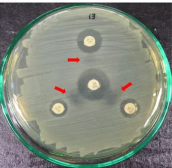
Twenty-nine strains shown synergic effects (clear zone) be- tween ticarcillin/clavulanic acid and the selected the third-generation cephalosporin antibiotics in the double disk synergy test (Fig. 1). Biochemical analyses of these 29 strains classified 16 strains as E. coli and three strains as K.
pneumoniae. They were all Gram-negative, oxidase-negative, and catalase positive.
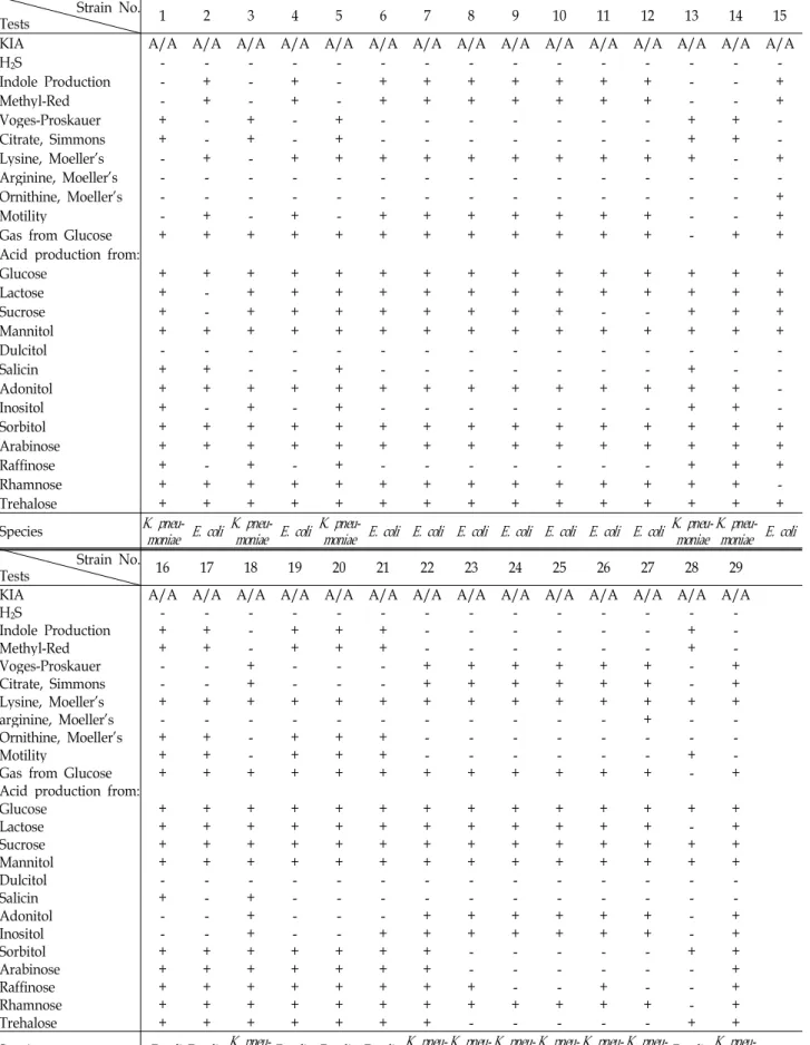
E. coli formed red plateaus on MacConkey agar and pro- duced acid in both slant and butt (A/A) on Kligler iron agar (KIA). All isolated E. coli strains were motile in BHI contain- ing 0.4% agar and were positive in both indole and meth- yl-red tests. Furthermore, all strains produced acids from glucose, mannitol, sorbitol, and trehalose but did not fer- ment dulcitol. The majority of the strains fermented lactose (88% of strains), sucrose (75%), adonitol (56%), arabinose (94%) rhamnose (88%), salicin (88%), and raffinose (63%) (Table 1). They could not use citrate as the sole carbon source
Fig. 1. Detection of the double-disk synergy test between the third generation of cephalosporins (ceftazidime, cef- triaxson, and cefotaxime) and ticacillin/clavulanic acid.
The distance of each of the third-generation antibiotics from ticacillin/clavulanic acid (center) is 25 mm. The ar- rows indicate synergic effects (clear zone) between ti- carcillin/clavulanic acid and the selected third-gen- eration cephalosporin antibiotics.
in Simmon's citrate agar and all strains were Voges-Proskauer negative. Most strains (94%) produced gas from glucose in Durham tubes. All strains were positive for lysine de- carboxylase, while only 66% of strains were positive for orni- thine decarboxylase and none of them were positive for argi- nine dihydrolase.
K. pneumoniae colonies on MacConkey agar were sticky, smoothly convex, and red centered with a clear border. Acid was produced in both slant and butt (A/A) on KIA. There was no motility in 0.4% agar medium, and both indole and methyl-red tests were negative. All of K. pneumoniaee strains used citrate as carbon source in Simmon's citrate agar and were Voges-Proskauer positive.
Conjugative transmission of plasmid-mediated ESBL
To identify plasmid-mediated ESBL transmission, 29 strai
ns resistant to ceftazidime (sodium azide
S) and E. coli J53
(sodium azide
R, ceftazidime
S) were used as the donors and
the recipient, respectively. Aliquots (0.1 ml) of a 1:10 BHI
dilution of each were spread on MacConkey agar containing
30 μg/ml ceftazidime and 50 μg/ml sodium azide, and incub
ated at 37
oC for 18 hr. Thirteen strains of K. pneu-
Table 1. Boichemical characteristics of the strains isolated from sewerage plant drain water at Kwanganri in Busan, Korea Strain No.
Tests 1 2 3 4 5 6 7 8 9 10 11 12 13 14 15
KIA A/A A/A A/A A/A A/A A/A A/A A/A A/A A/A A/A A/A A/A A/A A/A
H2S - - - -
Indole Production - + - + - + + + + + + + - - +
Methyl-Red - + - + - + + + + + + + - - +
Voges-Proskauer + - + - + - - - + + -
Citrate, Simmons + - + - + - - - + + -
Lysine, Moeller's - + - + + + + + + + + + + - +
Arginine, Moeller's - - - -
Ornithine, Moeller's - - - +
Motility - + - + - + + + + + + + - - +
Gas from Glucose + + + + + + + + + + + + - + +
Acid production from:
Glucose + + + + + + + + + + + + + + +
Lactose + - + + + + + + + + + + + + +
Sucrose + - + + + + + + + + - - + + +
Mannitol + + + + + + + + + + + + + + +
Dulcitol - - - -
Salicin + + - - + - - - + - -
Adonitol + + + + + + + + + + + + + + -
Inositol + - + - + - - - + + -
Sorbitol + + + + + + + + + + + + + + +
Arabinose + + + + + + + + + + + + + + +
Raffinose + - + - + - - - + + +
Rhamnose + + + + + + + + + + + + + + -
Trehalose + + + + + + + + + + + + + + +
Species
K. pneu-
moniae E. coli K. pneu-
moniae E. coli K. pneu-
moniae E. coli E. coli E. coli E. coli E. coli E. coli E. coli K. pneu- moniae K. pneu-
moniae E. coli
Strain No.Tests 16 17 18 19 20 21 22 23 24 25 26 27 28 29
KIA A/A A/A A/A A/A A/A A/A A/A A/A A/A A/A A/A A/A A/A A/A
H2S - - - -
Indole Production + + - + + + - - - + -
Methyl-Red + + - + + + - - - + -
Voges-Proskauer - - + - - - + + + + + + - +
Citrate, Simmons - - + - - - + + + + + + - +
Lysine, Moeller's + + + + + + + + + + + + + +
arginine, Moeller's - - - + - -
Ornithine, Moeller's + + - + + + - - - -
Motility + + - + + + - - - + -
Gas from Glucose + + + + + + + + + + + + - +
Acid production from:
Glucose + + + + + + + + + + + + + +
Lactose + + + + + + + + + + + + - +
Sucrose + + + + + + + + + + + + + +
Mannitol + + + + + + + + + + + + + +
Dulcitol - - - -
Salicin + - + - - - -
Adonitol - - + - - - + + + + + + - +
Inositol - - + - - + + + + + + + - +
Sorbitol + + + + + + + - - - + +
Arabinose + + + + + + + - - - +
Raffinose + + + + + + + + - - + - - +
Rhamnose + + + + + + + + + + + + - +
Trehalose + + + + + + + - - - + +
Species
E. coli E. coli K. pneu- moniae E. coli E. coli E. coli K. pneu- moniae K. pneu-
moniae K. pneu- moniae K. pneu-
moniae K. pneu- moniae K. pneu-
moniae E. coli K. pneu-
moniae
moniae and two strains of E. coli formed conjugants.
Biochemical tests clearly proved the identity of the con- jugants and the mother strain, E. coli J53 are the same (Table 2).
Antimicrobial susceptibility test
Fifteen of transconjugant-forming strains were tested us- ing eight kinds of antibiotics. One of two strains formed a transconjugant with E. coli and displayed resistance to seven antibiotics. The other strains were resistant to one or two antibiotics (Table 3). K. pneumoniae strains were also resistant to 2–7 antibiotics. Seven of the 13 strains were resistant to nalidixic acid, kanamycin, cephalothin, ampicillin, and ami- kacin (Table 3).
IEF points of transconjugant β-lactamase
Fifteen strains that formed transconjugants were tested for IEF point and the results were described as below. Two
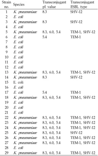
Table 2. Type and IEF of transconjugant ESBL Strain
no. Species Transconjugant
pI value Transconjugant ESBL type
1
K. pneumoniaee
8.3 SHV-122
E. coli
3
K. pneumoniaee
8.3 SHV-124
E. coli
5
K. pneumoniaee
8.3, 6.0, 5.4 TEM-1, SHV-126
E. coli
5.4 TEM-17
E. coli
8E. coli
9E. coli
10E. coli
11E. coli
12E. coli
13
K. pneumoniaee
8.3, 6.0, 5.4 TEM-1, SHV-1214
K. pneumoniaee
8.3 SHV-1215 E. coli 16
E. coli
17
E. coli
5.4 TEM-118
K. pneumoniaee
8.3, 6.0, 5.4 TEM-1, SHV-12 19E. coli
20
E. coli
21E. coli
22
K. pneumoniaee
8.3, 6.0, 5.4 TEM-1, SHV-12 23K. pneumoniaee
8.3, 6.0, 5.4 TEM-1, SHV-12 24K. pneumoniaee
8.3, 6.0, 5.4 TEM-1, SHV-12 25K. pneumoniaee
8.3, 6.0, 5.4 SHV-12 26K. pneumoniaee
8.3, 6.0, 5.4 TEM-1, SHV-12 27K. pneumoniaee
8.3, 6.0, 5.4 TEM-1, SHV-12 28E. coli
29
K. pneumoniaee
8.3, 6.0, 5.4 TEM-1, SHV-12Table 3. The pattern of multidrug resistance Species Resistance pattern Strain No.
K. pneumoniaee
Na, K, Te, Gm, Cf, Ap, An Na,K,Cf, Ap, An
Na, Te, Gm, Cf, Ap Na, K, Cf, Ap K, Cf, Ap Cf, Ap
29
26, 24, 22, 5, 23, 25, 27 17
13, 18 14 3
E. coli
Na, K, Te, Gm, Cf, Ap, AnCf, Ap
6 1
Abbreviations: An (amikacin 10 μg/ml), Ap (ampicillin 10 μg/
ml), C (chloramphenicol 30 μg/ml), Cf (cephalothin 30 μg/ml), Gm (gentamicin 10 μg/ml), K (kanamycin 30 μg/ml), Na (nalidixic acid 30 μg/ml), Te (tetracycline 30 μg/ml).
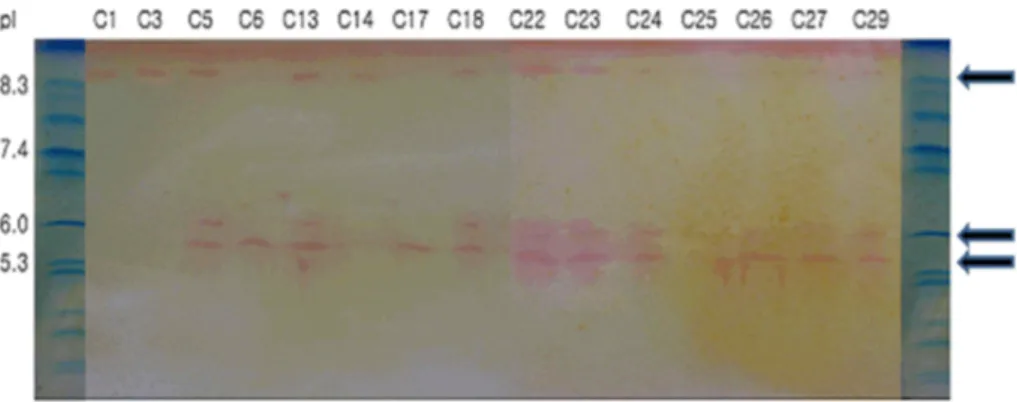
strains of E. coli (strain Nos. 6 and 17) formed a pI 5.4 prod- uct (Fig. 2). Thirteen of K. pneumoniae transconjugants were divided into two groups. One group contained three strains (Nos. 1, 3, and 14) having a pI 8.3 product and the other group contained 10 strains with products of pI 8.3, 6.0, and 5.4.
Isolation of plasmid and classification of ESBL genes by PCR
Two strains of E. coli (Nos. 6 and 17) displayed the same size of product (1,080 bp) as the TEM type. DNA sequence and protein analyses clarified that type of all E. coli trans- conjugants were TEM-1. On the other hand, electrophoresis of four K. pneumoniae strains (Nos. 1, 3, 14, and 25) detected products of approximately 780 bp indicative of SHV-12. The remaining nine strains produced both TEM and SHV types, in particular TEM-1 and SHV-12 (Figs. 3 and 4, Table 2).
Discussion
While ESBL-producing Enterobacteriaceae have been ex-
tensively studied and characterized in clinically-relevant set-
tings [21], only a few studies concerning environmental iso-
lates have been published [2,24]. However, presence of ESBL
strains in non-clinical environments is expected, since plas-
mid-mediated ESBL bacteria and their ESBL types have been
identified in slaughter houses, waste water, and river water
[14,15]. The present study was performed to isolate plas-
mid-mediated ESBL-producing strains and to classify the
ESBL types from sewage plant drain water at Kwanganri
in Busan. The particular sewage plant was built to pump
out sewage to the sea during periods of rainfall. Hence, we
collected water samples on the day following rain.
Fig. 2. IEF points of ESBL-producing transconjugants.
E. coli
(strain Nos. C6 and C17) formed a pI 5.4 product.K. pneumoniae
(Nos. C1, C3, and C14) having a pI 8.3 product and the rest of 10 strains with products of pI 8.3, 6.0, and 5.4.Fig. 3. PCR products (1,080 bp) of trans- conjugant TEM-type. M: molec- ular standard DNA ladder, C des- ignates the conjugant strains which are transmitted by ESBL plasmid of parental strains.
Fig. 4. PCR products of transconjugant SHV-type. Detected products of approximately 780 bp indicate the SHV-12 type of ESBL. M is the ab- breviation of molecular standard DNA ladder.
We focused on E. coli and K. pneumoniae, because both are representative plasmid-mediated ESBL-producing strains and also very important as opportunistic (E. coli) and noso- comial (K. pneumoniae) pathogens [9,11,23].
In Korea, TEM-52 is the dominant TEM type [10,20] and SHV-12 has been reported in clinical samples [11]. We pre- viously reported the isolation of TEM-52 and SHV-12 from slaughter house and river [14,15]. SHV-12 has a high proba- bility of being the dominant SHV type in Korea [15]. SHV-12 has a pI 8.2 and originated from SHV-1 with transpositions such as 35Leu→Gln, 238Gly →Ser, and 240Glu→Lys [19].
It is a nocosomial pathogen [10].
In the present study, 29 strains isolated by the double disk diffusion test were classified as strains of E. coli (n=16) and K. pneumoniae (n=13). All of K. pneumoniae isolates trans- ferred ESBL gene to transconjugant E. coli J53 in a plas- mid-mediated fashion. For E. coli, however, only two strains transferred ESBL gene(s) in lower number than K. pneumo- niae suggesting that the latter may more efficiently transfer plasmid-mediated ESBL genes than E. coli.
The study adds to the list of non-clinical sources of ESBL-producing strains. Their environmental presence may
pose a risk to humans and may spur the development of greater genetic diversity, especially concerning plasmid- mediated transmission.
Acknowledgment
This work was supported by Pukyong National University Research Fund in 2007 (No. 001200200708100).
References
1. Ambuler, R. P., A. F. W. Coulson, N. M. Frere, J. M.
Ghuysen, B. Joris, M. Forsman, R. C. Lebversque, G. Tiraby, and S. G. Waley. 1991. A standard numbering scheme for class A β-lactamases.
Biochem. J
. 276, 269-272.2. Brinas, L., M. Zarazaga, Y. Saenz, F. Ruiz-Larrea, and C.
Torres. 2002. β-lactamases in ampicillin-resistant
Escherichia coli
isolates from foods, humans, and healthy animals.Antimicrob. Agents Chemother
. 46, 3156-3163.3. Bush, K. 1989. Characterization of β-lactamases.
Antimicrob.
Agents Chemother
. 33, 259-263.4. Bush, K. and G. Jacoby. 1997. Nomenclature of TEM β- lactamases.
Antimicrob. Chemother
. 39, 1-3.5. Bush, K., G. A. Jacoby, and A. A. Medeiros. 1995. A func-
초록:하수처리수에서 분리된 장내세균의 광범위 베타락탐분해효소의 유형 김군도․이훈구*
(부경대학교 자연과학대학 미생물학과)
본 연구는 임상검체만이 아닌 주변 생활환경에도 광범위 베타 락탐분해효소를 생성하는 균주(extended-spec- trum β-lactamase, ESBL)가 존재하는지를 확인하고 만약 존재할 경우 그 균주를 분리하고 ESBL유형을 알아보기 위하여 실시되었다. 부산 광안리 하수처리 방류수에서 이중 디스크 확산 검사 결과 양성반응을 나타낸 29균주를 선별하였다. 이중 sodium azide에 내성을 가진 피전달 균주인 Escherichia coli J5에 교차접합이 이루어진 15균주를 대상으로 indole, methyl-red, Voges-Proskauer, Simmon's citrate 시험과 decarboxylase-dihydrolase 및 여러 종 류의 당 발효 시험 등 생화학 검사를 실시한 결과 Klebsiella pneumoniae(13균주)와 Escherichia coli(2균주)가 동정되 었다. 등전점, PCR, 유전자서열 분석을 실시하여 ESBL 유형을 결정하였다. Klebsiella pneumoniae의 ESBL 생성유 형은 SHV-12(4균주)와 SHV-12/TEM-1(9균주)의 2종류로 구분되었고, Escherichia coli의 ESBL 생성유형은 TEM-1(2균주)로 판명되었다.
tional classification scheme for β-lactamases and its correla- tion with molecular structure.
Antimicrob. Agents Chemother
. 39, 1211-1233.6. Ewing, W. H. 1986. The genus
Klebsiella
. In W.H. Ewing, Edwards and Ewing's Identification ofEnterobacteriaceae
. Elsevier, N.Y.7. Grimont, F., P. A. D. Grimont, and C. Richard. 1991. The genus
Klebsiella
, p. 2775-2796. The prokaryotes Vol. Ⅲ, 2nd ed. Springer-Verlag. N. Y.8. Jacoby, G. A. 1994. Genetics of extended-spectrum be- ta-lactamases.
Eur. J. Clin. Microbiol. Infect. Dis
. 13, 2-11.9. Jacoby, G. A. and I. Carreras. 1990. Activities of β-lactam antibiotics against
Escherichia coli
strains producing ex- tended-spectrum β-lactamases.Antimicrob. Agents Chemother
. 34, 859-862.10. Jeong, Y. S., J. C. Lee, H. Y. Kang, H. S. Yu, E. Y. Lee, C. H. Choi, S. H. Tae, Y. C. Lee, D. T. Cho, and S. Y. Seol.
2003. Epidemiology of nalidixic acid resisitance and TEM-1-and TEM-52-mediated ampicillin resistance of
Shigella sonnei
isolates obtained in Korea between 1980 and 2000.Antimicrob. Agents Chemother.
47, 3719-3723.11. Kim, J., Y. Kwon, H. Pai, J. W. Kim, and D. T. Cho. 1998.
Survey of
Klebsiella pneumoniae
strains producing ex- tended-spectrum β-lactamase: prevalence of SHV- 12 and SHV-2a in Korea.J. Clin. Microbiol
. 36, 1446-1449.12. Kim, Y. T., and H. K. Lee. 2000. Extended-spectrum β- lactamase (ESBL) typing of
Klebsiella pneumoniaee
isolated from clinical specimen in Pusan.Kor. J. Microbiol
. 36, 221-227.13. Kirby, R. 1992. Evolutionary origin of the class A and class C β-lactamases.
J. Mol. Evol
. 34, 345-350.14. Lee, H. K, 2006. Typing of extended-spectrum β-lactamase (ESBL) producing
Enterobacteriaceae
isolated from slaughter house in Pusan, Korea.Kor. J. Microbiol.
42, 125-130.15. Lee, H. K., H. J. Kim, and G. D. Kim. 2007. Typing of ex- tended-spectrum β-lactamases of
Escherichia coli
andKlebsiella pneumoniae
isolated from rivers in Pusan, Korea.Kor. J. Microbiol.
43, 116-123.16. Matthew, M., A. M. Harris, M. J. Marshall, and G. W. Ross.
1975. The use of analytical isoelectric focusing for detection and identification of β-lactamases.
Gen. Microbiol
. 88, 169-178.17. Medeiros, A. A. 1993. Nosocomial outbreaks of multi- resistant bacteria: extended - spectrum beta-lactamases have arrived in North America.
Ann. Intern. Med
. 119, 428-430.18. National Committee for Clinical Laboratory Standards.
1997. Performance standards for antimicrobial disk suscept- ibility tests. NCCLS document M2-A6. NCCLS, Wayne. PA.
19. Nueesch-Inderbinen, M. T., F. H. Kayser, and H. Haechlier.
1997. Survey and molecular genetics of SHV β-lactamases in
Enterobacteriaceae
in Switzerland: two novel enzymes, SHV-11 and SHV-12.Antimicrob. Agents Chemother
. 41, 943-949.20. Pai, H. J, S. Lyu, J. H. Lee, J. Kim, and Y. Kwon. 1999.
Survey of extended- spectrum β-lactamase in clinical iso- lates of
Escherichia coli
andKlebsiella pneumoniae:
prevalence of TEM-52 in Korea.J. Clin. Microbiol
. 37, 1758-1763.21. Philippon, A., R. Labia, and G. Jacobi. 1989. Extended-spec- trum β-lactamases (minireview).
Antimicrob. Agents Chemother
. 33, 1131-1136.22. Sambrook, J., F. E. Fritsch, and T. Maniatis. 1989. Molecular cloning laboratory manual, Vol. I. 1.25-1.28, Cold Spring Harbor Laboratory Press.
23. Silva, J., R. Gatoca, C. Auguilar, Z. Becerra, U. Garza- Ramos, M. Velázqez, G. Miranda, B. Leaños, F. Solórzano, and G. Echániz. 2001. Outbreak of infection with ex- tended-spectrum β-lactamase producing
Klebsiella pneumo- niae
in a Mexican hospital.J. Clin. Microbiol.
39, 3193-3196 24. Teshager, T., L. Domínguez, M. A. Moreno, Y. Saénz, C.Torres, and S. Cardeñosa. 2000. Isolation of an SHV-12 β -lactamase-producing