Isolation of an Invertebrate-type Lysozyme from the Body Wall of Spoon Worm, Urechis unicinctus
Hye Young Oh and Nam Gyu Park*
Department of Biotechnology, College of Fisheries Sciences, Pukyong National University, 45 Yongso-ro, Nam-gu, Busan 48513, Korea Received January 9, 2018 /Revised January 31, 2018 /Accepted March 12, 2018
Lysozymes are innate immune factors that play a critical role in the defense against pathogens in vari- ous invertebrate animals including spoon worms. In this study, an invertebrate-type lysozyme was isolated from the body wall of spoon worm, Urechis unicinctus. The acidified body wall extract was partially separated using a Sep-Pak C18 cartridge. Among the fractions, the materials that were eluted with 60% methanol/0.1% trifluoroacetic acid showed the most potent antimicrobial activity against Bacillus subtilis KCTC 1021. A series of high performance liquid chromatography (HPLC) steps were then utilized to isolate a single antimicrobial absorbance peak. The molecular weight of the anti- microbial peak was approximated using sodium dodecyl sulfate-polyacrylamide gel electrophoresis (SDS-PAGE), which was approximately 13 to 14 kDa. The partial primary structure of this anti- microbial protein that was analyzed, using LC-MS/MS, was CTGGRPPTCEDYAK (1611.69 Da).
Homology search of these fourteen residues, using the National Center for Biotechnology Information Basic Local Alignment Search Tool (NCBI BLAST), revealed that the isolated protein was similar to the invertebrate-type lysozymes described in other animals. Then, the antimicrobial and lysozyme en- zymatic (muramidase) activities of this protein were assessed. The isolated protein possessed anti- microbial activity and potent muramidase activity, which were comparable to those of hen egg white lysozyme. Therefore, the isolated protein was designated as Urechis unicinctus invertebrate-type lyso- zyme from the body wall, Uu-iLysb.
Key words : Antimicrobial protein, lysozyme, muramidase activity, purification, Urechis unicinctus
*Corresponding author
*Tel : +82-51-629-5867, Fax : +82-51-629-5863
*E-mail : [email protected]
This is an Open-Access article distributed under the terms of the Creative Commons Attribution Non-Commercial License (http://creativecommons.org/licenses/by-nc/3.0) which permits unrestricted non-commercial use, distribution, and reproduction in any medium, provided the original work is properly cited.
Journal of Life Science
2018 Vol. 28. No. 3. 300~306 DOI : https://doi.org/10.5352/JLS.2018.28.3.300서 론
해양생물체는 바다의 극단적 환경적 변화와 진화론적 다양 성에 기인하여 여러 생리활성 물질의 훌륭한 원천으로 여겨진 다[10, 11, 15]. 특히 해양 무척추동물의 경우, 척추동물의 복잡 한 면역체계에 비하여 간단한 방어시스템을 가지고 있다. 무 척추동물은 제대로 된 적응면역체계를 가지고 있지 않으며, 혈구와 같은 면역세포의 매개에 의한 세포성 면역 또는 혈장 의 체액성 면역과 같은 선천적 면역 반응에 의존하고 있다[2, 14]. 또한, 해양 무척추동물은 단순한 면역체계를 가지고 있음 에도 박테리아, 바이러스, 그리고 기생충의 공격으로부터 성 공적으로 방어하고 있다는 점에서 선천적 면역 인자를 연구하 는데 유용할 수 있다. 더욱이 이러한 선천적 면역 인자에 대한 연구는 고등 척추동물의 복잡한 면역 시스템을 이해하는 것에
도움이 될 뿐만 아니라 새로운 항균성 물질 개발의 선구물질 로서 활용될 수 있다.
개불(Urechis unicinctus)은 해양 무척추동물로서 중국, 러시
아, 일본, 그리고 한국의 해변에 서식하고 있다. 개불은 의충동
물문에 속하여 있으며, 조간대와 조하대의 부드러운 해양 토
층에 서식하는 여과섭식자이다[3, 5]. 이러한 여과섭식자들은
해양토층과 해수 등의 주변 환경을 삼켜 여과하는 과정을 통
하여 영양분을 섭취하는데, 개불의 경우 해양 토층에 U자 형
태 굴의 벽에 점액 성분의 그물망을 부착해 둔 뒤 이 망에
걸려드는 영양분을 섭취한다[3, 5, 19]. 이와 같은 영양분 섭취
방식은 개불이 영양분과 함께 여러 박테리아, 진균, 바이러스,
그리고 기생충 등 많은 병원균을 가지고 있는 해양 토층과
해수를 함께 삼키도록 한다. 따라서 개불은 이러한 병원체를
소화시키는 체계와 효과적인 면역체계를 가지고 있을 것이라
추측된다[14]. 개불에서는 신경성 펩타이드인 urechistachykin-
ins와 hemoglobin (UuHb-F-I)의 항균활성이 보고된 바 있으
나, 이 종에서 균에 대한 방어체계와 면역에 관여하는 구성
물질에 대한 충분한 연구는 진행되지 않았다[16, 23]. 따라서,
본 연구를 통하여 개불 체벽으로부터 항균성 단백질을 정제하
고자 하였다.
Fig. 1. Antimicrobial activity of U. unicinctus body wall extract fractions on Sep-Pak C18 cartridge.
재료 및 방법
실험동물
실험에 사용한 개불(Urechis unicinctus, 50마리)은 부산시 수영구 남천 해변시장에서 구입하여 13°C 멸균 해수 환경에서 3일간 순치하였고, 이후 체벽을 채집하여 액체질소로 즉시 동 결시킨 후 실험 사용까지 -75℃에 보관하였다.
시약 및 재료
HPLC-grade의 water (H
2O) 및 acetonitrile (CH
3CN)은 J.T.
Baker (NJ, USA)에서 구입하였고, Sep-Pak Vac C18 cartridge 는 Waters (MA, USA)에서 구입하였다. Tryptic soy broth (TSB)는 Merck (Darmstadt, Germany)에서 구입하였으며, Agarose (Low EEO Agar)는 Sigma (MO, USA)에서 구입하였 고, 그 이외 모든 시약은 특급을 사용하였다.
개불 체벽 조직 추출
개불의 체벽조직에 1% 초산 600 ml를 첨가하고 5분간 끓인 뒤 냉각하여 단백질분해효소의 활성을 저해하였다. T10 basic ULTRA-TURRAX dispenser (Speed#6; IKA, USA)를 사용하 여 조직을 분쇄한 뒤 원심분리(13,000x g, 40분, 4℃)하고 상층 액을 분리하였다. 이후 상층액을 Sep-Pak C18 cartridge (20 cc; Waters, USA)에 주입 한 후 0.1% trifluoroacetic acid (TFA) 를 포함한 10% 메탄올, 60% 메탄올, 그리고 100% 메탄올로 용출하여 10% 메탄올 용출액(Retained materials with 10%
methanol, RM10), 60% 메탄올 용출액(RM60), 그리고 100%
메탄올 용출액(RM 100)으로 분리하였다. RM10, RM60, 그리 고 RM100을 각 3 ml로 농축한 뒤 각 농축액의 1/500을 사용하 여 Bacillus subtilis KCTC 1021에 대한 항균활성을 확인하였다.
항균활성 측정
정제 과정 중 추출물과 각 분획, 그리고 정제된 물질의 항균 활성은 ultrasensitive radial diffusion assay (URDA)법을 사 용하여 측정하였다[22]. 정제 과정 중에는 B. subtilis를 사용하 여 항균활성을 확인하였으며, 정제된 물질의 항균활성은 B.
subtilis를 포함하여 2개의 그람 양성균과 2개의 그람 음성균에 실험하였다(B. subtilis, Staphylococcus aureus RN 4220, Escher- ichia coli D31, Edwardsiella tarda KCTC 12267). 먼저 균주를 적절한 온도에서 TSB에 16시간 배양한 후, 균의 농도가 10
8CFU/ml가 되도록 0.03% TSB가 포함된 10 mM phosphate buffer (PB, pH 6.57)를 사용하여 희석한다(84%T; Vitek Colorimeter #52-1210, Hach, USA). 희석된 균액 0.5 ml를 9.5 ml underlay gel solution (10 mM PB (pH 6.57), 0.03% TSB, 1% type I (low EEO) agarose)에 혼합하여 underlay gel에 포함된 균 농도가 5×10
6CFU/ml가 되도록 한 뒤 그리드가 있는 사각 플레이트에 부어 주어 평평하게 굳도록 한다. 그런
다음 약 2.4 mm 직경의 well을 1 mm 굵기의 underlay gel에 만들어 준다. 모든 샘플은 0.01% 초산 5 μl에 녹여준 뒤 각 well에 넣어준다(negative control, 0.01% 초산; positive con- trol, hen egg white lysozyme (HEWL)). 플레이트를 적절한 온도에서 3시간 동안 1차 배양한 후 위에 10 ml overlay gel (10 mM PB (pH 6.57), 6% TSB, and 1% type I (low EEO) agarose)을 부어 주어 2차 배양을 16시간 동안 적절한 온도에 서 시행하였다. 배양이 끝나면 각 well 주의 clear zone의 크기 (mm)를 측정하여 기록하였다.
항균활성 물질의 정제
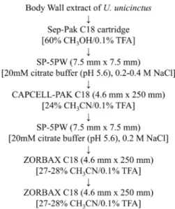
Sep-Pak C18 cartridge 분획 중 B. subtilis에 가장 강한 활성 을 보였던 RM60을 3 ml로 농축 시킨 후 SP-5PW 컬럼(10 μm, TSK-gel, 7.5×7.5 mm, Tosoh Co., Japan)에 주입하여 양이온 교환 고속 액체 크로마토그래피(high performance liquid chromatography, HPLC)를 시행하였다. 사용한 용매는 20 mM citrate buffer (pH 5.6)였고, 1 M NaCl의 농도에 따라 물질이 용출되었으며, 분리조건은 다음과 같다: 0 M NaCl (20 분), 0→1 M NaCl (50분), 유속 1 ml/min. 흡광도는 220 nm에 서 기록되었으며, 1분 마다 분획을 모았다. 그 후 각 분획의 일부를 동결건조 시켜 0.01% 초산에 녹인 후 B. subtilis에 대한 항균활성을 확인하였다. 첫번째 HPLC 분획 중 강한 항균활성 을 나타낸 분획을 모아 CAPCELL-PAK C18 컬럼(5 μm, 4.6×250 mm, Shiseido Co., Japan)에 주입하여 역상 HPLC를 시행하였다. 사용된 용매는 0.1% TFA를 포함하는 water와 acetonitrile이며, 분리조건은 5→65% acetonitrile (60분), 유속 1 ml/min였다. 흡광도는 220 nm에서 기록되었으며, 1분 마 다 분획을 모았다. 계속해서 분획의 항균활성을 측정하였으 며, 분획 중 강한 활성을 가지고 있었던 것을 모아 다시 SP-5PW을 사용하여 분리하였다. 사용한 용매는 처음 양이온 교환 HPLC와 같으며, 분리조건은 0 M NaCl (10분), 0→1 M NaCl (50분), 유속 1 ml/min였고, 분획은 피크별로 분취하였 다. 그 후 강한 활성이 있었던 분획을 ZORBAX 300SB C18 역상 컬럼(5 μm, 4.6×250 mm, Agilent, USA)에 주입하여 15→
35% acetonitrile (60분), 유속 1 ml/min 조건으로 용출하였다.
마지막으로 같은 컬럼에서 하나의 항균성 피크를 25→35%
acetonitrile (30분), 유속 1 ml/min 조건으로 정제하였다.
Fig. 2. Schematic isolation process of antimicrobial material from U. unicinctus body wall extract.
분자량 및 일차서열분석
정제한 물질의 대략적인 분자량을 15% polyacrylamide gel 에 로딩하여 sodium dodecyl sulfate-polyacrylamide gel elec- trophoresis (SDS-PAGE)를 시행하는 것으로 분석하였다. 그 후 Nano LC-MS/MS (ThermoScientific, MA, USA)를 사용하 여 정제한 물질의 일차서열을 분석하였다. 먼저 SDS-PAGE를 사용하여 물질을 분리한 후 in-gel trypsin digestion을 통해 물질을 절단하였다. 분해된 절편을 회수하여 동결건조한 후 분석한다. 분석에 사용된 컬럼은 Acclaim pepMap RSLC (75 μm×15 cm, nanoviper C18, Q-Exactive method, 100 Å, 2 μm) 였으며, 용매는 0.1% formic acid가 포함된 water와 acetoni- trile였다. LC-MS/MS를 통하여 얻은 부분적 아미노산 서열을 the national center for biotechnology information basic local alignment search tool (NCBI BLAST)를 사용하여 유사성이 있는 서열을 검색하였다.
라이소자임 활성 측정
정제된 단백질의 라이소자임 효소 활성을 라이소자임 활성 키트(Sigma-Aldrich, USA)의 Micrococcus lydeikticus를 분해하 는 능력을 분석하는 것으로 측정하였다. 먼저 M. lydeikticus를 optical density 450 nm에서 0.6-0.8 사이가 되도록 희석액을 만든 후, 큐벳에 800 μl씩 분주한다. 단백질을 녹인 용액 30 μl를 더해주어 최종 농도가 0.5 μg/ml가 되도록 맞추어 준 후 0, 1, 2, 3, 4, 5, 10, 20, 30, 40, 50, 60분에 optical density 450 nm에서 흡광도를 측정한다.
결과 및 고찰
개불 체벽으로부터 항균물질 추출 및 정제
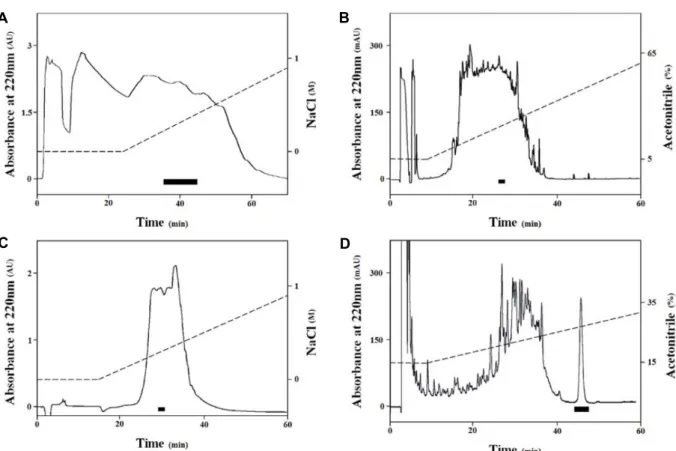
개불 체벽 추출물을 Sep-Pak C18 cartridge에 주입하여 얻 은 10%, 60%, 그리고 100% 메탄올 용출액(RM10, RM60, RM 100)의 B. subtilis에 대한 항균활성을 확인한 결과, RM10, RM60, 그리고 RM100에서 각 6.5 mm, 8.1 mm, 그리고 5.3 mm 크기의 clear zone을 관찰하였다. 가장 활성이 강했던 RM60을 사용하여 여러 단계의 HPLC 정제과정을 거쳐 하나 의 항균성 물질을 정제하였다(Fig. 2). 먼저 RM60을 농축한 뒤 SP-5PW를 사용하여 물질을 분리한 결과, 0.2-0.4 M NaCl로 용출된 분획들이 큰 항균활성을 보였다(Fig. 3A). 이 분획들을 CAPCELL-PAK C18 역상 컬럼을 통하여 분리하였으며, 그 결과 24% acetonitrile에 용출된 분획이 큰 항균활성을 나타내 었다(Fig. 3B). 계속해서 SP-5PW를 통해 이 분획을 다시 분리 하였으며, 0.2 M NaCl에 용출된 피크가 B. subtilis에 항균활성 을 나타내었다(Fig. 3C). 이를 ZORBAX C18 역상 컬럼을 사용 하여 분리한 결과 27-28% acetonitrile에서 항균 활성을 가진 피크를 분리하였다(Fig. 3D). 마지막으로 같은 컬럼에서 용출 조건을 바꾸어 27-28% acetonitrile에서 하나의 항균성 물질을 정제하였다(Fig. 4A).
항균물질의 분자량 및 일차서열분석
정제한 물질의 분자량을 알아보기 위해 SDS-PAGE를 하였 다(Fig. 4B). 15% polyacrylamide gel에서 정제한 물질은 15 kDa 크기의 표준물질보다 조금 더 아랫부분으로 이동하였으 므로, 물질은 13-14 kDa 정도의 분자량을 가지는 것으로 예측 되었다. 일차서열을 LC-MS/MS를 통해 분석한 결과, 크기가 1611.69 Da이 되는 절편의 서열을 분석하여 CTGGRPPTCE DYAK이라는 부분적 서열을 얻었다(Fig. 5). 이 서열을 NCBI BLAST를 통하여 분석해 본 결과, 이 항균성 물질은 다른 동물 들로부터 동정된 무척추동물형 라이소자임(invertebrate-type lysozyme)의 서열과 유사도를 가지고 있다는 것이 밝혀졌다.
연체동물인 Haliotis diversicolor에서 밝혀진 라이소자임(gene- bank accession number: AOX15707)과 92% 유사성이 있었으 며, 환형동물인 Eisenia fetida 에서 밝혀진 라이소자임(gene- bank accession number: AGJ83864)과 Hirudo medicinalis에서 밝혀진 라이소자임(genebank accession number: AAA96144) 과 85% 유사성이 있었다(Fig. 5). 이러한 유사성에 따라 본 연 구에서 개불 체벽으로부터 정제된 항균성 단백질을 개불 라이 소자임(Urechis unicinctus invertebrate-type lysozyme from body wall, Uu-iLysb)로 명명하였다.
Uu-iLysb의 항균 활성과 라이소자임 활성
개불 라이소자임(Uu-iLysb)의 활성을 확인하기 위하여 항
균활성 및 라이소자임 효소 활성실험을 진행한 결과, Uu-
iLysb는 활성확인에 사용된 4균주 모두에서 활성이 관찰되었
다(Fig. 6A). 가장 강한 활성은 B. subtilis와 E. coli에서 관찰되
었으며, 각 5.1 mm와 5.4 mm clear zone을 나타내었다. S.
A B
C D
Fig. 3. A series of HPLC steps used in the isolation process. The antibacterial activity of each fraction against B. subtilis was measured.
The black bar in each chromatogram indicates the fractions with strong antibacterial activity against B. subtilis, which were pooled for further purification process. A. First cation-exchange HPLC chromatogram of the body wall extract of U. unicinctus on SP-5PW. B. The reverse phase HPLC chromatogram of fractions pooled from A on CAPCELL-PAK C18. C. The cation-ex- change HPLC chromatogram of fractions pooled from B on SP-5PW. D. The reverse phase HPLC chromatogram of fractions pooled from C on ZORBAX 300SB C18.
A B
Fig. 4. A. The final HPLC chromatogram of the iso- lated antibacterial material. A single absorb- ance peak was eluted at 27-28 acetonitrile in 0.1% TFA on ZORBAX 300SB C18. B. The molecular weight of the isolated material migrated below 15 kDa on a 15% polyacry- lamide gel using SDS-PAGE.
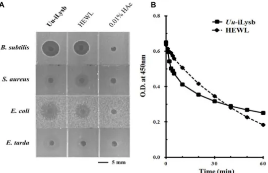
aureus와 E. tarda에서도 항균활성이 나타났으나 각 2.6 mm와 4.9 mm 크기의 clear zone이 흔적만 남은 형태로 관찰되었다.
또한 정제한 물질과 닭으로부터 정제된 c-type 라이소자임 (HEWL)을 같은 양으로 사용하여 4가지 균주에서 활성을 비교 한 결과, 비슷한 세기의 항균 활성이 나타났다. B. subtilis, E.coli, S. aureus, 그리고 E. tarda에서 각 4.4 mm, 3.7 mm, 2.6 mm, 그리고 3.0 mm 크기의 clear zone이 관찰되었으며, 개불
라이소자임과 마찬가지로 S. aureus와 E. tarda에서는 clear
zone의 흔적만 남은 형태로 관찰되었다. 라이소자임은 항균활
성 뿐만 아니라 muramidase 활성을 나타낸다고 알려져 있기
때문에 M. lydeikticus 기질을 사용하여 라이소자임 활성을 측
정하였다(Fig. 6B). 라이소자임 활성의 측정에 중요하게 생각
되는 처음 5분간은 HEWL보다 정제된 Uu-iLysb가 더 M. ly-
deikticus 기질을 더 많이 분해시키는 것으로 나타났다. 그러나,
Fig. 5. Homology search results of the partial amino acid sequence acquired through LC-MS/MS. The purified protein has high similarity with invertebrate-type lysozymes from other invertebrate animals: Haliotis diversicolor (genebank accession number:
AOX15707), Eisenia fetida(genebank accession number: AGJ83864), and Hirudo medicinalis (genebank accession number:
AAA96144).
A B
Fig. 6. A. The antimicrobial activity of Uu-iLysb (0.5 μg) and HEWL (0.5 μg) against four bacterial strains (Gram positive: Bacillus subtilis KCTC 1021, Staphylococcus aureus RN 4220; Gram negative: Escherichia coli D31, Edwardsiella tarda KCTC 12267). B.
The lysozyme activity of Uu-iLysb (0.5 μg/ml,
█) and HEWL (0.5 μg/ml,
◆).
40분 이후부터는 HEWL의 분해능력이 Uu-iLysb를 앞선 것으 로 관찰되었다.
라이소자임은 박테리아의 세포벽을 용해하는 효소로서 세 포벽의 주요 구성성분인 펩티도글리칸의 N-아세틸무람산 (NAM)과 N-아세틸글루코사민(NAG) 사이 β-1,4결합을 가수 분해하여 생물체의 방어체계에 참여하고 있을 것이라 생각된 다[13, 20]. 1922년 Fleming에 의해 처음 보고된 후 여러 동물 에서 라이소자임이 발견되고 있으며, 동물계에서 보고된 라이 소자임은 아미노산 서열 유사성과 3차 구조적 유사성에 따라 크게 세 가지(chicken-type, goose-type, invertebrate-type (i-type))로 분류되고 있다[1, 4]. 현재까지 연체동물, 극피동물, 선형동물, 환형동물, 반삭동물, 그리고 절지동물을 포함하는 많은 무척추동물에서 i-type 라이소자임이 보고되어 있다[6, 8, 9, 12, 17, 18, 21, 25, 27]. I-type 라이소자임은 대략 11-13 kDa 크기를 가지며, 다량의 시스테인 잔기가 이황화결합을
이루는 것이 특징이다. 보고된 라이소자임(muramidase) 활성 외에도 대부분의 i-type 라이소자임은 키틴분해 활성을 가지 고 있으며, Hm-ilys (Hirudo medicinalis, annelids)와 Vp-ilys (Venerupis philippinarum, mollusks), Ea-ilys (Eisenia andrei, annelids), 그리고 Pc-ilys2 (Procambarus clarkii, arthropods)를 포함하는 일부 i-type 라이소자임은 리신 곁사슬 아미노기(ε -lysine amino group)와 글루타민산 곁사슬 카르복실기 (glutamine γ-carboxamide group) 사이 이소펩타이드결합을 분해하는 활성을 가지고 있다[7, 12, 24, 26, 27].
본 연구에서 개불의 체벽으로부터 정제된 Uu-iLysb는 아미
노산 서열의 유사성과 라이소자임 활성을 가지고 있었다는
점에서 무척추동물형 라이소자임인 것으로 생각되며, 개불의
체벽에서 선천적 면역체계에 관여하여 중요한 역할을 하고
있을 것이라 여겨진다. 본 논문의 결과를 토대로 정확한 서열
을 얻기 위하여 추후 cDNA 클로닝을 진행할 예정이며, 재조
합 단백질을 생산하여 Uu-iLysb의 키틴분해활성과 이소펩타 이드 분해활성을 확인할 계획이다.
감사의 글
본 논문은 2016년 부경대학교 자율창의학술연구비(과제번 호; C-D-2016-0273)로 연구되었습니다.
References