Cloning, Expression, and Purification of a Lipase from Psychrotrophic Pseudomonas mandelii
Junsung Kim and Chang Woo Lee*
Department of Biomedical Science, Daegu University, Gyeongsan 712-714, Korea
Received February 8, 2012 /Revised March 13, 2012 /Accepted March 13, 2012A gene encoding a lipase, lipT, was cloned from the psychrotrophic bacterium Pseudomonas mandelii and sequenced. An open reading frame of 1,686 bp was found that encodes a polypeptide consisting of 562 amino acid residues. Sequence analysis revealed a Gly-His-Ser-Leu-Gly sequence, which match- es the consensus Gly-X-Ser-X-Gly motif conserved among lipolytic enzymes. The recombinant LipT protein was predominantly expressed as inclusion bodies in Escherichia coli and subsequently purified by nickel-chelate affinity chromatography. A small fraction of LipT was refolded, and the subsequent LipT exhibited substrate preferences for p-nitrophenyl butyrate (C4) and p-nitrophenyl octanoate (C8).
Key words : Lipase, Pseudomonas fluorescens, Pseudomonas mandelii, Psychrotrophic Bacterium
*Corresponding author
*Tel:+82-53-850-6464, Fax:+82-53-850-6469
*E-mail : [email protected]
Introduction
Lipases (EC 3.1.1.3) from cold-adapted bacteria play im- portant roles in industrial applications due to their high en- zymatic activity at low temperatures [9]. These applications include the use of immobilized lipases as additives in low-temperature laundry detergent formulations, the treat- ment of chilled dairy product in the food industry, and as catalysts for developing new therapeutic agents in the phar- maceutical industry [9,18].
Lipases belong to the α/β-hydrolase superfamily and con- sist of a single domain molecule [8]. Lipases act on esters of long-chain fatty acids which are insoluble in water, whereas esterases act on esters of short-chain fatty acids which are water soluble [3]. The lipase active site contains a catalytic triad consisting of serine, histidine, and aspartate residues [8]. Although lipases share a common catalytic mechanism and structure, they show low levels of sequence similarity at the amino acid level [6].
In this study, we utilized the genome sequence of Pseudomonas fluorescens Pf0-1 [17], to which the psychro- trophic bacterium Pseudomonas mandelii showed high homology. A gene coding for a lipase, lipT, was cloned from P. mandelii JR-1 using primers based on the non-coding re- gion sequences surrounding the P. fluorescens Pf0-1 lipase gene (UniProt ID: Q3KCS9). The recombinant LipT protein, which was mostly expressed in E. coli BL21 (DE3) as in-
clusion bodies, was purified using nickel-chelate affinity chromatography. After refolding, LipT exhibited substrate preferences for p-nitrophenyl butyrate (PNPB) and p-nitro- phenyl octanoate (PNPO).
Materials and Methods Materials
The TA cloning vector was purchased from Enzynomics (Korea). The pET28a expression vector was purchased from Novagen (USA). The HisTrap FF column was purchased from GE Healthcare (USA) and the esters for p-Nitrophenyl were purchased from Sigma (USA). All other reagents were obtained from Sigma unless noted otherwise.
Strain isolation and identification
P. mandelii JR-1 was isolated from natural mineral waters collected in Gyeongsan, Korea. Gram staining was per- formed as described previously [12]. The 16S rRNA sequenc- ing was carried out at Genotech (Korea). The 16S rRNA se- quence analysis of the isolated bacterium was carried out using an EzTaxon server [4].
Plate assay for lipase
Rhodamine B agar plates were prepared as described pre-
viously [10]. Holes of 3 mm diameter were punched into
the agar and the cavities formed were filled with either 10
μ l of bacterial culture or cell-free culture supernatant. Plates
were incubated overnight at 25°C, exposed to UV irradiation
(350 nm), and then photographed.
Gene cloning of lipT
The lipT gene was cloned from P. mandelii JR-1 by poly- merase chain reaction (PCR) in two steps. First, primers were designed based on the non-coding region sequences sur- rounding the lipase (Q3KCS9) in the P. fluorescens Pf0-1 genome. The forward primer was 5'-GACCACGGTGT GGGCTTGAC-3' and the reverse primer was 5'-GCTCACAACCAGAACGCCCC-3'. The resulting PCR product was subcloned into a TA vector and sequenced.
Second, the gene for lipT was amplified from the TA vector and subcloned into a pET28a vector. The forward primer used was 5'-GAGAGAtctagaAAGGAGATATACATGGGA CTGTTTGATTAC-3' (Xba I site in small letters, ribosome binding site sequence underlined and the 5'-end region of lipT in bold face type). The reverse primer used was 5'-GCGGCCGCaagcttCGCAAACGTGATGCCTG-3' (Hind III site in small letters and the 3'-end region of lipT in bold face type). A linker and His
6sequence (KLAAALEHHH HHH), which comes from a pET28a vector, were located on the C-terminus of LipT. The construct was confirmed by DNA sequencing.
Sequence analysis
A homology search was performed using BLAST (http://www.ncbi.nlm.nih.gov/BLAST/). Multiple se- quence alignments were performed using ClustalW (http://www.ebi.ac.uk/Tools/msa/clustalw2/).
Expression and purification of LipT
The gene for LipT with a C-terminal His
6sequence was transformed into E. coli BL21 (DE3). A single colony grown on an LB/kanamycin plate was selected for additional over- night growth at 37
oC, followed by inoculation into a 250 ml LB/kanamycin broth. At the mid-log phase (OD
600nm= 0.6~0.8) the growth temperature was lowered to 30
oC. After addition of 1 mM IPTG, the cells were grown for 4 more hours. The cells were harvested at 10,000× g for 5 min. The pellet was resuspended in Buffer A (20 mM Tris ․Cl, 0.
1 M NaCl, 5% glycerol, pH 8.0) followed by sonication at 4
oC. After centrifugation at 12,000× g for 10 min, the pellet was resuspended in Buffer A with 8 M urea. The imidazole concentration was adjusted to 5 mM in preparation for purifi- cation using nickel-chelate affinity column chromatography.
LipT was purified on an AKTA Explorer system (GE Healthcare) with a 1-ml HisTrap column using Buffer B (20 mM Tris․Cl, 0.1 M NaCl, 8 M urea, 70 mM imidazole, 5%
glycerol, pH 8.0). All purification steps were carried out at 4°C. The purified enzymes were frozen in N
2and stored at -80°C.
Enzyme assay
The substrate specificity of LipT for 0.4 mM p-nitrophenyl esters (C2 to C16) was measured in reaction buffer (100 mM Tris․Cl, 100 mM NaCl, 0.3% Triton X-100, pH 8.5). The ac- cumulation of p-nitrophenol was measured using a Shimadzu UV-160 spectrophotometer at 400 nm for 5 min at 25°C.
Nucleotide sequence accession number
The nucleotide sequence of the lipT gene from P. mandelii JR-1 has been deposited in GenBank under accession num- ber JQ284021.
Results and Discussion Strain identification and phylogenetic analysis
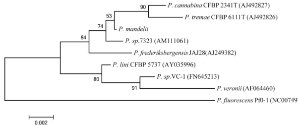
Taxonomical studies based on the 16S rRNA sequence re- vealed homology to the genus Pseudomonas (Fig. 1). Analysis using the EzTaxon server showed it possessed the highest level of similarity to Pseudomonas mandelii (100%), followed by Pseudomonas cannabina (99.53%) and Pseudomonas tremae (99.46%). A BLAST search of the NCBI database also con- firmed high degrees of similarity to P. mandelii strains. P.
mandelii is classified as belonging in the P. mandelii subgroup of the Pseudomonas fluorescens group [13].
Optimum growth temperature
As demonstrated by Gratia et al. [7], P. mandelii grew at 4°C but did not grow at 37°C. Although P. mandelii JR-1 is a psychrotrophic bacterium, its optimum growth rate was at 25~30°C (data not shown).
Investigation of lipase activity
To investigate P. mandelii lipase activity, we utilized agar
plates containing olive oil and rhodamine B as previously
demonstrated by Kouker and Jaeger [10]. Free fatty acids
released from the olive oil formed colored complexes with
rhodamine B, which is a basic dye. Upon UV irradiation,
orange fluorescence was observed for the P. mandelii culture
on the rhodamine B agar plates, whereas no fluorescence
was observed for E. coli (Fig. 2). The P. mandelii culture
supernatant also revealed orange fluorescence (data not
Fig. 1. Phylogenetic tree based on the 16S rRNA gene sequence of
P. mandelii
JR-1.P. mandelii
belongs to theP. mandelii
subgroup of theP. fluorescens
group [13].Fig. 2. Identification of lipase activity on a rhodamine B agar plate. After overnight incubation at 25°C, plates were ex- posed to UV irradiation at 350 nm.
shown). Our data indicated that P. mandelii produced lipase activity, of which, some was exhibited by extracellular lipases.
Cloning of the lipT gene
Generally, a bacterium expresses several genes of lipases and esterases, possibly reflecting a wide range of substrate specificities. We used two lipase-prospecting primers (OXF1-ACR1 and OXF1-ACR3), as reported by Bell et al. [2], to clone lipase genes from P. mandelii JR-1 (data not shown).
Although sequencing of the PCR products did not locate a lipase, a BLAST search revealed that the sequences had the highest degree of homology to those of P. fluorescens Pf0-1 from among the repository of completed microbial genomes (data not shown). Thus, the PCR primers were designed based on the non-coding region sequences surrounding four P. fluorescence pf0-1 lipases, found as lipase in the UniProt
database (UniProt ID: Q3KBP5, Q3KCS8, Q3KCS9, and Q3KIU1). A PCR product was amplified from the non-cod- ing region sequences surrounding a lipase (Q3KCS9), subcl- oned into a TA vector, and then sequenced. A 1,689-bp lipT gene amplified from the TA vector was subcloned into a pET28 vector. Surprisingly, both the lipT and the P. fluo- rescens Pf0-1 lipases had the same 1,689-bp sequence length.
Sequence analysis
The deduced LipT amino acid sequence comprised 562 amino acids residues. LipT contained a Gly-His-Ser-Leu-Gly sequence, a motif characteristic of the serine lipase family (Gly-X-Ser-X-Gly) [16] (Fig. 3). Sequence analysis also showed that LipT contains six motifs (Gly-Gly-X-Gly-X-Asp or Gly-X-X-Gly-X-Asp) which function as the C-terminal sig- nal for identification by ABC transporters [1,5] (Fig. 3). LipT showed 83% sequence similarity at the amino acid level to the P. fluorescens Pf0-1 lipase (Q3KCS9).
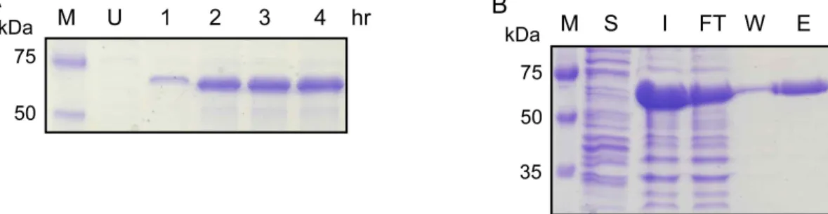
Expression and purification of LipT
An entire open reading frame of lipT with a C-terminal
His
6residues was constructed on a pET28a vector. LipT ex-
pression in E. coli BL21 (DE3) increased in a time-dependent
manner (Fig. 4A), mainly resulting in inclusion body
formation. Purification of LipT was carried out with 8 M
urea using a 1-ml HisTrap column. LipT was eluted at 70
mM imidazole, as shown in Fig. 4B, suggesting that it bound
weakly to the nickel resin. LipT was refolded by reducing
the urea concentrations, but only a small fraction of LipT
was refolded such that it resulted in lipase activity.
Fig. 3. Multiple amino acid sequence alignment. The amino acid sequence of LipT was compared with the
P. fluorescens
Pf0-1 lipase (Q3KCS9). The GXSXG motif (purple) of serine enzymes and the GGXGXD and GXXGXD motifs (cyan) for ABC transporters are denoted, respectively.Fig. 4. Expression and purification of LipT. (A) LipT was expressed in
E. coli
BL21 (DE3) upon 1 mM IPTG induction. U, uninduced.(B) Purification of LipT using nickel-chelate affinity chromatography. M, molecular weight marker; Lane S, soluble fraction after sonication; Lane I, Input from inclusion bodies; Lane FT, Flow-through; Lane W, Wash buffer (5 mM imidazole); Lane E, Elution buffer (70 mM imidazole). The calculated molecular weight of LipT was 59,055 Da and it migrated on a SDS gel as a 64 kDa protein.
Substrate specificity of LipT
Of all the p-nitrophenyl esters (C2 to C16) tested, LipT exhibited maximal hydrolysis with PNPB (C4) (Fig. 5). The
hydrolysis of PNPO (C8) was approximately 71% of that ob-
served for PNPB. LipT showed no enzymatic activity for oth-
er p-nitrophenyl esters (C2 and C12-16).
Fig. 5. Hydrolysis of
p
-nitrophenyl esters by LipT. The hydrol- ysis of thep
-nitrophenyl esters (C2 to C16) was ex- pressed as a percentage comparison to PNPB. There was no activity observed with C2 and C12 to C16 substrates.100%=1.4 μM/min.
A few other lipases demonstrated substrate preferences for medium-chain fatty acids which were similar to that of LipT. A lipase from a cold-adapted Psychrobacter sp. ex- hibited the highest hydrolytic activity with p-nitrophenyl caproate (C6), followed by PNPB [14]. Medium-chain acyl group p-nitrophenyl esters were also good substrates for psychrotrophic Pseudomonas sp. KB700A lipase (C10>C6>C4) [15]. A lipase from psychrotrophic Aeromonas sp. LPB4 fa- vored substrates containing medium chain acyl groups (C3 to C10) [11].
In conclusion, lipT, a gene coding for a lipase, was cloned from the psychrotrophic bacterium P. mandelii JR-1 and sequenced. The recombinant LipT protein was expressed in E. coli BL21 (DE3), mainly as inclusion bodies. LipT demon- strated substrate preferences for esters of medium-chain fat- ty acids (C4 and C8).
Acknowledgement
This work was supported by the Daegu University Research Grant, 2008 (to C. L.).
References
1. Ahn, J. H., Pan, J. G. and Rhee, J. S. 1999. Identification of the tliDEF ABC transporter specific for lipase in
Pseudomonas fluorescens
SIK W1.J. Bacteriol.
181, 1847-1852.2. Bell, P. J., Sunna, A., Gibbs, M. D., Curach, N. C., Nevalainen, H. and Bergquist, P.L. 2002. Prospecting for novel lipase genes using PCR.
Microbiology
148, 2283-2291.3. Chahinian, H. and Sarda, L. 2009. Distinction between ester- ases and lipases: comparative biochemical properties of se-
quence-related carboxylesterases.
Protein Pept. Lett.
16, 1149-1161.4. Chun, J., Lee, J. H., Jung, Y., Kim, M., Kim, S., Kim, B. K.
and Lim, Y. W. 2007. EzTaxon: a web-based tool for the identification of prokaryotes based on 16S ribosomal RNA gene sequences.
Int. J. Syst. Evol. Microbiol.
57, 2259-2261.5. Chung, C. W., You, J., Kim, K., Moon, Y., Kim, H. and Ahn, J. H. 2009. Export of recombinant proteins in
Escherichia coli
using ABC transporter with an attached lipase ABC trans- porter recognition domain (LARD).Microb. Cell Fact.
8, 11.6. Cygler, M., Schrag, J. D., Sussman, J. L., Harel, M., Silman, I., Gentry, M. K. and Doctor, B. P. 1993. Relationship be- tween sequence conservation and three-dimensional struc- ture in a large family of esterases, lipases, and related proteins.
Protein Sci.
2, 366-382.7. Gratia, E., Weekers, F., Margesin, R., D'Amico, S., Thonart, P. and Feller, G. 2009. Selection of a cold-adapted bacterium for bioremediation of wastewater at low temperatures.
Extremophiles
13, 763-768.8. Jaeger, K. E., Dijkstra, B. W. and Reetz, M. T. 1999. Bacterial biocatalysts: molecular biology, three-dimensional struc- tures, and biotechnological applications of lipases.
Annu.
Rev. Microbiol.
53, 315-351.9. Joseph, B., Ramteke, P. W. and Thomas, G. 2008. Cold active microbial lipases: some hot issues and recent developments.
Biotechnol. Adv.
26, 457-470.10. Kouker, G. and Jaeger, K. E. 1987. Specific and sensitive plate assay for bacterial lipases.
Appl. Environ. Microbiol.
53, 211-213.11. Lee, H. K., Ahn, M. J., Kwak, S. H., Song, W. H. and Jeong, B. C. 2003. Purification and characterization of cold active lipase from psychrotrophic
Aeromonas
sp. LPB 4.J. Microbiol.
44, 22-27.
12. Moyes, R. B., Reynolds, J. and Breakwell, D. P. 2009.
Differential staining of bacteria: gram stain.
Curr. Protoc.
Microbiol.
Appendix 3, Appendix 3C.13. Mulet, M., Lalucat, J. and Garcia-Valdes, E. 2010. DNA se- quence-based analysis of the
Pseudomonas
species.Environ.
Microbiol.
12, 1513-1530.14. Parra, L. P., Reyes, F., Acevedo, J. P., Salazar, O., Andrews, B. A. and Asenjo, J. A. 2008. Cloning and fusion expression of a cold-active lipase from marine Antarctic origin.
Enzyme Microb. Technol.
42, 371-377.15. Rashid, N., Shimada, Y., Ezaki, S., Atomi, H. and Imanaka, T. 2001. Low-temperature lipase from psychrotrophic
Pseudomonas
sp. strain KB700A.Appl. Environ. Microbiol.
67, 4064-4069.16. Rosenau, F. and Jaeger, K. 2000. Bacterial lipases from
Pseudomonas
: regulation of gene expression and mechanisms of secretion.Biochimie
82, 1023-1032.17. Silby, M. W. et al. 2009. Genomic and genetic analyses of diversity and plant interactions of
Pseudomonas fluorescens
.Genome Biol.
10, R51.18. Tutino, M. L., di Prisco, G., Marino, G. and de Pascale, D.
2009. Cold-adapted esterases and lipases: from funda- mentals to application.
Protein Pept. Lett.
16, 1172-1180.초록: Pseudomonas mandelii 의 lipase 유전자 클로닝, 발현 및 정제 김준성․이창우*
(대구대학교 의생명과학과)
내냉성 세균인