R E V I E W Open Access
Recent advances in the reconstruction of cranio-maxillofacial defects using
computer-aided design/computer-aided manufacturing
Ji-hyeon Oh
Abstract
With the development of computer-aided design/computer-aided manufacturing (CAD/CAM) technology, it has been possible to reconstruct the cranio-maxillofacial defect with more accurate preoperative planning, precise patient-specific implants (PSIs), and shorter operation times. The manufacturing processes include subtractive manufacturing and additive manufacturing and should be selected in consideration of the material type, available technology, post-processing, accuracy, lead time, properties, and surface quality. Materials such as titanium, polyethylene, polyetheretherketone (PEEK), hydroxyapatite (HA), poly-DL-lactic acid (PDLLA), polylactide-co-glycolide acid (PLGA), and calcium phosphate are used. Design methods for the reconstruction of cranio-maxillofacial defects include the use of a pre-operative model printed with pre-operative data, printing a cutting guide or template after virtual surgery, a model after virtual surgery printed with reconstructed data using a mirror image, and manufacturing PSIs by directly obtaining PSI data after reconstruction using a mirror image. By selecting the appropriate design method, manufacturing process, and implant material according to the case, it is possible to obtain a more accurate surgical procedure, reduced operation time, the prevention of various complications that can occur using the traditional method, and predictive results compared to the traditional method.
Keywords: Computer-aided design/computer-aided manufacturing, Three-dimensional imaging, Cranio-maxillofacial defect, Reconstructive surgical procedures, Custom implant, Patient-specific implant
Introduction
The reconstruction of complex cranio-maxillofacial defects is challenging due to the unique anatomy, the presence of a vital structure, and the variety of deficits [1, 2]. The re- construction of congenital or acquired cranio-maxillofacial defects due to congenital abnormalities, post-trauma, tumor resection, and infection requires both functional and esthetic considerations [3,4].
Computer-aided design (CAD) is the process of creating, modifying, analyzing, or optimizing a design using com- puter system. Computer-aided manufacturing (CAM) is the process of planning, managing, or controlling manu- facturing using computer system [5].
In the early 1960s, CAD was developed in the aircraft and automotive industries, and in the late 1960s, the supply of computer system for CAD was specialized [6].
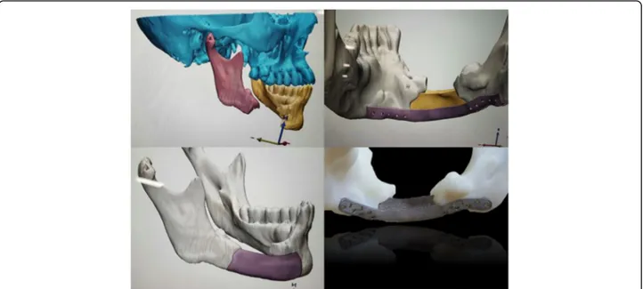
In the late 1980s, computer-controlled milling was used to produce prosthesis using three-dimensional (3D) im- aging data in computed tomography (CT) [7]. With the development of CAD/CAM technology, there have been increasing cases of restructuring cranio-maxillofacial de- fects to improve appearance and function with more accurate surgery and shorter operation times [8]. With CAD/CAM software, accurate pre-operative planning can be established, and surgeons can perform virtual ablation, plan osteotomy and reconstruction procedures, or create patient-specific implants (PSIs) [3,4,9] (Fig.1).
The advantages of CAD/CAM technology include im- proved accuracy of esthetic results, restoration of large and geometrically complex anatomical defects, reduction
Correspondence:[email protected]
Department of Oral and MaxilloFacial Surgery, Dental Hospital, Gangneung-Wonju National University, Gangneung, South Korea
Maxillofacial Plastic and Reconstructive Surgery
© The Author(s). 2018 Open Access This article is distributed under the terms of the Creative Commons Attribution 4.0 International License (http://creativecommons.org/licenses/by/4.0/), which permits unrestricted use, distribution, and reproduction in any medium, provided you give appropriate credit to the original author(s) and the source, provide a link to the Creative Commons license, and indicate if changes were made.
Oh Maxillofacial Plastic and Reconstructive Surgery (2018) 40:2 DOI 10.1186/s40902-018-0141-9
of operative times, more accurate fitting of implants, overcoming the disadvantages of autogenous bone grafts, and performing resection and reconstruction in one step [10, 11]. The 3D printing technique in the cranio- maxillofacial area surgery includes contour models that are accurate replicas of patient-specific anatomy, guides that are patient-specific templates that guide precise cut- ting and drilling, splints defined as the replica of the vir- tual post-operative position of the patient structure, and implants defined as three-dimensionally printed objects that are directly implanted in the patients [12,13].
In this paper, we will discuss the manufacturing pro- cesses using CAD/CAM, implant materials, the work- flow reconstructing the cranio-maxillofacial defects, and future directions of development.
Review
The manufacturing processes
The manufacturing processes include subtractive manu- facturing, which cuts off a piece of material to form the final shape, and additive manufacturing, which builds up the material by stacking [8]. Subtractive manufacturing, the traditional machining technique has the disadvan- tage in that it is difficult to make complicated shapes by computer numerical control (CNC) milling and there is a lot of material waste [14].
Additive manufacturing, known as rapid prototyping or 3D printing, has the advantage of being very sophisti- cated, with less material waste, faster production times, and the ability to produce complex structures [8]. There are several additive manufacturing processes, including binder jetting (BJG), direct metal laser sintering (DMLS), electron beam melting (EBM), laser engineered net
shaping (LENS), and fused deposition modeling (FDM) [15–17]. Figure2is a simplified schematic of the manu- facturing processes described in this paper.
BJG generally uses two materials: a powder material from which the part is made and a binder material that bonds between the powder materials (Fig. 3). It has the advantage that parts can be produced without support structures, but it has the disadvantage that post- processing takes more time than actual printing, result- ing in a significant increase in cost. In addition, the parts have rough microstructure and lower mechanical prop- erties than those produced by selective laser melting (SLM) or EBM because of the possibility of porosity and heat treatment [16].
DMLS, referred to using the terms SLM or selective laser sintering (SLS), uses a high-powered optic laser to fuse the metal powder to solid components based on a 3D CAD file and, similar to EBM, is built layer by layer [17]. Similar to BJG, a powder bed is used to create a 3D object. However, instead of using a spray solution, a laser is used to tie the powder particles together, and the laser is instructed to draw a specific pattern on the surface of the powder bed during the printing process [18]. When the first layer is completed, the roller sprin- kles a new layer of powder on top of the previous layer, pushes it flat, and then uses the laser to make the object layer by layer [19] (Fig. 4). DMLS have many advantages and disadvantages [16]. The advantages include the use of a wide range of materials, improved functionality, relatively low cost, and the production of ready-to-use near-net- shaped components. On the other hand, the disadvantages include relatively slow processes, size limitations, high power consumption, and high initial cost. In addition, the
Fig. 1 Pre-operative diagnosis, virtual surgery, and creation of patient-specific implants using CAD/CAM software
handling of the powder is tricky, the produced parts can have rough surfaces, and the brittle materials that cannot accommodate high internal stress during the manufactur- ing process can cause cracking of the parts.
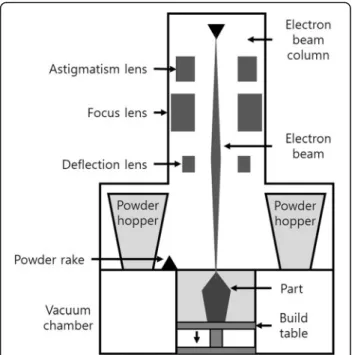
EBM is very similar to DMLS, but there is a slight dif- ference in that the parts are fabricated by melting the metal powder in a layer using an electron beam [17]
(Fig.5). In EBM, the cooling rate can be greatly reduced by increasing the temperature of the powder bed. Unlike DMLS, EBM has the ability to treat brittle materials that cannot be processed by DMLS because it slowly cools, avoiding solidification cracking of brittle materials. How- ever, it takes longer than DMLS and requires sufficient cooling time before removing parts from the substrate plate because the electron beams are used many times in the layers [16].
Similar to EBM and DMLS, LENS is used to fabricate metal parts directly from CAD solid models and has the difference in that metal powders are injected into the mol- ten pool generated by the condensed high-power laser beam [17]. The molten material line rapidly solidifies as the laser beam retreats, and after each layer is formed, the
laser head advances by one-layer thickness together with the powder feed nozzle, and a subsequent layer is created.
This is repeated several times until the entire object dis- played in the 3D CAD model is created [20] (Fig.6).
FDM is generally carried out with a polymer melted in a printer nozzle and arranged layer by layer. The mater- ial is melted and deposited at a defined location on the printing layer, and after the first layer is completed, the distance between the printing bed and the extruder noz- zle is increased and the second layer is printed on the first layer [21] (Fig.7).
There are various kinds of manufacturing processes.
The manufacturing process should be selected with con- sideration of the material type, available technology, post-processing, accuracy, lead time, properties, and sur- face quality [16].
Implant materials
The ideal material is biocompatible, easy to shape, high strength, non-toxic, inexpensive, durable, radiolucent,
Fig. 3 Binder jetting schematic Fig. 4 Direct metal laser sintering schematic
Fig. 2 Schematic of the manufacturing processes
Oh Maxillofacial Plastic and Reconstructive Surgery (2018) 40:2 Page 3 of 7
and lightweight [8,22]. However, no material satisfies these conditions [22–24]. Materials include non-resorbable ma- terials such as titanium, polyethylene, polyether ether ketone (PEEK), and hydroxyapatite (HA) and absorbable materials such as poly-DL-lactic acid (PDLLA), polylactide- co-glycolide acid (PLGA), and calcium phosphate.
Titanium is the metal of choice for manufacturing im- plants. It has the advantages of high strength, biocompati- bility, lightweight, corrosion resistance, and the potential for osseointegration [8,25,26]. However, it has the disad- vantage of causing scatter artifacts in CT scans [27].
Polyethylene includes porous polyethylene (PPE) and ultra-high molecular weight polyethylene (UHMW-PE).
PPE such as Medpor (Pufex Surgical Inc., College Park,
GA, USA) was used for reconstruction of the orbital floor and augmentation of the facial area [28]. PPE is very stable and easy to shape and has tissue ingrowth through its pores [29,30]. However, there is a possibility of infection [28]. UHMW-PE was used for reconstruc- tion of orbit or temporomandibular joint by making PSIs using CAD/CAM [31, 32]. Because of a solid structure, UHMW-PE can have a lower infection rate than PPE [32]. Polyethylene has the advantage of not producing artifact because of radiolucency in CT, but it also has a disadvantage that it is difficult to control implant pos- ition after surgery [3,32].
PEEK was used to reconstruct various craniofacial bone defects including cranioplasty [23, 33]. PEEK has similar strength and elasticity to bone and is easy to modify [34]. It is radiolucent in CT and offers more comfort to patients, with lower thermal conductivity and lighter weight than titanium. However, it had reports of infection and foreign body reaction [27].
HA is used as a biocompatible scaffolding material for bone tissue engineering [35]. It is osteoconductive and non-resorbable and shows tissue in-growth in the pres- ence of pores, with a strong capacity to bind both hard and soft tissues [36]. Pure HA is low in viscosity and dif- ficult to make complex shapes, but it can be overcome with custom-made HA using CAD/CAM [37,38].
As absorbable implants, PDLLA and PLGA are com- monly used, especially in pediatric craniofacial surgery [39]. However, foreign body reaction and the weakness of materials such as screw fracture have been reported [40, 41]. There are cases in which calcium phosphate implants have been used for the reconstruction of cranio-maxillofacial defects [42]. These printed calcium
Fig. 5 Electron beam melting schematic
Fig. 6 Laser engineered net shaping schematic
Fig. 7 Fused deposition modeling schematic
phosphate implants have good biocompatibility and suit- able biodegradation and are similar to the mineral phase of the bone, so they do not cause artifacts or interfer- ence seen in other metallic alloplasts in CT or MRI. In addition, calcium phosphate implants show less mechan- ical performance than titanium but are suitable as a scaf- fold for bone tissue growth and can be loaded with bioactive protein or antibiotics.
Workflow
The modeling software used for 3D printing includes Mimics (Materialise, Leuven, Belgium), SolidWorks (Dassault Systemes, Velizy-Villacoublay, France), Amira (FEI Visualization Sciences Group, Merignac, France), Rhino (Robert McNeel & Associates, Seattle, WA, USA), and SurgiCase CMF (Materialise, Leuven, Belgium). The printing software include ZPrinter and Projet (3D Sys- tems, Rock Hill, SC, USA) and Alaris (Objet Limited, Rehovot, Israel) [12].
PSIs can be constructed through a manufacturing process and can also be produced by shaping directly from a 3D printing skull model [3]. In general, CT scan images are converted into two-dimensional (2D) digital imaging and communications in medicine (DICOM) files and converted to the 3D stereolithography (STL) format using CAD software [1,8,43]. The design methods for the reconstruction of the cranio-maxillofacial defects are as follows.
i) After printing the skull model of the pre-operative form, pre-bending of the plate, fabrication of an onlay template by model surgery, or fabrication of the implant on the skull model [44–48]
ii) After performing virtual surgery, including resection and reconstruction in the pre-operative image, printing of a resection guide or fabrication of the template by printing the skull model with virtual surgery [49,50]
iii) After printing of the skull model reconstructed symmetrically using a mirror image of the unaffected side, pre-bending of the plate, using it as a template, or fabrication of the implant directly on the skull model [51–53]
iv) After reconstruction symmetrically using a mirror image of the unaffected side and design of the 3D implant to fit precisely to the defect, fabrication of the PSI by transferring the PSI data to CAM software [3,8,32,54–57]
Future directions
Digital workflows are time consuming and cannot be used for emergency procedures such as immediate post- traumatic surgery [10]. It takes from a few days to weeks to make the PSI outside the hospital [8]. However, with
the development of 3D printers, relatively inexpensive personalized 3D printers have been introduced and the accuracy has increased, making it possible to manufac- ture products inside the hospital, reducing the time re- quired. In addition, the development of professional CAD software familiar to surgeons and minimally inva- sive surgical procedures will provide predictable results.
Conclusion
By selecting the appropriate design method, manufactur- ing process, and implant material according to the case, it is possible to obtain a more accurate procedure, re- duced surgical time, the prevention of various complica- tions that can occur using the traditional method, and predictive results compared to the traditional method.
Acknowledgements Not applicable.
Funding Not applicable.
Availability of data and materials
The dataset supporting the conclusions of this article is included within the article.
Authors’ contributions
The author wrote the manuscript and approved the final manuscript.
Ethics approval and consent to participate Not applicable.
Consent for publication Not applicable.
Competing interests
The author declares no competing interests.
Publisher’s Note
Springer Nature remains neutral with regard to jurisdictional claims in published maps and institutional affiliations.
Received: 30 December 2017 Accepted: 16 January 2018
References
1. Rachmiel A, Shilo D, Blanc O, Emodi O (2017) Reconstruction of complex mandibular defects using integrated dental custom-made titanium implants.
Br J Oral Maxillofac Surg 55:425–427
2. Kim JW, Kim DY, Ahn KM, Lee JH (2016) Surgical implications of anatomical variation in anterolateral thigh flaps for the reconstruction of oral and maxillofacial soft tissue defects: focus on perforators and pedicles. J Korean Assoc Oral Maxillofac Surg 42:265–270
3. Rudman K, Hoekzema C, Rhee J (2011) Computer-assisted innovations in craniofacial surgery. Facial Plast Surg 27:358–365
4. Seok H, Kim MK, Kim SG (2016) Reconstruction of partial maxillectomy defect with a buccal fat pad flap and application of 4-hexylresorcinol: a case report. J Korean Assoc Oral Maxillofac Surg 42:370–374
5. Groover MP, Zimmers EW (1984) CAD/CAM: computer-aided design and manufacturing. Prentice-Hall, Englewood Cliffs; London
6. Smith EH (2013) Mechanical engineer’s reference book, 12th edn.
Butterworth Heinemann, Oxford, England; Boston
7. Toth BA, Ellis DS, Stewart WB (1988) Computer-designed prostheses for orbitocranial reconstruction. Plast Reconstr Surg 81:315–324
8. Owusu JA, Boahene K (2015) Update of patient-specific maxillofacial implant.
Curr Opin Otolaryngol Head Neck Surg 23:261–264
Oh Maxillofacial Plastic and Reconstructive Surgery (2018) 40:2 Page 5 of 7
9. Levine JP, Patel A, Saadeh PB, Hirsch DL (2012) Computer-aided design and manufacturing in craniomaxillofacial surgery: the new state of the art.
J Craniofac Surg 23:288–293
10. Peel S, Bhatia S, Eggbeer D, Morris DS, Hayhurst C (2017) Evolution of design considerations in complex craniofacial reconstruction using patient-specific implants. Proc Inst Mech Eng H 231:509–524
11. Kang SH, Kim MK, Lee JY (2016) Single-tooth dento-osseous osteotomy with a computer-aided design/computer-aided manufacturing surgical guide.
J Korean Assoc Oral Maxillofac Surg 42:127–130
12. Jacobs CA, Lin AY (2017) A new classification of three-dimensional printing technologies: systematic review of three-dimensional printing for patient- specific craniomaxillofacial surgery. Plast Reconstr Surg 139:1211–1220 13. Lee YC, Sohn HB, Kim SK, Bae OY, Lee JH (2015) A novel method for the
management of proximal segment using computer assisted simulation surgery: correct condyle head positioning and better proximal segment placement. Maxillofac Plast Reconstr Surg 37:21
14. Watson J, Hatamleh M, Alwahadni A, Srinivasan D (2014) Correction of facial and mandibular asymmetry using a computer aided design/computer aided manufacturing prefabricated titanium implant. J Craniofac Surg 25:1099–1101 15. Parthasarathy J (2014) 3D modeling, custom implants and its future
perspectives in craniofacial surgery. Ann Maxillofac Surg 4:9–18 16. Gokuldoss PK, Kolla S, Eckert J (2017) Additive manufacturing processes:
selective laser melting, electron beam melting and binder jetting-selection guidelines. Materials (Basel) 10:E672
17. Sidambe AT (2014) Biocompatibility of advanced manufactured titanium implants—a review. Materials (Basel) 7:8168–8188
18. Fina F, Goyanes A, Gaisford S, Basit AW (2017) Selective laser sintering (SLS) 3D printing of medicines. Int J Pharm 529:285–293
19. Otawa N, Sumida T, Kitagaki H et al (2015) Custom-made titanium devices as membranes for bone augmentation in implant treatment: modeling accuracy of titanium products constructed with selective laser melting.
J Craniomaxillofac Surg 43:1289–1295
20. Bandyopadhyay A, Krishna BV, Xue W, Bose S (2009) Application of laser engineered net shaping (LENS) to manufacture porous and functionally graded structures for load bearing implants. J Mater Sci Mater Med 20(Suppl 1):S29–S34
21. Fafenrot S, Grimmelsmann N, Wortmann M, Ehrmann A (2017) Three- dimensional (3D) printing of polymer-metal hybrid materials by fused deposition modeling. Materials (Basel) 10:E1199
22. Rubin JP, Yaremchuk MJ (1997) Complications and toxicities of implantable biomaterials used in facial reconstructive and aesthetic surgery: a comprehensive review of the literature. Plast Reconstr Surg 100:1336–1353 23. Zanotti B, Zingaretti N, Verlicchi A, Robiony M, Alfieri A, Parodi PC (2016)
Cranioplasty: review of materials. J Craniofac Surg 27:2061–2072 24. Aydin S, Kucukyuruk B, Abuzayed B, Aydin S, Sanus GZ (2011) Cranioplasty:
review of materials and techniques. J Neurosci Rural Pract 2:162–167 25. Acero J, Calderon J, Salmeron JI, Verdaguer JJ, Concejo C, Somacarrera ML
(1999) The behaviour of titanium as a biomaterial: microscopy study of plates and surrounding tissues in facial osteosynthesis. J Craniomaxillofac Surg 27:117–123
26. Schipper J, Ridder GJ, Spetzger U, Teszler CB, Fradis M, Maier W (2004) Individual prefabricated titanium implants and titanium mesh in skull base reconstructive surgery. A report of cases. Eur Arch Otorhinolaryngol 261:282–290
27. Shah AM, Jung H, Skirboll S (2014) Materials used in cranioplasty: a history and analysis. Neurosurg Focus 36:E19
28. Ridwan-Pramana A, Wolff J, Raziei A, Ashton-James CE, Forouzanfar T (2015) Porous polyethylene implants in facial reconstruction: outcome and complications. J Craniomaxillofac Surg 43:1330–1334
29. Wellisz T (1993) Clinical experience with the Medpor porous polyethylene implant. Aesthet Plast Surg 17:339–344
30. Frodel JL, Lee S (1998) The use of high-density polyethylene implants in facial deformities. Arch Otolaryngol Head Neck Surg 124:1219–1223 31. Haq J, Patel N, Weimer K, Matthews NS (2014) Single stage treatment of
ankylosis of the temporomandibular joint using patient-specific total joint replacement and virtual surgical planning. Br J Oral Maxillofac Surg 52:350–355 32. Kozakiewicz M, Szymor P (2013) Comparison of pre-bent titanium mesh
versus polyethylene implants in patient specific orbital reconstructions.
Head Face Med 9:32
33. Alonso-Rodriguez E, Cebrian JL, Nieto MJ, Del Castillo JL, Hernandez-Godoy J, Burgueno M (2015) Polyetheretherketone custom-made implants for
craniofacial defects: report of 14 cases and review of the literature.
J Craniomaxillofac Surg 43:1232–1238
34. O'Reilly EB, Barnett S, Madden C, Welch B, Mickey B, Rozen S (2015) Computed- tomography modeled polyether ether ketone (PEEK) implants in revision cranioplasty. J Plast Reconstr Aesthet Surg 68:329–338
35. Tesavibul P, Chantaweroad S, Laohaprapanon A et al (2015) Biocompatibility of hydroxyapatite scaffolds processed by lithography-based additive manufacturing. Biomed Mater Eng 26:31–38
36. Garagiola U, Grigolato R, Soldo R et al (2016) Computer-aided design/computer- aided manufacturing of hydroxyapatite scaffolds for bone reconstruction in jawbone atrophy: a systematic review and case report. Maxillofac Plast Reconstr Surg 38:2
37. Zhang L, Shen S, Yu H, Shen SG, Wang X (2015) Computer-aided design and computer-aided manufacturing hydroxyapatite/epoxide acrylate maleic compound construction for craniomaxillofacial bone defects. J Craniofac Surg 26:1477–1481
38. Figliuzzi M, Mangano FG, Fortunato L et al (2013) Vertical ridge augmentation of the atrophic posterior mandible with custom-made, computer-aided design/computer-aided manufacturing porous hydroxyapatite scaffolds.
J Craniofac Surg 24:856–859
39. Essig H, Lindhorst D, Gander T et al (2017) Patient-specific biodegradable implant in pediatric craniofacial surgery. J Craniomaxillofac Surg 45:216–222 40. Xue AS, Koshy JC, Weathers WM et al (2014) Local foreign-body reaction to commercial biodegradable implants: an in vivo animal study. Craniomaxillofac Trauma Reconstr 7:27–34
41. Wiltfang J, Merten HA, Becker HJ, Luhr HG (1999) The resorbable miniplate system Lactosorb in a growing cranio-osteoplasty animal model.
J Craniomaxillofac Surg 27:207–210
42. Klammert U, Gbureck U, Vorndran E, Rodiger J, Meyer-Marcotty P, Kubler AC (2010) 3D powder printed calcium phosphate implants for reconstruction of cranial and maxillofacial defects. J Craniomaxillofac Surg 38:565–570 43. Zhao L, Patel PK, Cohen M (2012) Application of virtual surgical planning
with computer assisted design and manufacturing technology to cranio- maxillofacial surgery. Arch Plast Surg 39:309–316
44. Danelson KA, Gordon ES, David LR, Stitzel JD (2009) Using a three dimensional model of the pediatric skull for pre-operative planning in the treatment of craniosynostosis—biomed 2009. Biomed Sci Instrum 45:358–363 45. Daniel M, Watson J, Hoskison E, Sama A (2011) Frontal sinus models and
onlay templates in osteoplastic flap surgery. J Laryngol Otol 125:82–85 46. Hatamleh MM, Cartmill M, Watson J (2013) Management of extensive frontal
cranioplasty defects. J Craniofac Surg 24:2018–2022
47. Azuma M, Yanagawa T, Ishibashi-Kanno N et al (2014) Mandibular reconstruction using plates prebent to fit rapid prototyping 3-dimensional printing models ameliorates contour deformity. Head Face Med 10:45 48. Lim CG, Campbell DI, Clucas DM (2014) Rapid prototyping technology in
orbital floor reconstruction: application in three patients. Craniomaxillofac Trauma Reconstr 7:143–146
49. Liu YF, Xu LW, Zhu HY, Liu SS (2014) Technical procedures for template- guided surgery for mandibular reconstruction based on digital design and manufacturing. Biomed Eng Online 13:63
50. Soleman J, Thieringer F, Beinemann J, Kunz C, Guzman R (2015) Computer- assisted virtual planning and surgical template fabrication for frontoorbital advancement. Neurosurg Focus 38:E5
51. Park SW, Choi JW, Koh KS, Oh TS (2015) Mirror-imaged rapid prototype skull model and pre-molded synthetic scaffold to achieve optimal orbital cavity reconstruction. J Oral Maxillofac Surg 73:1540–1553
52. Man QW, Jia J, Liu K, Chen G, Liu B (2015) Secondary reconstruction for mandibular osteoradionecrosis defect with fibula osteomyocutaneous flap flowthrough from radial forearm flap using stereolithographic 3-dimensional printing modeling technology. J Craniofac Surg 26:e190–e193
53. Yamada H, Nakaoka K, Horiuchi T et al (2014) Mandibular reconstruction using custom-made titanium mesh tray and particulate cancellous bone and marrow harvested from bilateral posterior ilia. J Plast Surg Hand Surg 48:183–190
54. Kim BJ, Hong KS, Park KJ, Park DH, Chung YG, Kang SH (2012) Customized cranioplasty implants using three-dimensional printers and polymethyl- methacrylate casting. J Korean Neurosurg Soc 52:541–546
55. Li J, Hsu Y, Luo E, Khadka A, Hu J (2011) Computer-aided design and manufacturing and rapid prototyped nanoscale hydroxyapatite/polyamide (n-HA/PA) construction for condylar defect caused by mandibular angle ostectomy. Aesthet Plast Surg 35:636–640
56. Rotaru H, Schumacher R, Kim SG, Dinu C (2015) Selective laser melted titanium implants: a new technique for the reconstruction of extensive zygomatic complex defects. Maxillofac Plast Reconstr Surg 37:1
57. Jo YY, Kim SG, Kim MK, Shin SH, Ahn J, Seok H (2017) Mandibular reconstruction using a customized three-dimensional titanium implant applied on the lingual surface of the mandible. J Craniofac Surg.https://doi.org/10.1097/
SCS.0000000000004119
Oh Maxillofacial Plastic and Reconstructive Surgery (2018) 40:2 Page 7 of 7