Relationship Between Lower Extremity Extensor Strength and Wall Squat Performance
Sung-hoon Jung1,2, PhD, PT, Ui-jae Hwang1,2, PhD, PT,
Jun-hee Kim1,2, BPT, PT, In-cheol Jeon4, PhD, PT, Oh-yun Kwon1,3,5, PhD, PT
1Kinetic Ergocise Based on Movement Analysis Laboratory
2Dept. of Physical Therapy, The Graduate School, Yonsei University
3Dept. of Physical Therapy, College of Health Science, Yonsei University
4Dept. of Physical Therapy, College of Health Science, Hoseo University
5Dept. of Ergonomic Therapy, The Graduate School of Health and Environment, Yonsei University
Abstract
1)Background: The wall squat exercise has been recommended for strengthening of the lower extremity muscles with maintaining lumbar lordosis. Although squat has been studied to be related to lower extremity extensor strength, the relationship between wall squat and lower extremity extensor strength unclear. Because squat and wall squat are biomechanically different, study on the relationship is needed.
Objects: The purpose of this study was to determine the lower extremity extensor strength associated with wall squat performance.
Methods: 74 healthy volunteers were recruited to participate in this study. The volunteers were measured hip and knee extensors strength and then performed wall squat exercise for maximum count.
Results: We found significant relationships between wall squat performance and hip extensor strength normalized by body weight, knee extensor strength normalized by body weight and the composite value.
In a regression analysis, hip extensor strength normalized by body weight explained 29% of the variation in wall squat performance in males and 35% in females.
Conclusion: These results demonstrate that hip extensor strength normalized by body weight is critical to wall squat performance in both sexes.
Keywords: Hip extensors; Isometric strength; Wall squat.
Introduction
The squat is a commonly used exercise to im- prove lower extremity strength in rehabilitation and fitness (Anderson et al, 1998; Cheatham et al, 2017;
Escamilla et al., 2001; Fry et al, 2003; Jung et al., 2017; McCurdy et al, 2005). Squat exercises lead to high activation in the knee extensor and hip ex- tensor, and are used to improve squat ability such as maximal squat strength (Aspe and Swinton, 2014;
Bazyler et al, 2014). Squat ability is required for weightlifters and basketball players, who must re-
peatedly perform squatting actions, and short track runners, speed skaters, and wrestlers, among others, who need to hold a squat position (Fry et al, 2003).
Since the squat is a weight-bearing exercise, it is commonly used by coaches and therapists to treat musculoskeletal disorders in order to effectively con- trol compensating motion and improve functional performance (Boling et al, 2006; Natri et al, 1998;
Witvrouw et al, 2004). However, the squat for a pro- longed period of time can cause excessive patellofe- moral forces and stresses, which can cause or even increase patellofemoral pain (Neumann, 2013).
Corresponding author: Oh-yun Kwon [email protected]
Additionally, during the squat, a lumbar flexion of 26.3˚ for men and 12.9˚ for women occurs (McKean et al, 2010). This repetitive flexion can lead to disc herniation and spondylolysis (Matsumoto et al., 2001;
Noyes et al, 1984; Paoli et al, 2009; Schoenfeld, 2010;
Vakos et al, 1994). Previous studies (Delitto and Rose, 1992; Kasim, 2007) suggested that the lumbar curve should be kept proper lordosis during a squat.
A partial weight-bearing exercise, wall squat, is rec- ommended to reduce excessive patellofemoral forces and maintain a lumbar lordosis without lumbar hy- perflexion during squatting.
The wall squat is a sliding down and up exercise performed against a wall. Because it is performed using partial body weight, it is an easy exercise for beginners to perform, and they can control patellofe- moral compression forces and stresses (Cho, 2013). It also minimizes excessive flexion and extension of the lumbar spine due to fixation of the lumbar curve against the wall. Wall squats involve a descending phase to a full squat position and an ascending phase to return to the initial position. It requires hip and knee flexion in the descending phase and hip and knee extension in the ascending phase. The rectus femoris as a hip flexor and knee extensor, as well as stabilizers at both a hip and knee, show increased muscle activity (77% of the maximal voluntary iso- metric contraction) during wall squats in the de- scending and ascending phases (Bevilaqua-Grossi et al, 2005). Also, the gluteus maximus can concentri- cally act as a hip extensor during wall squats in the ascending phase (Blanpied, 1999; Bolgla et al, 2014).
In previous studies, lower extremity muscles were observed when squat was performed twice (Ayotte et al, 2007) or multi times (Dionisio et al, 2008).
Although the maximal performance, such as repeti- tion maximum, is an important indicator of re- habilitation, the relationship between squat perform- ance for maximum count and lower extremity ex- tensor strength remains unclear. It is important to identify variables that affect squat performance for maximum count because the maximum repetition
ability is important in sport. Also the squat has been studied to be related to lower extremity extensor strength (Kritz et al, 2009; Schoenfeld, 2010).
Therefore, understanding the relationship between hip and knee extensor strength and wall squat perform- ance for maximum count (WSP) may help in the practice and evaluation of wall squat because squat and wall squat are biomechanically different. Thus, the purpose of this study was to determine the hip and knee extensor strength associated with WSP.
We hypothesized that hip and knee extensor strength would explain WSP.
Methods
Participants
A total of 74 healthy volunteers (47 males: age 23.5 ± 3.1 years; height 172.2 ± 17.1 ㎝; body weight 76.3 ± 13.2 ㎏ and 27 females: age 22.4 ± 1.4 years;
height 162.1 ± 4.5 ㎝; body weight 57.6 ± 8.5 ㎏) participated in this study. Participants were required to be free of metabolic, neuromuscular, and muscu- loskeletal disorders; have no history of back, knee, or ankle pain; and be without pain in any part of the body during wall squats. Before participating in the study, participants were informed of the study proce- dure and methods. This study was approved by the Yonsei University Wonju Institutional Review Board (approval number: 1041849-201702-BM-041-01).
Procedures
This study was divided into two sessions. First, participants were educated about wall squat ex- ercises, with a familiarization time of 5 min. Then, the isometric strength of the hip and knee extensor was measured. After 5 min of rest, participants per- formed as many wall squats as possible, and an ex- aminer counted successful WSP.
Wall squat
Each participant stood with his/her feet at should-
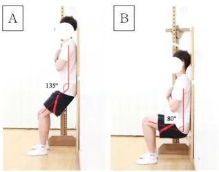
A B
Figure 1. Wall squat (A: Initial position, B:
Full squat position).
Figure 2. Strength measurement method.
er width apart and leaned against the wall. At this time, the knee was flexed to 135˚ and the dorsi- flexion of the ankle was 0˚ (Jung et al., 2017). The lumbopelvic region maintained a neutral position, al- lowing the lumbar curve to be flat on the wall (Figure 1A; initial position). A full squat position was defined as the hip flexed to 80˚ in the initial position maintaining a flat lumbar curve (Figure 1B).
The wall squat motion involved moving from the in- itial position to a full squat position for 5 s, main- taining that position for 5 s, and moving from the full squat position to the initial position for 5 s. This was counted as one cycle. The knee flexion angle between the initial and full squat positions was measured using the level and slope finder of the mo- bile phone application Clinometer (Plaincode Software Solutions, Stephanskirchen, Germany); a target bar was placed just below the ischial tuberosity in the full squat position (Figure 1B). Metronum Beats (Stonekick, Australia) was used to control the time spent descending, holding the full squat position, and ascending with a five-count metronome beat set at 60 beats/min. In order to unify the frictional force generated, the participants wore the same clothes and performed the wall squat.
Strength measurements
Hip and knee extensor strength were measured with Smart KEMA tension sensor (KOREATECH
Co, Ltd, Seoul, Korea). The cell had a measurement range of 0–1,960 N, with an accuracy of 4.9 N and a sampling rate of 10 ㎐. To measure hip extensor strength, participants flexed the knee to 90˚ in the prone position while the leg was slightly off to the side of the table. The thigh strap was fixed to the femur 2 ㎝ above the popliteal fossa. The examiner adjusted the length of the restraining belt at 5˚ of hip extension (Figure 2A). The examiner fixed the lumbar spine rotation of the participant during hip extension. The participant performed hip extension against a strap anchored by a glass suction cup for an maximal voluntary isometric contraction (MVIC) twice for 5 s each time. The participants sat upright on the edge of the therapeutic table to measure knee extensor strength at 90˚ hip and knee flexion. To fix the restraining belt, a glass suction cup was fixed on the floor and an ankle strap connected to the re- straining belt was fixed to the participant’s ankle.
The length of the restraining belt was adjusted so that the participants could reach 45˚ of knee ex- tension (Figure 2B). The participant performed a knee extension against the strap anchored by the glass suction cup for an MVIC twice for 5 s each time. The participants were shown how to stabilize themselves by holding on to the side of the table with their hands while sitting upright. Strength was analyzed by averaging the middle 3 s of each 5-s measurement. Strength was normalized by body weight (N/㎏). The intra-session reliability of the strength measurements was calculated using data from two trials for each participant. An intra-class
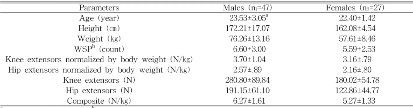
Parameters Males (nl=47) Females (n2=27)
Age (year) 23.53±3.05a 22.40±1.42
Height (㎝) 172.21±17.07 162.08±4.54
Weight (㎏) 76.26±13.16 57.61±8.46
WSPb (count) 6.60±3.00 5.59±2.53
Knee extensors normalized by body weight (N/㎏) 3.70±1.04 3.16±.79
Hip extensors normalized by body weight (N/㎏) 2.57±.89 2.16±.80
Knee extensors (N) 280.80±89.84 180.02±54.78
Hip extensors (N) 191.15±61.10 122.86±44.77
Composite (N/㎏) 6.27±1.61 5.27±1.33
amean±standard deviation,bwall squat performance for maximum count.
Table 1. Test results for all participants (N=74)
correlation coefficient (ICC) [3,1] model and 95% con- fidence intervals (CIs) were used to evaluate the in- tra-session reliability of each strength measurement.
Strength measurement during knee extension demon- strated excellent intra-session reliability (ICC[3,1]=.96, 95% CI: .926-.976); intra-session reliability was also good for hip extension measurement (ICC[3,1]=.90, 95% CI: .819-.940). And differences between male and female of muscle stiffness in the lower limbs can af- fect muscle strength (Blackburn et al, 2004; Granata et al, 2002; Harris-Hayes et al, 2009). Therefore, strength data was analyzed separately.
Statistical analysis
A power of 95% and a level of .05 were assumed, and the effect size (males: .36, females: .59) was cal- culated using Pearson’s correlation coefficient from squared multiple correlations (males: .265, females:
.372) using G*Power software to calculate the sample size. As a result of the power analysis, at least 46 males and 24 females were required. All statistical analyses were conducted using SPSS software ver.
22.0 (SPSS; IBM corp, Armonk, NY, USA). Pearson’s correlation and multiple regression analyses with stepwise selection were performed separately for each sex. Pearson’s correlation coefficients (r) were analyzed to examine the relationship between hip and knee extensor strength and WSP. Correlations were considered significant at p<.05. Multiple regression models with a stepwise selection were conducted to investigate which strength variables contributed most significantly to WSP for hip and knee extensor
strength, hip and knee extensor strength normalized by body weight, and a composite strength value (combined hip and knee extensor strength normalized by body weight) as independent variables, with WSP as the dependent variable. The determination co- efficient (adjusted R²) indicates variation in WSP that was explained by the regression variables.
Results
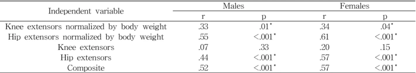
Results of count of WSP and hip and knee ex- tensor strength are summarized in Table 1. WSP was significantly related to hip extensor strength normalized by body weight (r=.55, p<.05), knee ex- tensor strength normalized by body weight (r=.33, p<.05), hip extensor strength (r=.44, p<.05), and the composite value (r=.52, p<.05) in males. WSP was significantly related to knee extensor strength nor- malized by body weight (r=.34, p<.05), hip extensor strength normalized by body weight (r=.61, p<.05), hip extensor strength (r=.57, p<.05), and the compo- site value (r=.57, p<.05) in females (Table 2).
Multiple regression analysis revealed that one varia- ble, hip extensor strength normalized by body weight, explained 30% of the variance in males with the fol- lowing model: Y=1.812+(hip extensor strength×.182), where the adjusted R² was .29 and standard error (SE) of the estimate (SEE) was 2.53 (Table 3). In females, hip extensor strength normalized by body weight explained 37% of the variance with the fol- lowing model: Y=1.412+(hip extensor strength×.190),
Independent variable Males Females
r p r p
Knee extensors normalized by body weight .33 .01* .34 .04*
Hip extensors normalized by body weight .55 <.001* .61 <.001*
Knee extensors .07 .33 .20 .15
Hip extensors .44 <.001* .57 <.001*
Composite .52 <.001* .57 <.001*
*p<.05.
Table 2. Correlations between independent variables and WSP
Model R R2 Adjusted R2 SE of the estimate
1 .55 .31 .29 2.53
Predictors: (constant), hip extensors/body weight.
Table 3. Stepwise linear regression model for males
Model R R2 Adjusted R2 SE of the estimate
1 .61 .37 .35 2.04
Predictors: (constant), hip extensors/body weight.
Table 4. Stepwise linear regression model for females
where the adjusted R² was .35 and SEE was 2.04 (Table 4). Figure 3, 4 is normal probability plot of regression standardized residual.
Discussion
Our study investigated the relationship between hip and knee extensor strength and WSP. The wall squat is important for activities and efficient exercise to increase hip and knee extensor strength. However, it has remained unclear which hip and knee extensor strengths are associated with WSP. This study dem- onstrated that hip extensor strength normalized by body weight is an important factor for WSP in males and females. These results may help in de- signing exercise programs to improve WSP in par- ticipants with poor performance.
Previous studies (Claiborne et al, 2006; Kim et al, 2015) of lower extremity strength during weight-bear- ing exercise have used strength normalized by body weight. The wall squat is a weight-bearing exercise;
therefore, body weight should be considered in wall squat studies. Here, hip extensor strength normalized by body weight had a higher Pearson’s correlation co- efficient (males: .55, females: .61) than did net hip ex-
tensor strength (males: .44, females: .57). We, there- fore, recommend designing wall squat exercise pro- grams considering hip extensor strength normalized by body weight, rather than net hip extensor strength.
The wall squat involves a descending phase to a full squat position (80˚ knee flexion) and an ascending phase to the initial position (135˚ knee flexion).
Previous studies (Blanpied, 1999; Chéron et al, 1997;
Dionisio, et al., 2008; Escamilla et al., 1998; Flanagan et al, 2003) showed that activation of the quadriceps as knee extensors was prominent during the descend- ing phase of the squat. During the ascending phase, the activation of the vastus medialis oblique was 55-66% of the MVIC; that of the gluteus maximus (GM) as a hip extensor was 56-86% of the MVIC (Ayotte, et al., 2007). During a wall squat, both phas- es must be performed; it is difficult to perform a squat successfully with only one of these muscle strengths. Therefore, a composite strength value that combined the strengths of the hip and knee extensors was considered as a factor related to WSP in this study. The composite strength value was significantly correlated with WSP (males: r=.55, p<.01; females:
r=.57, p<.01), although it was not included in the re- gression model.
Most squat exercise studies have focused on the
A B
Figure 3. Normal probability plot of regression standardized residual in female (A: Hip extensor/body weight, B: Knee extensor/body weight).
A B
Figure 4. Normal probability plot of regression standardized residual in male (A: hip extensor/body weight, B: knee extensor/body weight).
quadriceps. However, Bryanton et al. (2015) reported that the quadriceps and GM operate synergistically, and that the GM is the primary hip extensor during squat exercises. Also, during wall squatting, it de- scends in the descending phase in the direction of gravity, but in the ascending phase it should rise in the opposite direction of gravity against body weight.
Hence, hip extensors (e.g., the GM), which require great force in the ascending phase, might play an important role in successful WSP. In this study, hip extensor strength normalized by body weight was included in the regression model (males: adjusted R²=.29, females: adjusted R²=.35). This finding sug-
gests that hip extensor strength normalized by body weight plays an important role in WSP.
There are several limitations to this study. First, we considered only knee and hip extensors; we did not investigate the strength of other lower extremity muscles that may have affected the strength of the ankle joint, including ankle plantarflexors and dorsi- flexors (Macrum et al, 2012). Second, we did not in- vestigate the kinematics of the lower extremities during wall squats, although the ankle and hip joint angles were controlled. Additional studies should to be conducted to evaluate the kinematics of the lower extremities during wall squats to investigate asym-
metrical movement compared to that in other squat exercises. Third, this was a cross-sectional study.
The results show that WSP was correlated with hip extensor strength normalized by body weight. Thus, future studies should evaluate the increase in WSP after improving strengthening programs for the hip extensors normalized by body weight. Fourth, the present study measured the isometric strength.
Therefore, the results of this study did not consider torque. Finally, we could not control for the weight of the participants, although we controlled the extent of the wall slide by using the same wall.
Sport coaches and staff should assess and develop strength to enhance WSP. The present findings document the relationship between hip and knee ex- tensor strength and WSP. Our findings have im- portant practical applications for individuals who must perform repeated squats, including basketball players, weightlifters, wrestlers, and jumpers.
Because hip extensor strength normalized by body weight was the most influential variable affecting WSP among lower extremity extensor strength, hip extensor strength should be increased according to body weight to improve WSP.
Conclusion
The current findings suggest that hip extensor strength normalized by body weight is proportionally related with WSP. Thus, it could be concluded that hip extensor strength normalized by body weight is important factor for WSP in males and females.
Therefore, it is recommended that rehabilitation pro- grams designed for improving WSP should focus on strengthening hip extensor strength considering body weight.
Acknowledgements
The authors acknowledge financial and admin-
istrative support provided by Yonsei University Research Fund [2018-51-0001].
References
Anderson R, Courtney C, Carmeli E. EMG analysis of the vastus medialis/vastus lateralis muscles utilizing the unloaded narrow-and wide-stance squats. J Sport Rehabil. 1998;7(4):236-247.
Aspe RR, Swinton PA. Electromyographic and kinetic comparison of the back squat and overhead squat.
J Strength Cond Res, 2014;28(10):2827-2836.
https://doi.org/10.1519/JSC.0000000000000462 Ayotte NW, Stetts DM, Keenan G, et al.
Electromyographical analysis of selected lower extremity muscles during 5 unilateral weight- bearing exercises. J Orthop Sports Phys Ther.
2007;37(2):48-55. https://doi.org/10.2519/jospt.2007.
2354
Bazyler CD, Sato K, Wassinger CA, et al. The effi- cacy of incorporating partial squats in maximal strength training. J Strength Cond Res. 2014;
28(11):3024-3032. https://doi.org/10.1519/JSC.00000 00000000465
Bevilaqua-Grossi D, Felicio LR, Simões R, et al.
Electromyographic activity evaluation of the pa- tella muscles during squat isometric exercise in individuals with patellofemoral pain syndrome.
Revista Brasileira de Medicina do Esporte. 2005;
11(3):159-163.
Blanpied PR. Changes in muscle activation during wall slides and squat-machine exercise. J Sport Rehabil. 1999;8(2):123-134.
Blackburn JT, Riemann BL, Padua DA, and Guskiewicz KM. Sex comparison of extensibility, passive, and active stiffness of the knee flexors.
Clin Biomech (Bristol, Avon). 2004;19(1):36-43.
Bolgla L, Cook N, Hogarth K, et al. Trunk and hip electromyographic activity during single leg squat exercises do sex differences exist? Int J Sports Phys Ther. 2014;9(6):756-764.
Boling MC, Bolgla LA, Mattacola CG, et al.
Outcomes of a weight-bearing rehabilitation pro- gram for patients diagnosed with patellofemoral pain syndrome. Arch Phys Med Rehabil. 2006;
87(11):1428-1435. https://doi.org/10.1016/j.apmr.
2006.07.264
Bryanton MA, Carey JP, Kennedy MD, et al.
Quadriceps effort during squat exercise depends on hip extensor muscle strategy. Sports Biomech.
2015;14(1):122-138. https://doi.org/10.1080/14763141.
2015.1024716
Cheatham SW, Stull KR, Fantigrassi M, et al. Hip musculoskeletal conditions and associated factors that influence squat performance: A systematic review. J Sport Rehabil. 2018;27(3):263-273 https://doi.org/10.1123/jsr.2016-0246
Chéron G, Bengoetxea A, Pozzo T, et al. Evidence of a preprogrammed deactivation of the hamstring muscles for triggering rapid changes of posture in humans. Electroencephalogr Clin Neurophysioly.
1997;105(1):58-71. https://doi.org/10.1016/s0924-980x (96)96544-3
Cho M. The effects of modified wall squat exercises on average adults’ deep abdominal muscle thick- ness and lumbar stability. J Phys Ther Sci.
2013;25(6):689-692. https://doi.org/10.1589/jpts.25.
689
Claiborne TL, Armstrong CW, Gandhi V, et al.
Relationship between hip and knee strength and knee valgus during a single leg squat. J Appl Biomech. 2006;22(1):41-50.
Delitto R., Rose SJ. An electromyographic analysis of two techniques for squat lifting and lowering.
Phys Ther. 1992;72(6):438-448. https://doi.org/
10.1093/ptj/72.6.438
Dionisio VC, Almeida GL, Duarte M, et al. Kinematic, kinetic and EMG patterns during downward squatting. J Electromyogr Kinesio. 2008;18(1):
134-143. https://doi.org/10.1016/j.jelekin.2006.07.010 Escamilla RF, Fleisig GS, Zheng N, et al.
Biomechanics of the knee during closed kinetic chain and open kinetic chain exercises. Med Sci
Sports Exerc. 1998;30(4):556-569. https://do- i.org/10.1097/00005768-199804000-00014
Escamilla RF, Fleisig GS, Zheng N, et al. Effects of technique variations on knee biomechanics dur- ing the squat and leg press. Med Sci Sports Exerc. 2001;33(9):1552-1566. https://doi.org/10.1097/
00005768-200109000-00020
Flanagan S, Salem GJ, Wang MY, et al. Squatting exercises in older adults: kinematic and kinetic comparisons. Med Sci Sports Exer. 2003;35(4):
635-643. https://doi.org/10.1249/01.MSS.0000058364.
47973.06
Fry AC, Smith JC, Schilling BK. Effect of knee po- sition on hip and knee torques during the bar- bell squat. J Strength Cond Res. 2003;17(4):
629-633.
Granata KP, Wilson SE, and Padua DA. Gender dif- ferences in active musculoskeletal stiffness. part I. quantification in controlled measurements of knee joint dynamics. J Electromyogr Kinesiol.
2002;12(2):119-126.
Harris-Hayes M, Sahrmann SA, and Van Dillen LR.
Relationship between the hip and low back pain in athletes who participate in rotation-related sports. J Sport Rehabil. 2009;18(1):60-75.
Jung SH, Kim MH, Hwang UJ, Kim JH, Kwon OY.
(2017). Comparison of Knee Extensor and Hip Extensor Strength According to Wall Squat Performance. Physical Therapy Korea. 24(1):
79-85.
Kasim, P. Optimizing squat technique. Strength and Conditioning Journal. 2007;29(6):10-13.
Kim SH, Kwon OY, Park KN, et al. Lower ex- tremity strength and the range of motion in re- lation to squat depth. J Hum Kinets. 2015;45(1):
59-69. https://doi.org/10.1515/hukin-2015-0007 Kritz M, Cronin J, Hume P. The bodyweight squat: A
movement screen for the squat pattern. Strength
& Conditioning Journal. 2009;31(1):76-85.
Macrum E, Bell DR, Boling M, et al. Effect of limit- ing ankle-dorsiflexion range of motion on lower extremity kinematics and muscle-activation pat-
This article was received September 16, 2019, was reviewed September 16, 2019, and was accepted November 8, 2019.
terns during a squat. J Sport Rehabil. 2012;
21(2):144-150.
Matsumoto H, Suda Y, Otani T, et al. Roles of the anterior cruciate ligament and the medial collat- eral ligament in preventing valgus instability. J Orthop Sci. 2001;6(1):28-32.
McCurdy KW, Langford GA, Doscher MW, et al.
The effects of short-term unilateral and bilateral lower-body resistance training on measures of strength and power. J Strength Cond Res.
2005;19(1):9-15. https://doi.org/10.1519/14173.1 McKean MR, Dunn PK, Burkett BJ. The lumbar and
sacrum movement pattern during the back squat exercise. J Strength Cond Res. 2010;24(10):2731- 2741. https://doi.org/10.1519/JSC.0b013e3181e2e166 Natri A, Kannus P, JÄrvinen M. Which factors pre-
dict the long-term outcome in chronic patellofe- moral pain syndrome? A 7-yr prospective fol- low-up study. Med Sci Sports Exerc. 1998;
30(11):1572-1577. https://doi.org/10.1097/00005768- 199811000-00003
Neumann DA. Kinesiology of the musculoskeletal system-e-book: foundations for rehabilitation:
Elsevier Health Sciences. 2013
Noyes F, Butler D, Grood E, et al. Biomechanical analysis of human ligament grafts used in knee-ligament and reconstructions. J. Bone Joint
Surg. Am. 1984;66(3):344-352.
Paoli A, Marcolin G, Petrone N. The effect of stance width on the electromyographical activity of eight superficial thigh muscles during back squat with different bar loads. J Strength Cond Res. 2009;23(1):246-250.
Schoenfeld BJ. Squatting kinematics and kinetics and their application to exercise performance. J Strength Cond Res. 2010;24(12):3497-3506.
https://doi.org/10.1519/JSC.0b013e3181bac2d7 Vakos J, Nitz A, Threlkeld A, et al. Electromyographic
activity of selected trunk and hip muscles during a squat lift. Effect of varying the lumbar posture.
Spine (Phila Pa 1976). 1994;19(6):687-695. https://
doi.org/10.1097/00007632-199403001-00008
Witvrouw E, Danneels L, Van Tiggelen D, et al. Open versus closed kinetic chain exercises in patellofe- moral pain: a 5-year prospective randomized study. Am J Sports Med. 2004;32(5):1122-1130.
https://doi.org/10.1177/0363546503262187