Res. Plant Dis. 16(3) : 232-237 (2010)
© The Korean Society of Plant Pathology
채소(가지, 알타리무, 슈가로프)에 발생한 토마토반점위조바이러스 ( Tomato spotted wilt virus) 발생과 병징 특성
조점덕·김진영1·김정수2*·최홍수2·최국선 국립원예특작과학원, 1경기도농업기술원, 2국립농업과학원
Occurrence and Symptoms of Tomato spotted wilt virus on Egg Plant, Whole Radish and Sugar Loaf in Korea
Jeom-Deog Cho, Jin-Young Kim1, Jeong-Soo Kim2*, Hong-Soo Choi2 and Gug-Seoun Choi National Institute of Horticultural & Herbal Science, R.D.A. Suwon 441-440, Korea
1Gyunggi Province Agricultural Research and Extension Service, Hwasung 445-972, Korea
2National Academy of Agricultural Science, R.D.A. Suwon 441-440, Korea (Received on August 2, 2010; Accepted on September 29, 2010)
Tomato spotted wilt virus (TSWV) was occurred on the three vegetables of egg plant (Solanum melongena), whole radish (Raphanus acanthiformis) and sugar loaf (Cichorium intybus) at Anyang area infested with TSWV. Whole radish was produced the symptoms of necrotic spots on the leaves, and necrosis and malformation on the roots by TSWV. Egg plant was induced the symptoms of typical multiple ring spots on the leaves and necrotic rings on the fruits. Sugar loaf was infected severely with the typical symptoms of ring spots on the leaves and stunt. The three isolates of TSWV could infect locally on the indicator plants of Chenopodium amaranticolor, C. quinoa and Nicotiana debney, and systemically on N. glutinosa, N.
benthamiana and Datura stramonium. Two TSWV isolates from egg plant and sugar loaf were very similar in virulence. However, the virulence of TSWV from whole radish was very different as local infection on 5 Nicotiana species including N. tabacum ‘Xanthi NC’.
Keywords : Egg plant, Sugar loaf, Tomato Spotted Wilt Virus, Whole radish
토마토반점위조바이러스(Tomato spotted wilt virus;
TSWV)는 Bunyaviridae Topovirus의 대표 바이러스로서 1919년 호주에서 Brittlebank가 처음 보고하였으며(우리나 라에서는 2003년 충남 예산의 파프리카에서 한 개의 식 물체가 감염된 이후에 2004년 경기도 안양 지역에서 크 게 발생되었다(김 등, 2004). 그 후 2009년까지 경기, 충 남, 전북, 전남, 경남, 강원도에서 23개 시군에 지속적으 로 확산되고 있다(최 등, 2010). TSWV는 전 세계적으로 발생하여 피해를 주고 있어 국가간 지역간 이동금지 바 이러스로 지정되어 관리되고 있다. TSWV는 이와 같이 지리적 분포가 넓고 기주 범위도 단자엽 및 쌍자엽 식물
의 82과 900여종으로 매우 넓다. 특히 토마토, 고추, 상 추, 국화 등 주요 재배작물에 피해가 50~90%의 수량감 소를 초래하고 있으며(Boiteux 등, 1993; Cho 등, 1986, 1989; van Zijl 등, 1986) 우리나라에서는 경기도 안양의 토마토, 충남 당진의 고추 및 국화의 경우 발생 하우스 의 모든 식물체가 감염되는 피해가 발생하였다. TSWV 가 대 발생한 하우스에서 토마토를 대상으로 매개충인 총채벌레의 발생생태 중 토양에서 번데기를 거치는 생활 환을 차단하여 TSWV를 방제하고자 토양소독방법을 개 발하여 85% 이상의 TSWV의 방제효과를 거두었다(Kim 등, 2009). 한편 TSWV의 신속한 진단을 위한 진단기술 개발은 핵산 분리과정 없이 유전자 진단을 할 수 있는 TSWV VC/RT-PCR을 개발하여 현장에서 활용하고 있다 (조 등, 2006). 우리나라에서 대 발생 지역인 경기 안양
*Corresponding author
Phone) +82-31-290-0430, Fax) +82-31-290-0406 Email) [email protected]
DOI: 10.5423/RPD.2010.16.3.232
채소류에 발생한 토마토반점위조바이러스 발생과 특성 233 과 충남 당진에서 감염된 채소 작물의 종류와 병징 특성
에 대하여 토마토, 고추, 시금치 등 14개 작물에 대하여 일차 보고된 바 있으며(조 등, 2005), 화훼 작물에서는 국 화, 봉선화, 과꽃 등 4개 작물에서 발생과 병징 특성을 보고하여 모두 19개 채소, 화훼작물에서 보고하였다(조 등, 2005; 김 등, 2006; 정 등, 2006). 본 논문에서는 채소 작물 14종 이외에 감염 작물로 조사된 가지, 알타리무 및 슈가로프 3종에 대한 발생과 병징 특성에 대하여 보고하 고자 한다.
재료 및 방법
감염 식물 채집. 경기도 안양 지역에서 TSWV의 전 형적인 원형반점 병징 등 바이러스병 증상을 보이는 것 을 육안으로 조사하였으며, 감염 작물은 가지(Solanum melongena), 알타리무(Raphanus acanthiformis), 슈가로프 (Cichorium intybus) 3종이었다.
유전자 진단. 유전자 진단(VC/RT-PCR)에 사용한 TSWV 의 특이 프라이머는 증폭산물이 777 bp인 ncp-F:
5'-ATGTCTAAGGTTAAGCTCAC-3', ncp-R: 5'-TCAA GCAAGTTCTGCGAGTT-3'이었으며 VC/RT-PCR 조건은 48oC, 45분; (94oC, 2분; 94oC, 30초; 50oC, 30초; 72 oC, 90 초) 35회; 72oC, 7분이었다(조 등, 2006). 증폭된 생산물 을 1.2% agarose gel에서 100V 전압에서 40분 전기영동 한 후 증폭산물을 염색하여(SafeViewTM) 관찰하였다(조 등, 2005).
생물검정. 바이러스 병징을 나타내는 식물체를 채집하 여 식물체 무게의 4배 0.01 M 인산완충액(pH 7.0)과 함 께 유발을 이용하여 마쇄한 다음 즙액을 600 mesh 카보 랜덤을 뿌린 3~4엽기의 어린 건전한 지표식물에 인공 접 종하였으며, 접종 후 즉시 수도물로 깨끗이 세척하였다.
완충액에 마쇄하는 단계부터 접종단계까지 4oC를 유지하 여 실시하였다. 지표식물은 붉은 명아주 등 18종의 식물 을 이용하였고 접종 후 2~3주 동안 병징 발현 여부를 조 사하였다.
결과 및 고찰
TSWV 분리주 획득 및 병징 특성. 경기도 안양 관양 동 지역의 TSWV가 발생하고 있는 농가에서 채집한 가 지(Solanum melongena), 알타리무(Raphanus acanthiformis), 슈가로프(Cichorium intybus)의 3종의 작물에 감염된 바이 러스 분리주의 유전자 진단 및 생물검정 결과 모두 TSWV 이었다. TSWV에 감염된 알타리무는 식물체가 대부분 고
사하였다(Fig. 1A). 잎은 TSWV의 전형적인 병징인 원형 반점이 나타났으며 원형반점의 크기와 모양은 다양하였 다(Fig. 1B). 원형반점은 가장자리가 황갈색을 띤 괴저 반 점이었으며 잎이 전체적으로 황색 고사하였다. 뿌리는 가 로와 세로로 주름무늬가 형성되어 쭈글쭈글하였으며 기 형이었다(Fig. 1C). 이 주름무늬는 모두 갈색으로 조직이 괴사하였으며 뿌리 표피뿐만 아니라 내부까지 모두 조직 이 갈변 고사하여(Fig. 1D) 바이러스 병징으로 인한 직접 적 피해가 매우 컸다.
TSWV에 가장 병징 피해가 심한 토마토와 고추(조 등, 2005)와 같은 가지과 작물인 가지의 경우에도 병징이 매 우 심하게 나타났다. 어린 잎에서는 작은 원형반점이 무 수히 생겨 잎 전체가 황화 고사하였다(Fig. 1E). 그러나 비교적 노화된 잎에서는 TSWV의 전형적인 원형반점이 나타났다. 이 원형반점은 잎의 엽맥을 경계로 하지 않고 크기가 다양하게 커져 가면서 큰 원형반점을 형성하였다 (Fig. 1F). 과실에서는 원형반점이 크게 변하면서 조직이 고사하여 부분적으로 과실이 괴저로 변하였으며(Fig. 1G), 고사하지 않은 부분은 큰 반점이 움푹 들어가 과일이 마 르는 증상과 함께 기형이 되었다(Fig. 1H). 슈가로프는 엽 채류로서 잎을 이용하는 채소이지만 TSWV에 감염되면 잎이 전개가 되지 않고 식물체 전체가 위축되어 땅 바닥 에 붙어서 생육을 하지 못하였다(Fig. 1I). TSWV에 감염 되어 약간의 생육이 된 식물체의 잎에서는 전형적인 원 형반점이 나타났으며 식물체가 전체적으로 황화 괴저로 변화하였다(Fig. 1J, K).
생물적 특성. 가지, 알타리무, 슈가로프에서 분리한 TSWV 순수 분리주의 지표식물에서의 반응은 Table 1에 서 보는 바와 같다. 알타리무에서 분리한 TSWV의 생물 적 특성을 조사하기 위하여 Chenopodium quinoa 등 12 종의 지표식물에 인공 접종한 결과 7종은 국부 감염되었 으며, Nicotiana glutinosa 등 5종의 지표식물에서는 전신 감염되었다. 흰 명아주(C. quinoa)에서는 큰 원형반점(Fig.
2A), N. glutinosa의 접종엽에는 원형반점이 나타났으며 (Fig. 2B) 상엽은 괴저로 고사하였다(Fig. 2C). 가지에서 분리한 TSWV는 N. glutinosa를 포함한 19종의 지표식물 에 접종한 결과 15종의 지표식물에 전신 감염하였으나 명 아주(C. quinoa)와 담배(N. debney)에서는 국부 감염이었 고 Tetragonia expansa는 감염되지 않았다. N. glutinosa (Fig. 2D)와 N. rustica(Fig. 2E)에서는 접종엽과 상엽에 모 두 괴저반점과 잎 괴저 병징이 나타났다. 특히 독말풀 (Datura stramonium) 접종잎에서는 전형적인 다중 원형반 점이 나타났으며(Fig. 2F) 식물체가 기형이 되었다. 슈가 로프에서 분리한 TSWV는 C. quinoa 등 16종의 지표식
Fig. 1. Almost all plants of whole radish infected with TSWV died in natural field (A). Leaf of whole radish produced various shapes and sizes of the typical ring spots (B). On roots, TSWV produced malformation and necrosis on surface (C) and inner tissues (D). The infected roots were produced severe wrinkles horizontally and compactly. Chlorotic spots and ring spots were produced on the newly developed leaf (E) and typical ring spots were induced on the older leaf (F) in eggplant. The infected fruits of egg plant were necrotized (G) and followed by dent surface with drying (H). In sugar loaf, all plants were stunted severely (I). The leaves of sugar loaf were produced yellowing with necrosis (J) and the small chlorotic spots and necrotic ring spots were produced (K).
채소류에 발생한 토마토반점위조바이러스 발생과 특성 235
물에 인공접종한 결과 명아주(C. quinoa, C. amaranticolor) 와 N. debney에서는 국부 감염이었으며, N. glutinosa 등 13종의 지표식물에서는 전신 감염이었다. 슈가로프 분리 주는 C. quinoa(Fig. 2G)와 N. rustica(Fig. 2H)에서 매우 큰 원형반점이 나타났으며 이 후 병징이 잎 괴저로 진전 되었다. 또한 N. glutinosa(Fig. 2I), N. tabacum ‘Xanthi NC’(Fig. 2J) 그리고 N. tabacum ‘Ky-57’(Fig. 2K)에서는 접종엽에 원형반점은 잘 나타나지 않고 괴저반점이 나타 났으며 병징이 진전됨에 따라 식물체는 잎 괴저와 기형 을 보였다.
알타리무, 가지 및 슈가로프에서 분리한 TSWV 분리주 는 대체로 지표식물에서의 병원성이 비슷하였지만, 알타 리무에서 분리한 TSWV는 N. tabacum ‘Xanthi NC’ 등 5 종에서 국부 감염되어 병원성이 상이하였다. 또한 N. tabacum
‘Xanthi NC’ 등 4종과 Tetragonia expansa의 경우 알타리 무에서 분리한 TSWV는 국부감염되었으나 가지에서 분 리한 TSWV는 감염이 되지 않았으며, Physalis floridana
에서는 가지에서 분리한 TSWV는 전신 감염이었으나 알 타리무에서 분리한 TSWV는 감염이 되지 않아 동일한 발 생지역인 안양지역 내에서도 병원성변이가 매우 다양하 게 일어나고 있었다. 우리나라에서 발생한 TSWV 8개의 분리주의 생물적 특성을 보면(조 등, 2009), 감염 식물체 와 발생 지역에 따라서 분리주의 병원성이 전신 감염과 국부 감염 또는 무병징의 병원성 차이가 나타났으며 앞 으로 분리주별 병원성과 관련된 유전적 진화연구가 보다 심도 있게 연구가 이루어져야 할 것이다. 특히 가지, 알 타리무, 슈가로프 3종의 식물은 안양지역에서 모니터링 한 결과 2004년부터 2006년에는 감염이 확인되지 않았으 나 2007년에 TSWV의 감염이 확인되어, 상습 발생지의 경우 TSWV와 기주식물과 매개충과의 상호작용에 의한 병원성 변이가 발생한 것으로 추정되므로(Whitfield, 2005) 이에 대한 유전적, 진화적 연구가 더 필요할 것으로 생각 되며, TSWV는 기주범위가 매우 넓은 바이러스이므로 계 속적인 모니터링이 필요한 것으로 생각된다.
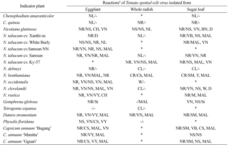
Table 1. Biological characteristics of Tomato spotted wilt virus isolated from eggplant, whole radish and sugar loaf on the indicator plants by mechanical inoculation
Indicator plant Reactionsa of Tomato spotted wilt virus isolated from Eggplant Whole radish Sugar loaf
Chenophodium amaranticolor NL/- * NL/-
C. quinoa NL/- NR/- NR/-
Nicotiana glutinosa NR/NS, CH, VN NS/NS, NL NR/NS, VN, BN, D
N. tabacum cv. Xanthi nc NR/D NL/- NR/VB, NS, MAL
N. tabacum cv. White Burly NS/NS, NR, NL * NR/MAL, VN
N. tabacum cv.Samsun NN NR/VN, NR, NS, MAL * *
N. tabacum cv. Samsun NR, VN/NR, MAL NL/- NR/VN, NR
N. tabacum cv. Ky-57 * NR, VN/NS, MAL NR/NS, MAL, VN
N. debneyi NR/- CL/- CL/-
N. benthamiana NR, VN/MAL, NR CR/Ch, MAL CR/SM, Y, MAL
N. occidentalis NR, VN/NS, VN, MAL W/- *
N. clevelandii NR, VN/NS, MAL, VN CL/- NR/VN, NS, W, D
N. rustica NR, VN/VY, CH * NR/M, MAL
Gomphrena globosa NR/St -/MAL VN, NS/St
Tetragonia expansa -/- CL/- *
Datura stramonium NR, VN/VY, MAL NR/VN, MAL NR/SM, MAL
Physalis floridana NS, VN/CS, VY -/- *
Capsicum annuum ‘Bugang’ NR/CS, MAL, VN * NR/SM, VB, CS, MAL
C. annuum ‘Manitta’ NR/VY, MAL * NS/NS
C. annuum ‘Gguari’ NR/CS, VY, MAL * NR/SM, NS, MAL
aBN, bud necrosis; CR, chlorotic ring; CL, chlorotic local; D, death; NS, necrotic spot; NL, necrotic local; MAL, malformation; NR, necrotic ring; NS, necrotic spot; St, stunt; SM, severe mosaic; VN, vein necrosis; VY, vein yellowing; W, wilt; Y, yellowing; *, not tested; -, no symptom;
Inoculated leaf/Upper leaf.
Fig. 2. TSWV isolate from whole radish induced large necrotic ring spots in the inoculated leaf of Chenopodium quinoa (A) and Nicotiana glutinosa (B). The whole radish isolate killed N. glutinosa with leaf necrosis (C). TSWV isolate from egg plant produced necrotic ring spots on the inoculated and upper leaf of N. glutinosa (D) and N. rustica (E). Multiple ring spots were produced on the inoculated leaf of Datura stramonium (F). TSWV isolate from sugar loaf induced large necrotic ring spots on the inoculated leaf of C.
quinoa (G) and N. rustica (H). Systemic infection were occurred with necrotic spots on the inoculated leaf of N. glutinosa (I), N. tabacum
‘Xanthi NC’ (J) and N. tabacum ‘Ky-57’ (K).
채소류에 발생한 토마토반점위조바이러스 발생과 특성 237
요 약
Tomato spotted wilt virus(TSWV)의 상습 발생지인 안 양지역에서 가지, 알타리무, 슈가로프에서 TSWV가 발생 하였다. 알타리무에서는 잎에 괴저반점과 뿌리에 괴저 병 징이 나타났다. 가지에서는 잎에 전형적인 다중 원형반점 을 나타냈으며 열매에 심한 괴저를 나타냈다. 슈가로프에 서는 잎에 전형적인 원형반점과 심한 위축 병징이 나타 났다. 가지, 알타리 무, 슈가로프에서 분리한 TSWV의 생 물적 특성은 흰 명아주, 붉은 명아주, 담배(N. devney)에 서는 국부 감염이었으며, 담배(N. glutinosa, N. benthamiana) 와 독말풀(D. stramonium)에서는 전심감염이었다. 가지와 슈가로프에서 분리한 TSWV는 병원성이 유사하였으나, 알타리 무에서 분리한 TSWV는 N. tabacum ‘Xanthi NC’
등 5종의 담배에서 국부 감염되어 병원성이 매우 상이하였다.
참고문헌
Brittlebank, C. C. 1919. Tomato diseases. J. Agric. 17: 231-235.
Cho, J. D., Kim, J. S., Kim, J. Y., Choi, G. S. and Chung, B. N.
2009. Biological characteristics and nucleotide relationships in Korean Tomato spotted wilt virus isolates. Plant Pathol. J. 25:
26-37.
Boiteux, L. S., Nagata, T., Dutra, W. E. and Fonseca, M. E. N.
1993. Sources of resistance to tomato spotted wilt virus (TSWV) in cultivated and wild species of Capsicum.
Euphytica 67: 89-94.
Cho, J. D., Kim, J. S., Kim, J. Y., Kim, J. H., Lee, S. H., Choi, G.
S., Kim, H. R. and Chung, B. N. 2005. Occurrence and symptoms of Tomato spotted wilt virus on vegetables in Korea. Res. Plant Dis. 11: 213-216.
Cho, J. D., Kim, J. S., Kim, H. R., Chung, B. N. and Ryu, K. H.
2006. Convenient nucleic acid detection for Tomato spotted
wilt virus: Virion captureed/RT-PCR (VC/RT-PCR). Res.
Plant Dis. 12: 139-143.
Cho, J. J., Mau, R. F. L., German, T. L. Hatman, R. W., Yudin, L.
S., Gonsalves, D. and Provvidneti, R. 1989. A multidisciplinary approach to management of Tomato spotted wilt virus in Hawai. Plant Dis. 73: 375-383.
Cho, J. J., Mau, R. F. L., Gonsalves, D. and Mitchell, W. C. 1986.
Reservoir weed host of Tomato spotted wilt virus. Plant Dis.
70: 1014-1017.
Choi, H. S., Lee, S. H., Kim, M. K., Kwak, H. R., Kim, J. S., Cho, J. D. and Choi, G. S. 2010. Occurrence of virus diseases on major crops in 2009. Res. Plant Dis. 16: 1-9.
Chung, B. N., Park, H. S., Jung, J. A. and Kim, J. S. 2006.
Occurrence of Tomato spotted wilt virus in chrysanthemum in Korea. Plant Pathol. J. 22: 230-234.
Goldbach, R. and Peters, D. 1994. Possible causes of the emergence of tospovirus diseases. Seminars of Virology 5:
113-120.
Kim, J. H., Choi, G. S., Kim, J. S. and Choi, J. K. 2004.
Characterization of Tomato spotted wilt virus from paprika in Korea. Plant Pathol. J. 20: 297-301.
Kim, J. Y., Cho, J. D., Kim, J. S., Hong, S. S., Lee, J. G., Choi, G.
S. and Lim, J. W. 2009. Reduction of Tomato spotted wilt virus on table tomatoes in greenhouses by soil fumigation. Plant Pathol. J. 25: 151-156.
Kim, J. S., Cho, J. D., Kim, J. Y., Lee, S. H., Chung, B. N. and Kim, J. H. 2006. Occurrence and symptoms of Tomato spotted wilt virus on ornamental plants in Korea. Res. Plant Dis. 12:
148-151.
Whitfield, A. E., Ullman, D. E. and German, T. L. 2005.
Tospovirus-thrips interactions. Annu. Rev. Phytopathol. 43:
459-489.
van Zijl, J. J. B., Bosch, S. E. and Coetzee, C. P. J. 1986. Breeding tomatoes for processing in South Africa. Acta Hort. 194: 69- 75.