online©ML Comm
-
160
- 대한두경부종양학회지제 25 권 제 2 호 2009
경부 기도 전방에 발생한 유피낭종 1예
청주성모병원 이비인후과,1 병리학과2
김윤환1·오장근1·신향미2·김영생1
= Abstract =
Dermoid Cyst Anterior to the Cervical Trachea
Yoon Hwan Kim, MD1, Jang-Gun Oh, MD1, Hyang-Mi Shin, MD2, Young Saeng Kim, MD1 Department of Otolaryngology-Head & Neck Surgery1 and Pathology,2 Cheongju Saint Mary’s Hospital, Cheongju, Korea
Dermoid cysts are benign neoplasm that is derived from both ectoderm and mesoderm. They may be found at various sites of the body, but are extremely rare to occur in the lower neck. We present the case of a 22-year old woman with a midline cystic mass anterior to the cervical trachea. The mass was excised under local anesthesia.
Histopathologically the diagnosis was confirmed as a dermoid cyst.
KEY WORDS:Dermoid cyst·Neck·Trachea.
서 론
유피낭종은 발생 초기과정에서 외배엽 및 중배엽세포로 부터 발생하는 선천성 양성종양이다. 신체의 어느 부위에서도 발생할 수 있으나 두경부에서의 발생은 약 7%로 드문 편이 다. 두경부에서는 외측 눈썹 1/3 부위가 가장 호발 부위이며, 80% 이상이 안와, 구강, 비강 순으로 발생한다. 그러나 경 부 하부에서의 유피낭종은 매우 드물어 국내외 문헌 고찰에 서 보고된 바가 없다. 최근 저자들은 갑상선 하부의 경부 기 도 전방에서 발생한 유피낭종을 치험하였기에 이를 문헌 고 찰과 함께 보고하는 바이다.
증 례
22세 여자 환자가 약 1년 전부터 서서히 커지는 전경부 종물을 주소로 내원하였다. 신체검진에서 종물은 흉골상절 흔(suprasternal notch)에서 약 3cm 상부의 정중부에 위치
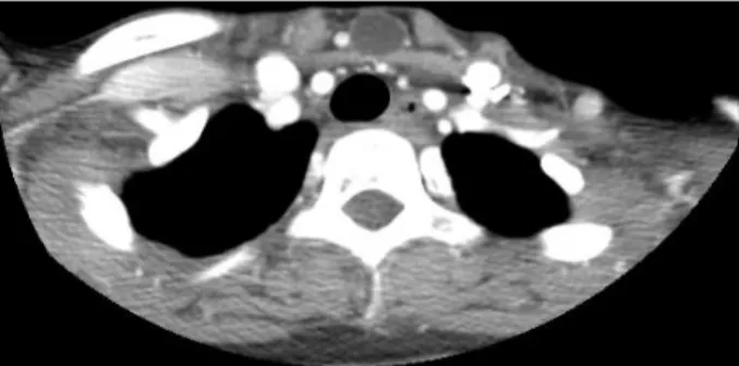
해 있었다. 부드럽고 무통성 이었으며 연하 시 가동성은 없었 다. 경부 초음파 검사에서 경부 기도 전방에 주변과 경계가 명 확하고 내부에는 혼합 에코도를 보이는 원형의 종물이 관찰 되었다. 조영 증강 전산화 단층 촬영에서는 내부에 균일한 음 영을 보이는 낭성 종물이 관찰되었으며, 경부 림프절 비대 소 견은 관찰되지 않았다(Fig. 1).
국소 마취하에 종물을 제거하였다. 흉골상절흔 약 3cm 상 방에 수평의 피부절개 후 종물을 노출시키기 위해 설골하근을 박리하였다. 박리 중 낭종이 파열되었으며 낭종은 치즈 같은 물 질과 모낭으로 채워져 있었다(Fig. 2).
조직검사에서 낭종은 피지선, 한선, 모낭 등의 피부부속기 를 포함한 중층편평상피로 이루어진 유피낭종으로 진단되었 다(Fig. 3). 술 후 1년째까지 특별한 재발 소견 없이 외래에 서 추적관찰 중이다.
고 찰
유피낭종은 발생 3~4주차에 첫 번째와 두 번째 새궁의 정중부가 융합하는 동안 발생하는 상피세포 잔유물로부터 유 래되는 것으로 보고되고 있다.1) 외배엽에서 유래된 편평상피 와 피지선, 한선, 모낭 등의 피부 부속기를 포함한다.
교신저자:김영생, 360-568 충북 청주시 상당구 주중동 589-5 청주성모병원 이비인후과
전화:(043) 219-8183·전송:(043) 212-5001 E-mail:foreverlife2000@hanmail.net
-
161
- 두경부의 유피낭종은 비교적 드물며 대부분 정중부에서 발 생하나 때로는 경부의 측부에서 발생하기도 한다.2,3) 주로 외측 눈썹 부위와 비배부의 정중앙 부위에서 발생하지만 경부 하부 에서도 발생할 수 있다.4) 문헌 고찰상 경부 하부의 유피낭종은 배아세포의 정중부 융합선과 관련된 갑상선, 후두하부(suboc- cipital), 흉골상부에서 보고된 적이 있으나, 갑상선 하부의 경 부 기도 전방의 유피낭종은 거의 보고된 적이 없다.1,5)유피낭종은 천천히 자라며 전형적으로 벽이 얇고 경계가 분 명하다. 유피낭종은 일반적으로 20대나 30대에서 발견되지만 광범위한 연령대에서 나타날 수 있으며, 성별에 따른 발생 빈 도의 차이는 없다.6)
경부 하부에 발생한 유피낭종은 같은 부위에서 발생할 수 있는 낭종성 병변과 감별진단을 요하는 데, 갑상설관낭종, 낭 성 림프관종(cystic hygroma), 갑상선 결절, 이소종(chor- istoma), 기관식도 낭종(tracheoesophageal cyst), 경부 기관지 낭종(cervical bronchogenic cyst), 이소성 흉선 종 괴(ectopic thymic cyst), 그리고 지방종 등이 있다.7) 이 중 갑상설관낭종은 경부의 상부 뿐 아니라 하부에서도 가장 흔 히 발생하며 수술 방법이 유피낭종과 다를 수 있기 때문에 감 별하는 것이 중요하다. 갑상설관낭종과 달리 유피낭종은 설골
과 연관성이 없으므로 삼키거나 혀를 내밀 때 움직이지 않는 다. 그러나 확진은 조직 병리학적 소견에 의해 이루어진다.
초음파, 전산화단층촬영, 자기공명영상 등의 영상학적 검사가 술 전 진단에 도움을 줄 수 있으며, 미세 침 흡인을 통한 세포 검사 및 배양도 유용할 수 있다.8)
유피낭종은 비록 양성이지만 크기의 증가 및 이차 감염의 위험성, 정확한 조직학적 평가를 위해 수술적으로 완전 절제 가 필요하다. 악성으로의 진행은 매우 적지만 편평세포암종으 로의 암종성 변이가 보고된 바 있다.9,10) 완전 절제 후에는 재발 이 흔하지 않지만 낭종의 불완전한 제거나 술 중 파열이 있는 경우에는 재발할 수 있다.11)
중심 단어:
유피낭종·경부·기관.References
1) Pryor SG, Lewis JE, Weaver AL, Orvidas LJ. Pediatric dermoid cyst of the head and neck. Otolaryngol Head Neck Surg. 2005; 132:938-942.
2) Rosen D, Wirtschafter A, Rao VM, Wilcox TO Jr. Dermoid cyst of the lateral neck: A case report and literature review. Ear Nose Throat J. 1998;77:125-132.
3) Gorur K, Talas DU, Ozcan C. An unsual presentation of neck der- moid cyst. Eur Arch Otorhinolaryngol. 2005;262(4):353-355.
4) McAvoy JM, Zuckerbraun L. Dermoid cysts of the head and neck in children. Arch Otolaryngol. 1976;102(9):529-531.
5) Zhang XY, Ishihara T, Ono T. Dermoid cyst at the suprasternal no- tch: An adult case. Scand J Plast Reconstr Surg Hand Surg. 2005; 39(1):57-59.
6) Taylor BW, Erich JB, Dockerty MB. Dermoids of the head and neck. Minn Med. 1966;49(10):1535-1540.
7) Som PM, Sacher M, Lanzieri CF, Solodnik P, Cohen BA, Reede DL, et al. Parenchymal cysts of the lower neck. Radiology. 1985;157 (2):399-406.
8) Acree T, Abreo F, Smith B, Bagby J, Heard J. Diagnosis of der- Fig. 1. Contrast-enhanced CT image shows a thin-walled, well-cir-
cumscribed, and cystic mass anterior to the cervical tra- chea.
Fig. 2. Operative finding:The cyst was ruptured during dissection.
It was filled cheesy material and hair follicles.
Fig. 3. The cyst is lined by epidermal and dermal tissues contain- ing skin appendages(H&E, ×100).
-
162
-moid cyst of the floor of the mouth by fine-needle aspiration cyt- ology: A case report. Diagn Cytopathol. 1999;20(2):78-81.
9) Devine JC, Jones DC. Carcinomatous transformation of a sub- lingual dermoid cyst. A case report. Int J Oral Maxillofac Surg.
2000;29:126-127.
10) Mosby EL, Robertson GR, Sugg WE Jr. Compound dermoid cyst of the floor of the mouth. J Oral Surg. 1974;32(8):601-603.
11) Blenkinsopp PT, Rowe NL. Recurrent dermoid cyst of the floor of the mouth. Br J Oral Surg. 1980;18(1):34-39.