Chlorogenic acid 및 인동등 ethyl acetate 분획의 비장 및 흉선 세포에서의 유전자 발현 분석을 통한 면역조절효과
하태광
#, 이영철 *
상지대학교 한의과대학 본초학교실
Immunomodulatory effects of chlorogenic acid and ethyl acetate fraction from Lonicera japonica on cytokine gene expression profiles in spleen and thymus
Tae-Kwang Ha
#, Young-Cheol Lee
*Department of Herbology, College of Oriental Medicine, Sangji University Wonju 220-702, Republic of Korea
ABSTRACT
Objective: Lonicera japonica contains anti complementary polysaccharides and polyphenolic compound. Among these polyphenolic substances, chlorogenic acid is the major active component of this plant. However, the immunological mechanisms for these activities, have not been elucidated, nor the active components. To clarify immunomodulatory effects of those we examined the relationship between the activity of CD8+ T cell-mediated lysis and the frequency of cytokine profiles in spleen, thymus (especially IFN-γ, IL-4, GM-CSF etc.) expressing CD8+ T cells activated by IL-2.
Methods:To study immunomodulatory effects ethyl acetate fraction from Lonicera japonica , chlorogenic acid on cytokine gene expression from spleen, thymus cells, RT-PCR was performed after quantitative normalization for each gene by a densitometry using β-actin gene expression. A modified standard
51Cr-release assay was used to measure cytotoxic activities of cytotoxic T cells. Spleen, thymus cells from NOD mice were stained with CD3, CD4, CD44, CD69 in staining buffer and analyzed by two color flow cytometry.
Results:We showed that ethyl acetate fraction from Lonicera japonica in combination with IL-2 resulted in a significant enhancement of PCR products for IFN-γ, IL-4, IL-10, GM-CSF, IL-6 and cytotoxtic CD8+ T cell proportion in spleen and thymus T cells in NOD mice. This suggests that IFN-γ, IL-6 like IL-4 may be acting as a regulatory rather than proinflammatory cytokine.
Conclusions:In conclusion, based on the results of the present study which showed that ethyl acetate fraction from Lonicera japonica and chlorogenic acid upregulating cytokine gene expression in spleen and thymus, we are tempted to speculate that some of the therapeutic efficacies such as anti-diabetic activity of Lonicera japonica are due to the immunomodulatory its ethylacetate fraction and chlorogenic acid.
Key words:Immunomodulation, Lonicera japonica thumb., chlorogenic acid, cytokines, CD8+
1. Introduction
Lonicera japonica Thunb., family Caprifoliaceae, Korean name “Indongdeung”, is one of the medicinal plants widely used in Korea as an immunomodulational
herb. Lonicera japonica Thunb. has been used to treat urinary disorders, fever and headache. It has been known as an anti-inflammatory agent in Korea from ancient times and is used widely for upper respiratory tract infections, diabetes mellitus and rheumatoid arthritis
1).
*교신저자:이영철. 강원도 원주시 우산동 상지대학교 한의과대학 본초학교실.
․ Tel:033-730-0672. ․ E-mail:lyc072@sangji.ac.kr.
․ 접수:2011년 4월 27일 ․ 수정:2011년 6월 2일 ․ 채택:2011년 6월 10일
This plant has been shown to display a wide spectrum of biological and pharmacological activities such as antibacterial, antiviral
2), antioxidant
3). Lonicera japonica contains anticomplementary polysaccharides and polyphenolic compound. It is known to contain loganin, an iridoid glucoside, as the main constituent as well as other derivatives of secologanin
4). The polyphenolic compounds inhibit the platelet aggregation, thromboxane biosynthesis and hydrogen peroxideinduced endothelial injury
5). Recently, the 95%
ethanol extract from the leaves of Lonicera japonica has been demonstrated to reduce HIV loading and possess immunomodulating properties
6).
Among these polyphenolic substances chlorogenic are representative. Chlorogenic acid is the major active component of this plant. Chlorogenic acid (1,3,4,5-tetrahydroxycyclohexane carboxylic acid 3- (3,4-dihydroxycinna-mate)), of which chemical structure is shown in Fig. 1, is a kind of polyphenol derivative which widely exists in higher plants like fruits, vegetables, black teas, and some traditional Chinese medicines
7). It not only has the functions of antioxidation, of inhibiting hypertension, and of stimulating the flowering of plants, but also affects the activity of trypsin, amylase, and other enzymes.
Chlorogenic acid is a phenolic acid formed by esterification of caffeic and quinic acids, is present as 50.3% of the total phenolics identified in potato peel extracts and exerts antioxidant and antimicrobial activities
8-9). Additionally, it has been claimed to modulate the in vitro activity of G-6-PASE
10). In previous studies, it was found that intravenous infusion of chlorogenic acid to insulin-resistant, obese Zucker (fa/fa) rats improved (P b.05) glucose tolerance by 22%
11). It might be that chlorogenic acid can regulate glucose metabolism by modulating G-6-PASE activity in vivo.
Helper T (Th) cells have been divided into two subclasses, Th1 and Th2, according to the profile of cytokine secretion
12). Th1 cells produce mainly IFN-γ, IL-2, TNF-a and Th2 cells IL-4, IL-5 and IL-6.
Several other cytokines are secreted by both Th1 and Th2, including, IL-3, GM-CSF, IL-10, and IL-13.
IFN-γ and IL-12 suppress IL-13 production, and IL-13 reciprocally suppresses IL-12 and IFN- γ production
13). IL-10 down-regulates IFN-γ. Currently immunity to infection, diabetes mellitus, cancer, asthma and rheumatoid arthritis is thought to be controlled by distinct type 1 (Th1) and type 2 (Th2) subpopulations of T cells, as discriminated on the
basis of cytokine secretion and function.
Recently, we have been screening for natural reagents from plants and fruits on the basis of the Korean traditional herb medicines and medical formulae. Several medicinal herbs have shown to promote immunity in different ways.
In the present study, we examined the immunomodulatory activity of ethyl acetate fraction from Lonicera japonica , chlorogenic acid on spleen and thymus cells. We found that ethyl acetate fraction from Lonicera japonica and chlorogenic acid activated several parameters of cytokines gene expression such as IFN-γ, IL-4, GM-CSF, IL-6, IL-2, IL-10 expression of co-stimulatory molecules, cytolytic activity and blood glucose level in NOD mice. The active component of Lonicera japonica appeared to be ethyl acetate fraction and chlorogenic acid. The fact that Lonicera japonica contains immunomodulatory polyphenolic substances may explain some of the therapeutic efficacies of Lonicerajaponica , which has been used in folk medicine to treat various diseases including cancer, diabetes mellitus, rhreumatoid arthritis and so on.
2. Materials and methods
2.1. Plant material and preparation of extracts
The sample of Lonicera japonica were purchased from Buyeo-gun agriculture technology center (Buyeo, Korea). The voucher specimens are deposited in our laboratory (Department of Herbology, College of Oriental Medicine, Sanji University Wonju 220-702, Republic of Korea). Chlorogenic acid were purchased from Sigma-Aldrich (Sigma-Aldrich,Korea). Ethylacetate (EtOAc), 1-butanol (BuOH), chloroform (CHCL3) were purchased from Merck (Darmstate, Germany). Water was prepared in a Milli-Q water purification system (Millipore, Bedford, MA,USA). All other chemicals and reagents were of analytical grade and used without further purification.
Caulis et folium of Lonicerajaponica (1200 g) were extracted with 1.5 L of boiled distilled water at 100℃
for 2 hrs. The extracts were concentrated to water
extracts approximately 100 g and it (100 g) was
successively extracted with ethyl acetate (EtOAc),
1-butanol (BuOH), chloroform (CHCL3) and water
(H2O). Then, the extracts were filtered and evaporated
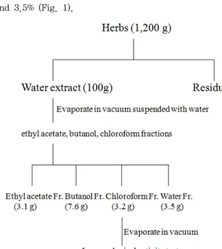
on a rotatory evaporator(Rotary evaporator, BUCHI B-480, Switzerland) and finally dried by a freeze drier (Freezedryer, EYELAFDU-540, Japan) to yield the extracts each fractions. The yield(w/w) of ethyl acetate (EtOAc), 1-butanol (BuOH), chloroform (CHCL3) and water (H2O) extracts were about 3.1, 7.6, 3.2 and 3.5% (Fig. 1).
Fig. 1. Schematic diagram for partial fractions from Lonicera japonica
Extraction and Isolation. The dried plant material (1.2 kg) was extracted with distilled water and concentrated. The resulting syrup (100 g) was subsequently partitioned with CHCl3, EtoAC, butanol fractions.
Fig. 2. The chemical structure of chlorogenic acid
2.2. Animals and blood glucose determination in NOD mice
Four to five-week-old male C57BL/6 mice were obtained at Daehan Biolink Co. LTD. (Eumsung, RepublicofKorea). NOD mice were obtained from the KRIBB (Korea Research Institute of Bioscience and
Biotechnology (Daejeon, Republic of Korea)and kept in our animal facilities under specific pathogen-free conditions. Mice were treated with anti-CD3 mAbs (5
㎍/day for 5 consecutive days i.v.) and EtOAc fraction (100 mg/kg p.o) from 8 weeks of age. In males, spontaneous diabetes begins to appear by 14 weeks of age. Mice were screened for blood glucose levels at least once a week after animals were 8 weeks old.
Blood glucose analysis was performed every week using a Lifescan One Touch Profile Meter (Issy les Mouline aux, France) with one drop of blood sampled from the tail. A 1.0g/lcalibration standard solution was assayed at least every samples. In this study, attention was focused on any sign of glycemic disturbance. The threshold for positivity was set at the mean of the pretest values plus two standard errors. Mice were considered diabetic when glucose levels reached 250mg/dl.
All animal procedures were conducted in accordance with the guidelines of the Institutional Animal Care and Use Committee, Korea Research Institute of Bioscience and Biotechnology (Daejeon, Republic of Korea).
2.3. RT-PCR
To study immunomodulatory effects ethyl acetate fraction from Lonicera japonica , chlorogenic acid on cytokine gene expression from spleen, thymus cells, RT-PCR was performed after quantitative normalization for each gene by a densitometry using β-actin gene expression. Briefly, total cellular RNA was extracted by using RNAzolB(Tel-Test, Friendswood, TX) according to the manufacturer's instruction. Aliquots(3g) of total RNA were transcribed into cDNA at 37°C for 1h in a total volume of 20㎕ with 2.5 U of moloney murine leukemia virus reverse transcriptase (Roche, Mannheim, Germany). Reverse transcribed cDNA samples were added to a PCR mixture consisting of 10x PCR buffer, 0.2 mM dNTP, 0.5 U Taq DNA polymerase (Takara, Tokyo, Japan), and 10 pmol of primers for each gene.
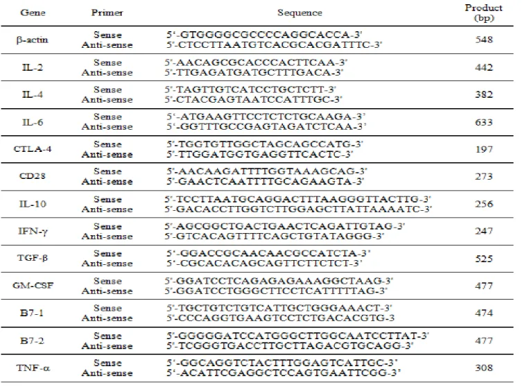
The primer sequences are as shown in table 1.
Amplifications were performed with 25 cycles for β
-actin and 30 cycles for the others. The amplification
profile included denaturation at 95°C for 1 min,
primer annealing at 55°C for 1 min, and extension
at 72°C for 1 min, followed by an additional
extension step at 72°C for 10 min. PCR products
were electrophoresed and visualized by ethidium
bromide staining.
Table 1. Primer sequences for RT-PCR
2.4.
51Cr-release assay
A modified standard
51Cr-release assay was used to measure cytotoxic activities of cytotoxic T cells as previously described
14). Briefly, as a single-cell suspension of target cells, B16/F10 tumor cells were prepared and incubated for 48 hrs, and then labeled
51
Cr. As a effector cells, mouse T cells from healthy C57BL/6 mice we retransferred to 24-well plates with coating with anti-CD3mAb(0.5㎍/well) and incubated for 48 hrs after being stimulated in medium supplemented with EtOAc fraction from Lonicera japonica and rIL-2(10ng/㎖) and thymus T cells were placed at the indicated effector:target cell ratios in a 96-well roundbottom microtiter tissue culture plate.
51
Cr-labeled target cells (1x10
4cells per well) were added to the wells in triplicate and incubated for 6 h in a 37-C, 5% CO2- humidified incubator. After incubation, 100 ul of the culture supernatant was collected, and radioactivity was detected by g counting. Results are expressed as the percentage of the specific release based on the formula % specific
release = (experimental release - spontaneous release)/(maximum release - spontaneous release) x 100. All data represent the standard deviation of at least three different determinants and were compared using Student's unpaired t-test.
2.5. Antibodies and flow cytometric analysis.
All antibodies(CD3, CD4, CD44, CD69) for flow cytometric analysis were purchased from Becton Dickinson (BD) PharMingen (SanDiego,CA). Spleen, thymus cells from NOD mice were stained with the indicated antibodies in staining buffer(PBS containing 1% FBS and 0.01% NaN3) for 10 min on ice, and analyzed by two color flow cytometry on a FACScan using Cell Quest software(BD Biosciences, MountainView, CA).
2.6. Statistical Analysis.
For statistical analysis of data, P-values were
analyzed using a unpaired Student's t-test software program (Startview 5.1; Abacus Concepts, Berkeley,CA).
Results were considered statistically significant when P values were*p<0.05,**p<0.01,***p<0.001.
3. Results
3.1. mRNA expression of cytokines in splenocytes treated with ethyl acetate, butanol and chloroform fractions from Lonicera japonica
As shown in Fig. 3, the mRNA for β-actin was detectable in spleen cells treated with RPMI-1640 media (Lane 1), ethyl acetate fractions (Lane 2, 3), butanol fractions (Lane 4, 5), and chloroform fractions (Lane 6, 7) respectively. PCR products for IL-10, IFN-γ, TNF-a, IL-4 and TGF-b1, amplified from spleen cell RNA preparations. Especially, PCR products for IL-10, IFN-γ were increased in ethyl acetate fractions (Lane 2, 3) compared with control groups (Lane 1). The results demonstrates that components in ethyl acetate fractions significantly affected these cytokines mRNAs expression of spleen cells.
Fig. 3. mRNA expression of cytokines in splenocytes treated with EtOAc fraction from Lonicera japonica
The mice spleen cells were incubated in the presence of EtOAc fraction of Lonicera japonica for 6 hrs before the isolation of total RNA. Total RNA(3 ㎍/
㎖) was used in RT reactions. cDNA was amplified (30 cycles) using primer pairs to IL-10, IFN-γ, TNF-α, IL-4, TGF-β1 and β-actin. PCR products were visualized by ethidium bromide on 2% agarose gels.
Lane M, 100bp DNA marker; lane 1, only cells(Splenic cells); lane2, EtOAc fraction from Lonicera japonica (100㎍/㎖); lane 3, EtOAc fraction from Lonicera japonica (10㎍/㎖); lane 4, butanol
fraction from Lonicera japonica (100 ㎍/㎖); lane 5, butanol fraction from Lonicera japonica (10㎍/㎖);
lane 6, chcl
3fraction from Lonicera japonica (100㎍/
㎖); lane 7, chcl
3fraction from Lonicera japonica (10
㎍/㎖).
3.2. mRNA expression of cytokines in thymus treated with ethyl acetate, butanol and chloroform fractions from Lonicera japonica
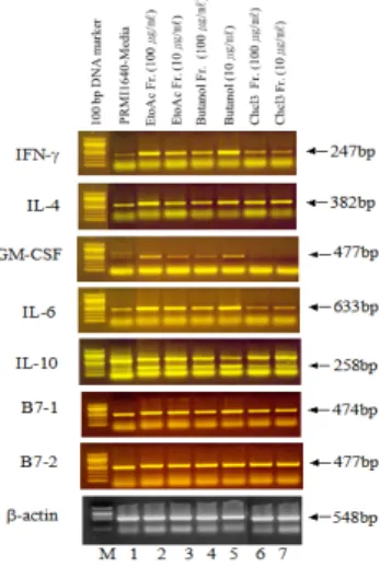
As shown in Fig. 4, the mRNA for β-actin was detectable in thymus cells treated with RPMI-1640 media (Lane 1), ethyl acetate fractions (Lane 2, 3), butanol fractions (Lane 4, 5), and chloroform fractions (Lane 6, 7) respectively. PCR products for IFN-γ, IL-4, GM-CSF, IL-6, IL-10, B7-1 and B7-2 amplified from thymus cell RNA preparations. The relative expression of above genes was higher in the thymus of ethyl acetate fraction group and in both spleen, when compensated with that of the b-actin genes. Especially, PCR products for IFN-γ, IL-4, GM-CSF, IL-6 were increased in ethyl acetate fractions (Lane 2, 3) compared with control groups (Lane 1). The results demonstrates that components in ethyl acetate fractions significantly affected these cytokines mRNAs expression of thymus cells.
Fig. 4. mRNA expression of cytokines in thymus treated with EtOAc fraction from Lonicera japonica
The mice thymus cells were incubated in the presence of EtOAc fraction from Lonicerajaponica for 6 hrs before the isolation of total RNA. Total RNA(3
㎍/㎖) was used in RT reactions. cDNA was amplified (30 cycles) using primer pairs to IFN-γ, IL-4, GM-CSF, IL-6, IL-10, B7-1, B7-2, and β-actin.
PCR products were visualized by ethidium bromide on
2% agarose gels. Lane M, 100bp DNA marker; lane
1, only cells (Spleen cells); lane2, EtOAc fraction
from Lonicera japonica (100 ㎍/㎖); lane 3, EtOAc fraction from Lonicera japonica (10 ㎍/㎖); lane 4, butanol fraction from Lonicera japonica (100 ㎍/㎖);
lane 5, butanol fraction from Lonicera japonica (10 ㎍ /㎖); lane 6, chloroform (CHCl
3) fraction from Lonicera japonica (100 ㎍/㎖); lane 7, chloroform (CHCl
3) fraction from Lonicera japonica (10 ㎍/㎖)
3.3. mRNA expression of cytokines in thymus co-stimulated with EtOAc fraction from Lonicera japonica and anti-CD3mAb
As shown in Fig. 5, the mRNA for β-actin was detectable in thymus cells treated with RPMI-1640 media (Lane 1), CD3 (Lane 2) only, CD3 in the presence of PMA (100 ng/㎖) and ionomycin (1 ㎍/ml), or treated with EtoAc fractions (100, 50, 10, 1 ㎍/㎖
), chlorogenic acid (1, 0.1, 0.01, 0.001 ng/㎖) respectively. PCR products for IFN-γ, IL-4, GM-CSF and IL-2 amplified from thymus cell RNA preparations. Especially, PCR products for IFN-γ, IL-4 were increased in ethyl acetate fractions (100, 50, 10 ㎍/㎖ ), chlorogenic acid (1 ng/㎖) compared with control groups (Lane 2). The results demonstrates that components in ethyl acetate fractions, chlorogenic acid significantly affected these cytokines mRNAs expression of thymus cells.
Fig. 5. mRNA expression of cytokines in thymus co-stimulated with EtOAc fraction from Lonicera japonica and anti-CD3mAb
Thymus cells were transferred to 24-well plates coated with anti-CD3mAb (0.5 ㎍/well) and stimulated in medium supplemented with PMA (100 ng/㎖), ionomycin (1 ㎍/ml), EtOAc fraction from Lonicera japonica and chlorogenic acid and then cultured with RPMI1640 media for 6 hours before the isolation of total RNA. Total RNA(3 ㎍/㎖) was used in RT reactions. cDNA was amplified (30 cycles) using
primer pairs to IFN-γ, IL-4, GM-CSF, IL-2 and β -actin. PCR products were visualized by ethidium bromide on 2% agarose gels.
3.4 B16/F10 tumor target cells killed by rIL-2 and EtoAc fraction-activated mouse T cells
The use of cytotoxicity assays is of central importance in characterizing the function of T lymphocytes, and the
51Chromium(Cr)-release assay is the most widely practiced method for analyzing the lytic activity of a cytotoxic T-cell (CTL) culture.
Cytotoxic T cell activities were measured by testing lytic activity against melanoma targets that differed in the relative amounts of MHC Class I expression.
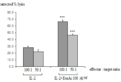
Cytotoxic T cells were highly effective in lysing melanoma targets at all effector versus target (E:T ) ratios (Fig. 6). Tumor targets were lysed at all E:T ratio. The relatively weak ability to lyse cells on the part of 50:1 (E:T) ratio is likely explained by a low percentage of cytotoxic T cells in the preparation from thymus T cells.
Fig. 6. Lytic activity of mouse T lymphocytes following rIL-2 incubation
As a single-cell suspension of target cells, B16/F10 tumor cells were prepared and incubated for 48 hrs, and then labeled
51Cr. As a effector cells, mouse T cells from healthy C57BL/6 mice were transferred to 24-well plates with coated with anti-CD3mAb(0.5㎍
/well) and incubated for 48 hrs after being stimulated
in medium supplemented with EtOAc fraction from
Lonicera japonica and rIL-2 (10 ng/㎖).
51Cr labeled
target cells were added to replicates wells containing
effector cells and incubated 6hrs. After 6hrs
incubation, the culture supernatants(100㎕) were
dispensed to the wells of 96-well microtiter plates
with replicates of three wells for each effector cell
concontration. The
51Cr release were measured in γ
-scintillation counter (LKB,U.S.A) 1min/sample as
described in materials and methods. Statistically
significant value compared with control data by T
test(
***p<0.001).
3.5 Blood glucose level in NOD mice treated with EtOAc fraction from Lonicera japonica and anti-CD3 mAbs
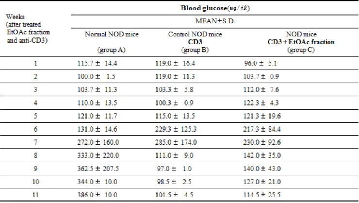
Mice were considered diabetic as a positive blood glucose when glucose levels reached 250mg/dl. The blood glucose level of NOD mice were increased from 14 weeks of age (Table 2, Fig. 7). At 18 weeks of age, 90% of NOD normal mice were positive. In the
EtOAc fraction from Lonicerajaponica -treated groups, an decrease in the blood glucose was seen between 15 and 18 weeks of age. Blood glucose levels were lower in the EtOAc fraction from Lonicerajaponica and anti-CD3 mAbs treated groups than the untreated mice. Although the blood glucose level of mice was also lower in two experimental groups than the untreated mice, statistical significance was not attained.
Table 2. Blood glucose in NOD mice treated with EtOAc Fraction from Lonicera japonica and anti-CD3 mAb compared to control NOD mice (group B) and normal mice (group A) Group A(Normal group):It was treated with RPMI-1640 media.
Group B(Control group):It was treated with anti-CD3 mAb (5 ㎍/day for 5 consecutive days i.v.)
Group C(Samples group):It was treated with anti-CD3 mAb (5 ㎍/day for 5 consecutive days i.v.) and EtOAc fraction.
Experimental NOD treatment groups are compared with control NOD mice Mean of S.D. triplicate plates. P-value:Stastistically significant value as compared with data of control group by T-test.
Fig. 7. Blood glucose level in NOD mice treated with EtOAc fraction from Lonicera japonica and anti-CD3 mAb compared to control NOD mice.
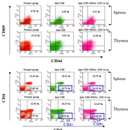
3.6 Inhibitory effect of ethyl acetate fraction from Lonicera japonica on spleen, thymus surface marker
Effects of ethyl acetate fraction from Lonicera
japonica on leukocyte subsets in spleen and thymus,
there were marked change in numbers of CD8+ T
cells compared to control cells (Fig. 8). Ethyl acetate
fraction and anti-CD3 mAb treated group resulted in
further increase in CD4-CD8+ T cells (by 0.4% in
spleen, 0.5% in thymus), whereas CD44+CD69+ T
cells were reciprocally decreased in both anti-CD3
only, ethyl acetate fraction and anti-CD3 mAb treated
groups (Fig. 8).
Fig. 8 EtOAc fraction from Lonicera japonica and anti-CD3 mAb down-regulates CD44+CD69+, CD3+CD8+ expression of spleen and thymus T cells in NOD mice.