279
Time-course response of epichlorohydrin on epididymal histopathology in rats
Kang-Hyeon Kim, In-Sik Shin, Jeong-Hyeon Lim, Sung-Hwan Kim, Na-Hyeong Park, Changjong Moon, Sung-Ho Kim, Dong-Ho Shin, Jong-Choon Kim*
Animal Medical Center, College of Veterinary Medicine, Chonnam National University, Gwangju 500-757, Korea (Accepted: December 14, 2009)
Abstract : This research aimed to investigate the time-course effect of epichlorohydrin (ECH) on epididymal histopathology in Sprague-Dawley rats. Twenty-four male rats were randomly assigned to four groups with 6 rats in each group and were administered a single oral dose of ECH (70 mg/kg) or its vehicle. Six animals each were sacrificed on days 0 (control), 1, 2, and 7 after treatment. During the study period, clinical signs, body weights, reproductive organ weights, testicular spermatid count, epididymal sperm count, motility and morphology, and histopathology were examined. No treatment- related effects on body weights and reproductive organ weights were noted at any time point. On the contrary, sperm motility decreased slightly on days 1 and 2 after treatment and then decreased significantly on day 7 after treatment. The first signs of histological changes were the appearance of cell debris in the ducts and vacuolization of the epithelial cells observed in the proximal caput epididymis on day 1 after treatment. The incidences and grades of the histological changes including cell debris in the ducts, epithelial vacuolization, oligospermia, and epithelial disruption increased on day 2 and then decreased slightly on day 7 after treatment. These results show that a single oral dose of 70 mg/kg ECH to male rats results in cell debris in the ducts and vacuolization of the epithelial cells in the proximal caput epididymis, followed by reversible oligospermia, epithelial disruption, and decreased sperm motility.
Keywords : epichlorohydrin, histopathology, rats, reproductive dysfunction, sperm
Introduction
Epichlorohydrin (ECH) is an organochlorine compound with an irritating and chloroform-like odor that is used in the production of various synthetic materials, including epoxy resins, glycerin, coatings, adhesives, paints, varnishes, insecticides, and many other products [18]. Due to its increased production and wide-spread use, the potential risk caused by ECH in humans has steadily increased, which may result in severe health impacts [8, 18].
Exposure to ECH can occur by inhalation, ingestion, and eye or skin contact in the general population, as well as in workers with specific occupations. ECH is rapidly and extensively absorbed following ingestion, inhalation, and dermal contact [7]. The toxicity of ECH has been extensively studied in short and long term animal studies over the past several decades. Previous
studies demonstrated that ECH is an anti-fertility agent that acts both as an epididymal toxicant and an agent capable of directly affecting sperm motility [3, 9, 20].
However, limited data have been published on histopathological alterations of reproductive organs in rats. Moreover, there have been conflicting findings regarding the histopathological changes of epididymis in the literature. Hahn [9] reported that male rats treated with 15 mg/kg/day for 12 days showed loss of fertility within the first week of exposure, but no effects were seen on the histopathology of the reproductive organs. John et al . [12] also reported that inhalation exposure of ECH to male rats for 10 weeks resulted in a dose-related infertility at 93.4 and 189.0 mg/m
3air, but microscopic examination did not reveal any abnormalities in the reproductive organs. On the contrary, Cooper et al. [3] reported that five oral doses of 20 mg/kg or one single oral dose of 100 mg/kg to
*Corresponding author: Jong-Choon Kim
College of Veterinary Medicine, Chonnam National University, Gwangju 500-757, Korea
[Tel: +82-62-530-2827, Fax: +82-62-530-2809, E-mail: [email protected]]
male rats produces large retention cysts in the ductuli efferentes and proximal caput of the epididymidis, suggesting severe histological alterations in the reproductive organs.
In this study, therefore, we investigated the potential adverse effects of ECH on epididymal histopathology after a single oral administration in male rats. Although the most likely route of exposure is by inhalation, the oral route of exposure was selected for this study because there is a possibility of oral and dermal absorption.
Materials and Methods
Animals
Ten-week old male Sprague-Dawley rats were purchased from Orient-Bio (Seoul, Korea) and used after one week of quarantine and acclimatization. The animals were maintained in a room at 23
±3
oC, with a relative humidity of 50
±10%, under a controlled 12- h light/dark cycle. All rats were housed two per cage in stainless-wire cages and were allowed sterilized tap water by ultraviolet irradiation and fed on commercial rodent chow (Samyang Feed, Korea) ad labium . The Institutional Animal Care and Use Committee of Chonnam National University approved the protocols for the animal study, and the animals were cared for in accordance with the Guidelines for Animal Experiments of Chonnam National University.
Test chemical and treatment
ECH was purchased from Aldrich (USA). The test chemical was dissolved in corn oil (Sigma-Aldrich, USA) before administration. The dosage volume was 10 mL/kg and was calculated according to the body weight on the day of administration (designated day 0). The rats were given a single oral dose of vehicle or ECH.
Experimental groups and dose selection
Twenty-four male rats were assigned randomly into two experimental groups: one treatment group receiving 70 mg/kg ECH (n = 18) and a vehicle control group (n = 6). The dose was selected on the basis of toxicity studies as described earlier [3, 20].
Observation of animals
All rats were observed daily for clinical sings
throughout the treatment period and abnormal signs were recorded individually for type, observation day/
time and duration. Body weights were measured at the initiation of treatment and on days 1, 2, and 7 of test.
Necropsy and organ weight
Six rats each in the treatment group were sacrificed on days 1, 2, and 7 of test using anesthetic ether. All rats in the control group were sacrificed on day 7 of test. Complete gross postmortem examinations were performed on all terminated animals. The gross weights of the testis, epididymis, prostate gland, and seminal vesicle were measured and then their relative organ weights were calculated as the ratio between organ weight and body weight.
Histopathological examination
The right testis and right epididymidis were taken and fixed with Bouin fixative and 10% neutral buffered formalin solution, respectively. The tissues were routinely processed, embedded in paraffin and sectioned at 3-5 µm. These sections were stained with Hematoxylin-Eosin for histopathologic examination and then examined microscopically.
Sperm examination
Sperm analysis was conducted as previously described [2, 13]. At the scheduled necropsy, the testes and epididymides were removed and weighed. The left testis was homogenized with 12 mL of distilled water for the sperm head counts. The sperm suspension was placed into a hemacytometer (Neubauer, Germany) and the number of spermatids was counted using a light microscope (Leica, Germany). The left cauda epididymis was homogenized with 10 mL of physiological saline to determine the sperm counts. The number of sperm was counted as in the testis. For the motility measurements, the sperm was obtained from the ductus deferens, placed in Hanks’ balanced salt solution (pH 7.2; Sigma Chemical, USA) containing 5 mg/mL bovine serum albumin (Sigma Chemical, USA) and maintained at 37
oC. Motility was observed using a microscope with a stage warmer (Microwarm plat;
MDF-10, Japan). The sperm morphology was also
examined using optical microscopy of the sperm
smears (sperm suspension containing 1% Eosin Y)
collected from the left ductus deferens. Two hundred
sperm per animal were evaluated for head and tail
defects by light microscope. The sperm morphology was classified as normal, small head, amorphous head, two heads/tails, straight hook, excessive hook, folded tail, short tail, and no tail.
Statistical analyses
All values were expressed as mean
±SD. Variables such as body weights, organ weights, and spermatid count were subjected to one-way ANOVA, and Dunnett's multiple comparison test [4] was conducted when analytic results were significant. The percentages of motile sperm and sperm abnormalities were analyzed by the Kruskal-Wallis nonparametric ANOVA [15], followed by multiple comparisons using Dunnett’s test.
The clinical signs, necropsy findings and histopatho- logical findings were represented as frequencies and were subjected to the Fisher’s exact probability test [5]
when necessary. The difference was considered
statistically significant when p < 0.05 or p < 0.01.
Results
Clinical signs and body weights
Throughout the study period, no treatment-related
Table 2. Relative organ weights of male rats treated with epichlorohydrin
Items Control Days after treatment (70 mg/kg)
Day 1 Day 2 Day 7
No. of male rats 6 6 6 6
Body weight (g) 410.9 ± 12.52 373.9 ± 18.11 387.2 ± 17.97 403.8 ± 16.71 Testis: left (%) 0.42 ± 0.017 0.50 ± 0.039 0.51 ± 0.075 0.46 ± 0.037
right (%) 0.42 ± 0.052 0.52 ± 0.051 0.52 ± 0.065 0.44 ± 0.030
Epididymis: left (%) 0.15 ± 0.008 0.16 ± 0.020 0.15 ± 0.011 0.16 ± 0.035 right (%) 0.16 ± 0.019 0.15 ± 0.010 0.15 ± 0.002 0.17 ± 0.016 Seminal vesicles (%) 0.14 ± 0.023 0.14 ± 0.028 0.14 ± 0.028 0.15 ± 0.033
Prostate (%) 0.31 ± 0.066 0.38 ± 0.060 0.37 ± 0.034 0.35 ± 0.036
Values are presented as means ± SD.
Table 1. Body weight of male rats treated with epichlorohydrin
Items Epichlorohydrin (mg/kg/day)
0 70
Day 0 390.9 ± 15.35 (6) 391.4 ± 16.44 (18) Day 1 387.9 ± 16.81 (6) 373.3 ± 17.30 (18) Day 2 387.7 ± 13.63 (6) 380.8 ± 17.55 (12) Day 7 410.9 ± 12.54 (6) 403.8 ± 16.71 (6) Values are presented as means ± SD (g). Numbers in paren- thesis are animals tested.
Table 3. Sperm analysis of male rats treated with epichlorohydrin
Items Control Days after treatment (70 mg/kg)
Day 1 Day 2 Day 7
No. of rats 6 6 6 6
Sperm heads in testis (×10
6) 252.7 ± 23.59 234.6 ± 16.24 234.9 ± 17.80 236.3 ± 18.13 Sperm in epididymis (×10
6) 189.9 ± 16.52 182.7 ± 16.12 172.1 ± 22.12 166.1 ± 16.99 Sperm motility (%) 85.8 ± 2.22 59.4 ± 16.07 58.2 ± 12.36 38.8 ± 35.95
*Sperm abnormalities (%) 8.0 ± 1.22 15.2 ± 16.48 8.6 ± 1.52 24.8 ± 25.72
Amorphous head 0.8 ± 0.45 1.0 ± 0.71 0.4 ± 0.55 0.2 ± 0.45
Two heads/tails 0.2 ± 0.45 0.2 ± 0.45 0.4 ± 0.55 0.2 ± 0.45
Excessive hook 0.0 ± 0.00 0.0 ± 0.00 0.2 ± 0.45 0.0 ± 0.00
Straight hook 0.6 ± 0.89 0.2 ± 0.45 1.6 ± 1.34 1.2 ± 0.84
Folded tail 3.0 ± 2.92 4.6 ± 2.61 4.6 ± 2.19 1.2 ± 1.30
No tail 3.4 ± 2.41 9.2 ± 14.04 1.4 ± 1.14 22.0 ± 26.09
Values are presented as means ± SD.
*Significant difference at
p< 0.01 level compared with the control group.
death was observed in any animals treated with 70 mg/
kg ECH. However, treatment-related clinical signs, as evidenced by the increased incidence of nasal discharge (n = 6), soft feces (n = 5), decreased locomotor activity (n = 12), ataxia (n = 3), and piloerection (n = 7), were found in the treatment group. On the contrary, there were no statistically significant differences in body weights and organ weights between the vehicle control and treatment groups (Tables 1 and 2).
Sperm analysis
As shown in Table 3, sperm motility was significantly decreased on day 7 of test in the treatment group when compared with the control group. Sperm abnormality was slightly higher on day 7 in the treatment group than that in the control group, but no significant difference was detected compared with the control group.
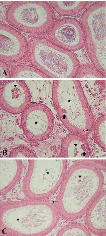
Histopathological findings
The results of the histopathological examination were summarized in Table 4. Histopathological changes were observed only in the proximal caput epididymis of the treatment group. The first signs of histological damage induced by ECH administration were the appearance of cell debris in the ducts and
vacuolization of the epithelial cells observed in the proximal caput epididymis on day 1 of test. The incidences and grades of the histological changes including cell debris in the ducts, epithelial vacuolization, oligospermia, and epithelial disruption increased on day 2 of test and then decreased slightly on day 7 of test (Fig. 1).
Discussion
Treatment-related clinical signs were observed transiently on days 0 and 1, whereas they were not found in any of the animals treated with ECH after day 2 of test. These findings may be attributed to mucus and gastrointestinal irritation effects of ECH because ECH is a chemical that acts as an eye, skin, and nasal irritant [6]. It was previously reported that acute respiratory irritation with hemorrhages and severe edema occurs in rats after inhalation or oral application [14, 16]. On the other hand, ECH administration did not adversely affect body weights and reproductive organ weights in rats.
The significant decrease of sperm motility observed in the treatment group on day 7 of test is consistent with the results of previous studies [17, 19]. Toth et al . [19] demonstrated that a 23-day repeated oral dose Table 4. The incidence and severity of histopathological lesions in epididymis of male rats treated with epichlorohydrin
Findings Grade Control Days after treatment (70 mg/kg)
Day 1 Day 2 Day 7
Cell debris in duct
−
6 1 0 4
+ 0 5 1 2
++ 0 0 4 0
+++ 0 0 1 0
Vacuolization of epithelial cell
−
6 2 2 2
+ 0 4 2 4
++ 0 0 2 0
+++ 0 0 0 0
Oligospermia
−
6 6 4 5
+ 0 0 2 1
++ 0 0 0 0
+++ 0 0 0 0
Epithelial disruption
−