DOI : 10.3341/jkos.2008.49.9.1532
키드 증후군의 안과적 증상과 조직학적 특징
정재훈1․전연숙1․이수현2․정행선2․김재찬1
중앙대학교 의과대학 안과학교실1, Modern Cell & Tissue Technologies2
목적: Keratitis-ichthyosis-deafness (KID) 증후군은 신생혈관 각막염, 홍반성피부각화증, 감각신경성 난청의 주 요 3징후를 동반하는 선천성 외배엽 이상형성 질환이다. 안과적으로 눈꺼풀의 과각화, 속눈썹 소실, 건성각결막염, 각 막미란과 궤양, 각막 신생혈관 생성 및 혼탁 등이 동반될 수 있고 경미한 정도에서 시력 상실까지 발생할 수 있다고 보 고되었다. 이에 심한 안구 증상을 동반한 키드 증후군 1예의 안과적 증상과 각막 병변의 조직학적 특징을 보고하고자 한다.
증례요약: 양안 각막 혼탁과 드문 눈썹, 눈꺼풀의 과각화 등의 소견을 보이는 5세 남아가 전신 피부 과각화, 무모증, 감각신경성 난청이 있고 GJB2 유전자 돌연변이가 확인되어 키드 증후군으로 진단하였다. 보존적인 치료를 하였으나 호전이 없어 표층 각막절제술 및 양막이식을 하여 증상이 호전되었다. 각막 병변은 비정상적인 각막상피 분화, 상피층 의 connexin 26의 발현 결여, 바닥막의 부분적인 결손 등의 특징이 있었다.
<대한안과학회지 2008;49(9):1532-1538>
<접수일 : 2008년 1월 14일, 심사통과일 : 2008년 5월 14일>
통신저자 : 김 재 찬
서울시 용산구 한강로3가 65-207 중앙대학교 용산병원 안과 Tel: 02-748-9838, Fax: 02-792-5076 E-mail: jck50ey@kornet.net
* 본 논문의 요지는 2007년 대한안과학회 제98회 추계학술대회 에서 포스터로 발표되었음.
Keratitis-ichthyosis-deafness (KID) 증후군 은 1981년 Skinner et al1에 의해 최초로 보고된 신 생혈관 각막염, 홍반성피부각화증, 감각신경성 난청의 주요 3징후를 동반하는 선천성 외배엽 이상형성 질환이 다. 이 증후군 환자들에게서 다양한 상피 조직들의 세포 간 신호 전달 조절의 주 틈새 이음 단백질인 connexin 26을 발현하는 GJB2 유전자의 돌연변이가 보고되었다.2-6 Connexin 26은 진피, 각막상피, 속 귀 등의 외배엽 조직들에 존재하기 때문에 키드 증후군은 주요 3징후 외에 반흔성 탈모, 손톱과 발톱 이상형성, 치아 이상, 세균 및 진균 감염의 위험성 증가, 편평상피 세포 암의 위험성 증가와 같이 전신에 걸쳐 다양한 임 상양상이 나타날 수 있다.7-10 이 증후군은 안과적으로 눈꺼풀의 과각화, 속눈썹 소실, 건성안, 윤부 줄기 세포 결핍에 의한 각막미란과 궤양, 파누스 형성, 각막 신생 혈관 생성 및 혼탁 등이 동반될 수 있으며 그 정도는 눈
부심과 같이 경미한 정도에서 시력 상실에 이르기까지 다양하게 보고되었다.11-13
지금까지 키드 증후군의 각막 병변에 대한 조직학적 접근이 이루어지지 않았고, 국내에는 각막염을 동반한 키드 증후군의 보고가 있으나14 안과적 특성에 대한 자 세한 보고가 없었기에 본원에서 경험한 심한 안과적 증 상을 동반한 5세 키드 증후군 환아의 임상양상과 각막 병변의 조직학적 소견에 대해 문헌고찰과 함께 보고하 는 바이다.
증례보고
5세 남아가 밝은 데서 눈을 찡그리고, 물체를 가까이 보려 하며 양안 각막의 혼탁을 주소로 본원 안과에 내 원하였다.
환아는 임신 38주에 제왕절개로 출생하였으며 출생 체중은 3 Kg이었다. 영아 때부터 전신 피부의 각화가 심하여 피부과 진료상 어린선인 줄 알고 지내던 중 피 부 각화가 심해지고 무모증, 머리와 등을 제외한 곳에 땀이 안 나는 증상이 있고, 치아 이상 및 난청, 지능 및 인지 기능 저하 등이 있어 3세경 소아과, 이비인후과, 피부과 등에서 진료를 보았고, 4세 때 환자 및 친부모 에 대해 유전학적 검사를 시행하였다. 유전학 검사상 가족력은 없고, GJB2 유전자(13q11)의 exon 11에 서 점상 돌연변이[D50N]가 확인되어 키드 증후군이
= 증례보고 =
Figure 1. Clinical appearance of a 5-year-old boy with KID syndrome. (A) Frontal view of face showed scarring alopecia and hyperkeratotic skin lesions. (B) Palmoplantar hyperkeratosis.
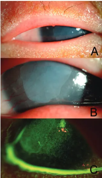
Figure 2. Slit lamp photographs of the right eye of a pateint with KID syndrome before surgery. (A) Hyperkeratic lid lesions and sparse lashes. (B) Gray-white elevated scarring opacity was observed on the central cornea. (C) Epithelial erosions were observed with fluorescein stain.
전신에 걸쳐서 피부의 과각화 증상이 있었으며 특히 두 피와 손, 발바닥에서 심한 증상이 있었다(Fig. 1).
안과 검사상 나안시력이 우안 안전수지 및 좌안 0.06~0.08 정도였고, 교정이 불가능 하였다. 양측 눈 썹과 속눈썹이 거의 없는 상태로 눈꺼풀 피부의 각화가
국소 스테로이드, 30% 자가 혈청 점안 및 눈물점 마개 삽입술 등 보존적인 치료를 하였으나 외안부 질환의 호 전은 없었으며, 우안은 각막혼탁 병변이 진행하여 전신 마취 하에 우안 표층 각막절제술 및 양막이식술을 시행 하였다.
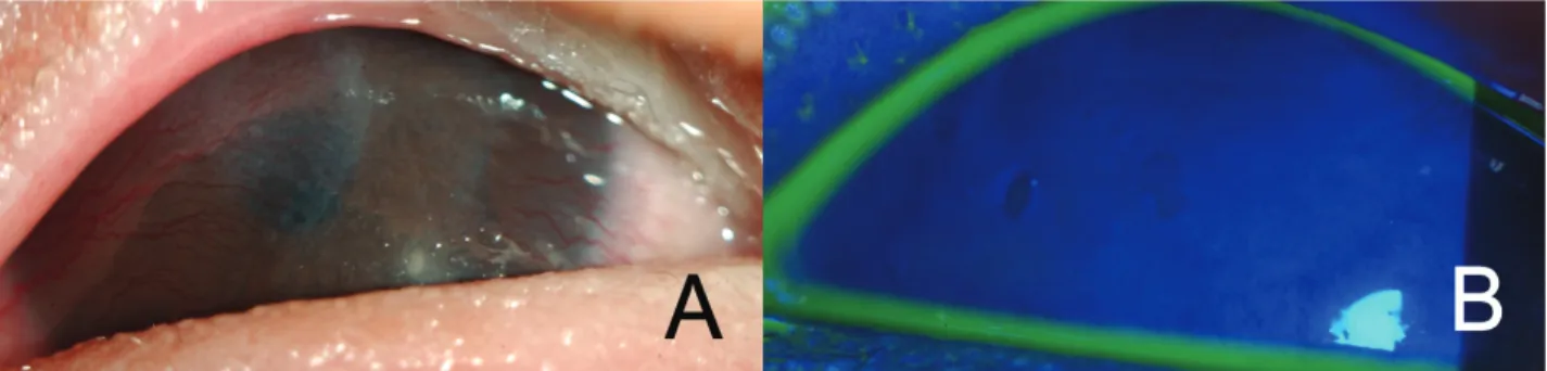
Figure 3. Slit lamp photographs of right eye after surgery. Lamellar keratectomy and amniotic membrane transplantation were done. (A) Central scarring opacity was regressed at post operative 4months. (B) A complete epithelization was achieved.
Figure 4. Histopathology of the corneal lesion. High-power magnification showed the characteristic squamous cell hyperplasia and partial destruction of Bowman’s layer (arrow). (hematoxylin-eosin stain, ×400).
수술 후에도 인공누액과 30% 자가 혈청 점안 및 Tretinoin 0.025% 연고를 안검에 도포하는 보존적 치료를 병행하였고, 수술 후 한달 째 수술 전에 비해 우 안 시력이 0.02로 향상되고, 각막의 혼탁이 감소하여 홍채와 동공, 전방의 관찰이 가능하였으며 완전한 각막 상피화(Fig. 3)를 관찰할 수 있었다. 눈을 찡그리거나 물체를 가까이서 보려 하는 증상이 호전된 것을 부모에 의해 확인할 수 있었고, 수술 후 4개월 경과 관찰까지 특별한 합병증 없이 잘 유지되고 있었다.
고 찰
본 증례는 피부, 청력, 안구 증상을 동반하고 유전학 검사를 통해 확진된 전형적인 키드 증후군으로 기존의 증례가 피부 증상이 전형적인 것에 비해, 양안 모두 윤 부 줄기세포 결핍으로 인해 안구 증상과 합병증이 심하 였다. 보존적인 치료에 한계가 있어 수술적 치료를 시 도하여 증상의 호전이 있었고, 인공누액과 자가 혈청
점안 및 Tretinoin 0.025% 연고를 안검에 도포하 는 지속적인 보존 치료로 외안부를 안정화 시킬 수 있 었다.
수술 시 절제한 각막 병변 조직 검사상 편평상피세포 의 과증식 및 부분적인 보우만층 파괴 소견(Fig. 4) 이, 면역조직화학 검사상 각막상피 조직에 특이적인 cytokeratin 3과 편평상피, 샘상피의 바닥세포층, 근 육상피세포, 중피세포에 특이적인 cytokeratin 5의 강한 양성 염색소견이 관찰되었으며 각막을 제외한 각 질화 상피세포에 특이적인 cytokeratin 10과 내장의 비각질화 상피세포에 특이적인 cytokeratin 13에 양 성 염색소견이 관찰되었다(Fig. 5). 정상 각막 조직에 서는 발현되지 않는 cytokeratin 5, 10, 13의 양성 염색소견으로 비정상적인 각막 분화를 확인할 수 있었 다. 각막상피가 분화하면서 발현되는 틈새이음(gap junction) 단백인 connexin의 면역조직화학 검사 를 통해 바닥세포 위 층에서 정상적으로 발현되는 connexin 43의 양성 염색 소견에 비해 connexin 26 이 발현되지 않는 것을 확인할 수 있었고(Fig. 6), 바 닥막의 주요 구성 성분인 collagen Ⅳ와 laminin 5의 부분적인 염색 결손 소견(Fig. 7)으로 각막 병변의 국 소적인 바닥막 결손을 확인할 수 있었다.
키드 증후군은 선천성 외배엽 이상형성 질환이다. 분 자 유전학 분석을 통하여 connexin 26을 발현하는 GJB2 유전자의 돌연변이가 키드 증후군 환자들에서 보고되었고,2-5 드물지만 세 가족의 예에서 상염색체 우 성의 유전성향이 보고되기도 하였다.11,15,16 피부조직에 서 다량으로 발견되며 틈새이음을 형성하는 connexin 의 발현 장애는 키드 증후군 환자들에게서 나타나는 과 각화증, 홍색 각피성 판, 반흔성 대머리, 손, 발톱 이상 형성, 치아 이상 등의 증상과 관련이 있고 이차적으로 바이러스, 세균, 진균 등의 피부 감염 위험성이 증가될 수 있다.9 Connexin 26은 속 귀 조직에서 털 세포의 칼륨 이온의 재흡수에 중요한 역할을 하는 것으로 추정 되며 이는 키드 증후군 환자들의 감각신경성 난청과 관
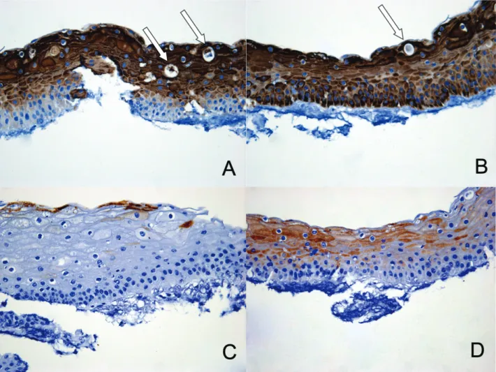
Figure 5. Immunohistochemical staining for cytokeratin of the corneal lesion of KID syndrome. It shows strong immunopositivity to cytokeratin 3 in the suprabasal layer (A), to cytokeratin 5 in all epithelial layer (B). Hyperkeratosis of the cornea shows special features of the keratin pearl formations (arrows). It shows immunopositivity to cytokeratin 10 in superficial layer (C), to cytokeratin 13 in suprabasal & superficial layer (D) (Original magnification ×400 in all of the photomicrographs).
련이 있다.17 또한 이 증후군 환자들의 편평상피암 발생 율이 11% 정도로 보고되어 connexin 26의 종양 억제 자 기능도 제기되었다.18
Caceres-Rios et al에 따르면 피부와 청력질환이 확인된 키드 환자 61명 중 95%에서 안구 증상이 있었 고, 혈관성 각막염 환자는 79%정도로 나타났으며8, Wilson et al에 의하면 키드환자 35명 중 84%에서 각막이상이 발견되었다고 한다.10 이처럼 각막염이 모 든 환자에 있어 질병 초기에 두드러진 증상이 아니고, 어린선이 모든 환자의 주된 피부 병변이 아니기 때문에 키드라는 용어 대신 선천성 외배엽 질환으로서 kera todermatous ectodermal dysplasia (KED)라는 용어가 제안되기도 하였다.8 실제로 보고된 키드 증후 군의 안과적인 증상은 없거나 경미한 정도8에서 이 증 례에서처럼 시력에 심각한 문제를 초래하는 경우까지
다양하게 나타날 수 있다.12,13
안구 조직 중 각막상피, 눈물샘, 눈꺼풀 피부 및 모낭 등에 connexin 26이 존재하는 것으로 보고되었으며
5,19-21
키드 증후군의 안구 증상은 다음 2가지 기전에 의해 설명할 수 있을 것이다. 첫째, 가장 특징적인 안구 증상은 건성각결막염으로 추정한다.12,22 눈물샘은 connexin 26과 32로 구성된 틈새 이음으로 되어 있으 며, 이 배열은 정상적인 샘 분비 기능에 필수 적이다.
눈꺼풀 피부와 모낭에도 connexin 26이 존재하여 각 질세포의 분화, 이주, 발현 등에 관여하고, E-cadhe rin과 상호 작용으로 눈썹의 성장에 관여하고 있다.20 그러므로 비정상적인 connexin이 발현되면 눈물샘, 눈꺼풀 피부와 모낭의 과각화 진행으로 정상 기능이 상 실되어 눈물분비가 되지 않고 마이봄샘의 분비가 손상 되어 건성각결막염을 유발하는 것으로 보인다.19 안구
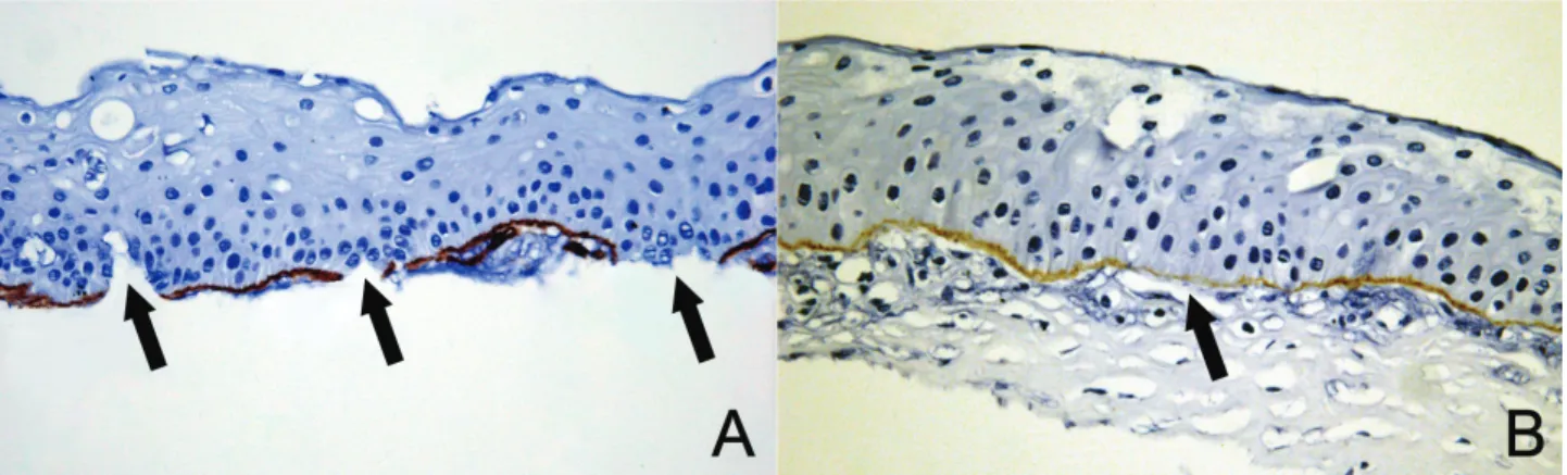
Figure 6. Immunohistochemical staining for connexin of the corneal lesion of KID syndrome. It shows positive immune staining to connexin 43 in the suprabasal and middle layer (A, Original magnification ×400), but negative immune staining to connexin 26 (B, Original magnification ×400).
Figure 7. Immunohistochemical staining for basement membrane of the corneal lesion of KID syndrome. There are partial absences of the staining in the basement membrane (arrows) to collagen Ⅳ (A, Original magnification ×400) and to laminin 5 (B, Original magnification ×400).
증상이 경미한 키드 증후군 환자들은 정상적인 눈물 띠 를 유지하고, 눈물막 파괴 검사나 쉬르머 검사가 정상 인 것으로 보고되고 있어13 심각한 안구 합병증의 예방 에 눈물 분비 기능을 유지하는 것이 필수적인 것으로 추정된다.
둘째, 윤부 줄기 세포 결핍은 Gicquel et al11에 의 해 보크트 배열이 결여된 두 명의 키드 환자에서 보고 되었다. 세포 전환이 느려지고 각막상피 세포층이 취약 해져 표층 각막염이 발생하게 되고, 눈물샘과 눈꺼풀 피부, 눈썹의 과각화로 인한 건성각결막염이 윤부 줄기 세포 결손을 악화시켜 각막의 결막화와 파누스 형성, 재발적이고 지속적인 각막미란, 각막궤양과 신생혈관 형성 등의 심한 안구 합병증을 일으킬 수 있다.
키드 증후군은 피부, 청력, 안구에서 상피 세포의 과 각화로 인한 다양한 증상과 합병증을 나타내는 선천성 외배엽 질환으로 동반되는 안구 증상의 정도는 다양할 수 있으나 본 증례에서처럼 심각한 안구 증상을 동반할 수 있다. 인공누액 점안과 스테로이드 및 싸이클로스포 린 A를 함유한 항염증 안약 점안으로 일부에서 성공적
인 치료 사례가 보고되었으나11,12,22,23
피부 증상 치료 에 비해 안구 증상 치료에 대해 대규모 연구가 진행되 지는 않았으며, 수술적인 시도로는 파누스 제거,10 각막
이식,11,16,23 표층 각막절제술과 양막이식술 및 동종 윤
부 이식술, 표층 각막이식술13 등이 보고 되었으나 결과 는 모두 성공적이지 못하였다. 따라서 본 질환이 의심 되는 임상증상이 나타나는 환자들은 유전학적 검사를 통해 확진하고 적절한 안과 검사를 통해 합병증을 예방 할 수 있는 치료가 병행되어야 할 것으로 생각된다.
참고문헌
1) Skinner BA, Greist MC, Norins AL. The keratitis, ichthyosis, and deafness (KID) syndrome. Arch Dermatol 1981;117:285-9.
2) Alvarez A, del Castillo I, Pera A, et al. De novo mutation in the gene encoding connexin-26 (GJB2) in a sporadic case of keratitis-ichthyosis-deafness (KID) syndrome. Am J Med Genet 2003;117:89-91.
3) Richard G, Rouan F, Willoughby CE, et al. Missense
ichthyosis-deafness syndrome. J Invest Dermatol 2002;118:724-7.
6) Simon AM, Goodenough DA. Diverse functions of vertebrate gap junctions. Trends Cell Biol 1998;8:477-83.
7) Langer K, Konard K, Wolff K. Keratitis, ichthyosis and deafness (KID)-syndrome:report of three cases and a review of the literature. Br J Dermatol 1990;122:689-97.
8) Caceres-Rios H, Tamayo-Sanchez L, Duran-Mckinster C, et al.
Keratitis, ichthyosis, and deafness (KID syndrome): review of the literature and proposal of a new terminology. Pediatr Dermatol 1996;13:105-13.
9) Gilliam A, Williams ML. Fatal septicemia in an infant with keratitis, ichthyosis, and deafness (KID) syndrome. Pediatr Dermatol 2002;19:232-6.
10) Wilson GN, Squires RH Jr, Weinberg AG. Keratitis, hepatitis, ichthyosis, and deafness:report and review of KID syndrome.
Am J Med Genet 1991;40:255-9.
11) Gicquel JJ, Lami MC, Catier A, et al. Limbal stem cell deficiency associated with KID syndrome, about a case [in French]. J Fr Ophtalmol 2002;25:1061-4.
12) Sonoda S, Uchino E, Sonoda KH, et al. Two patients with severe corneal disease in KID syndrome. Am J Ophthalmol 2004;137:181-3.
13) Messmer EM, Kenyon KR, Rtittinger O, et al. Ocular manifestations of keratitis-ichthyosis-deafness (KID) syndrome.
Ophthalmology 2005;112:1-6.
14) Kim L, Lee DH. A Case of Keratitis, Ichthyosis and Deafness
17) Kikuchi T, Kimura RS, Paul DL, et al. Gap junction systems in the mammalian cochlea. Brain Res Brain Res Rev 2000;32:163-6.
18) Madariaga J, Fromowitz F, Phillips M, Hoover HC Jr.
Squamous cell carcinoma in congenital ichthyosis with deafness and keratitis. A case report and review of the literature. Cancer 1986;57:2026-9.
19) Walcott B, moore LC, Birzgalis A, et al. Role of gap junctions in fluid secretion of lacrimal glands. Am J Physiol Cell Physiol 2002;282:501-7.
20) Wiszniewski L, Limat A, Saurat JH, et al. Differential expression if connexins during stratification of human keratinocytes. J Invest Dermatol 2000;115:278-85.
21) Hernandez Galindo EE, Theiss C, Steuhl KP, Meller D. Gap junctional communication in microinjected human limbal and peripheral corneal epithelial cells cultured on intact amniotic membrane. Exp Eye Res 2003;76:303-14.
22) Derse M, Wannke E, Payer H, et al. Successful topical cyclosporine. A in the therapy of progressive vascularising keratitis in keratitis-ichthyosis-deafness (KID) syndrome (Senter syndrome) [in German]. Klin Monatsbl Augenheilkd 2002;219:383-6.
23) Carey AB, Burke WA, Park HM. Malignant fibrous histiocytoma in keratosis, ichthyosis, and deafness syndrome. J Am Acad Dermatol 1998;19:1124-6.
=ABSTRACT=
Ocular Manifestations and Histologic Characteristics of Keratitis-Ichthyosis-Deafness (KID) Syndrome
Jae Hoon Jeong, M.D.
1, Yeoun Sook Chun, M.D., Ph.D.
1, Soo Hyun Lee, Ph.D.
2, Haeng Sun Jeong, Ph.D.
2, Jae Chan Kim, M.D., Ph.D.
1Department of Ophthalmology, Chung-Ang University Yongsan Hospital1, Seoul, Korea Modern Cell & Tissue Technologies2, Seoul, Korea
Purpose: Keratitis-ichthyosis-deafness (KID) syndrome is a congenital ectodermal disorder presenting the triad of vascularizing keratitis, erythrokeratoderma, and sensorineural deafness. Ocular manifestations such as hyperkeratinization of the eyelids, loss of eyelashes, keratoconjunctivitis sicca, corneal erosions, ulceration, neovascularization, and scarring opacity may be absent or mild, but if present and severe, they can lead to major visual loss. We report a patient with KID syndrome with severe ocular manifestations and the histologic characteristics of a corneal lesion.
Case summary: A 5-year-old boy was referred to the Ophthalmology Department for bilateral hyperkeratinization of eyelids, bare eyelashes, and corneal opacity. He showed hyperkeratotic skin lesions and sensorineural hearing loss. Molecular analysis showed a mutation in the GJB2 gene and confirmed the diagnosis of KID syndrome.
Initial conservative treatment did not preserve ocular surface integrity, and instead it was maintained by surgical procedures including superficial lamellar keratectomy with amniotic membrane transplantation. The histologic characteristics of corneal lesions are abnormal epithelial differentiation, absence of connexin 26 expression, and partial destruction of the basement membrane.
J Korean Ophthalmol Soc 2008;49(9):1532-1538
Key Words: Histologic characteristics, KID syndrome, Ocular manifestations
Address reprint requests to Jae Chan Kim, M.D., Ph.D.
Department of Ophthalmology Chung-Ang university Yongsan Hospital
#65-207 Hangangro 3-ga, Yongsan-gu, Seoul 140-757, Korea Tel: 82-2-748-9838, Fax: 82-2-792-6295, E-mail: jck50ey@kornet.net