www.krspine.org
The Effects of Sagittal Spino-Pelvic Alignment on the Clinical Symptoms of Thoracolumbar Kyphosis
in Osteoporotic Patients
Whoan Jeang Kim, M.D., Dae Geon Song, M.D., Jae Won Lee, M.D., Shann Haw Chang, M.D., Kun Young Park, M.D., Yong Ho Kim, M.D., Sang Beom Ma, M.D., Won Sik Choy, M.D.
J Korean Soc Spine Surg 2019 Sep;26(3):76-83.
Originally published online September 30, 2019;
https://doi.org/10.4184/jkss.2019.26.3.76
Korean Society of Spine Surgery
SMG-SNU Boramae Medical Center, 20, Boramae-ro 5-gil, Dongjak-gu, Seoul 07061, Korea Tel: +82-2-831-3413 Fax: +82-2-831-3414
©Copyright 2017 Korean Society of Spine Surgery pISSN 2093-4378 eISSN 2093-4386
The online version of this article, along with updated information and services, is located on the World Wide Web at:
http://www.krspine.org/DOIx.php?id=10.4184/jkss.2019.26.3.76
This is an Open Access article distributed under the terms of the Creative Commons Attribution Non-Commercial License (http://
creativecommons.org/licenses/by-nc/4.0) which permits unrestricted non-commercial use, distribution, and reproduction in any medium, provided the original work is properly cited.
Journal of Korean Society of
Spine Surgery
The Effects of Sagittal Spino-Pelvic Alignment on the Clinical Symptoms of Thoracolumbar Kyphosis in Osteoporotic Patients
Whoan Jeang Kim, M.D., Dae Geon Song, M.D., Jae Won Lee, M.D., Shann Haw Chang, M.D., Kun Young Park, M.D., Yong Ho Kim, M.D., Sang Beom Ma, M.D., Won Sik Choy, M.D.
Department of Orthopedic Surgery, College of Medicine, Eulji University, Daejeon, Korea
Study Design: Retrospective study.
Objectives: The purpose of this study was to assess the effect of sagittal spino-pelvic alignment on the clinical symptoms of thoraco- assess the effect of sagittal spino-pelvic alignment on the clinical symptoms of thoraco- the effect of sagittal spino-pelvic alignment on the clinical symptoms of thoraco- effect of sagittal spino-pelvic alignment on the clinical symptoms of thoraco- clinical symptoms of thoraco- symptoms of thoraco- lumbar kyphosis (TLK; T10-L2 Cobb’s angle >20°) in osteoporotic patients.
Summary of Literature Review: Few studies have investigated the clinical symptoms and radiological features of TLK caused by de- TLK caused by de- caused by de- generative changes. There is also controversy over whether clinical symptoms will deteriorate in patients with TLK or which treatment should be chosen according to the degree of TLK.
Materials and Methods: From May 2005 to May 2016, we reviewed 75 patients who were diagnosed with TLK (T10-L2 Cobb’s angle
>20°) and osteoporosis. Patients were excluded from the study if they had neurological symptoms, underlying spinal disorders, or unstable vertebral fractures. Fifty patients with TLK due to an osteoporotic vertebral compression fracture (group F) and 25 patients with senile TLK (group S) were assessed by clinical symptoms and radiological parameters. Thoracolumbar kyphosis angle and sagittal vertical axis (SVA) were also analyzed. Clinical symptoms were assessed using a visual analogue scale (VAS) and the Oswestry Disability Index (ODI).
Results: There were no significant differences in clinical symptoms (VAS, ODI) and radiological parameters between groups F and S, or according to the degree of TLK (20°-30°, 30°-40°, >40°). Clinical symptoms were signifi cantly more severe in patients with sagittal imbal- TLK (20°-30°, 30°-40°, >40°). Clinical symptoms were signifi cantly more severe in patients with sagittal imbal- LK (20°-30°, 30°-40°, >40°). Clinical symptoms were signifi cantly more severe in patients with sagittal imbal- (20°-30°, 30°-40°, >40°). Clinical symptoms were signifi cantly more severe in patients with sagittal imbal- >40°). Clinical symptoms were signifi cantly more severe in patients with sagittal imbal- 40°). Clinical symptoms were signifi cantly more severe in patients with sagittal imbal- ). Clinical symptoms were signifi cantly more severe in patients with sagittal imbal- linical symptoms were signifi cantly more severe in patients with sagittal imbal- were signifi cantly more severe in patients with sagittal imbal- were significantly more severe in patients with sagittal imbal- ance (SVA >5 cm) than in those with sagittal balance.
Conclusions: Sagittal imbalance is a more important factor affecting clinical symptoms than the cause or the degree of TLK. Therefore, sagittal imbalance should be considered in the management of TLK in osteoporotic patients.
Key Words: Thoracolumbar kyphosis, Osteoporosis, Sagittal spino-pelvic alignment, Sagittal imbalance
Received: August 6, 2019 Revised: August 7, 2019 Accepted: September 3, 2019 Published Online: September 30, 2019 Corresponding author: Dae Geon Song, M.D.
ORCID ID: Dae Geon Song: https://orcid.org/0000-0001-5849-7695 Whoan Jeang Kim: https://orcid.org/0000-0002-1263-4567 Department of Orthopedic Surgery, College of Medicine, Eulji University, Dunsan-dong, Seo-gu, Daejeon, Korea
TEL: +82-42-611-3280, FAX: +82-42-611-3283 E-mail: jaystar4u@naver.com
서론
흉요추부 후만증(thoracolumbar kyphosis)은 다양한 원인에 의해 발생할 수 있으며 주된 발생 원인으로는 선천성 연골 무형 성증, 강직성 척추염 등 선행 질환에 의한 후만증, 발달성 후만 증, 결핵 및 화농성 척추체염 등 감염성 질환에 의한 후만증, 골 절 후에 발생하는 외상성 후만증, 퇴행성 후만증 등을 꼽을 수 있다.1-4) 흉요추부 후만증 환자는 척추가 굽게되고(stooping) 두 경부가 하방 전위(downward tilt)되어 전방 주시에 제한이 발생 한다. 또한 무게 중심이 전방으로 이동 되므로 요통이 유발되고 일상 생활에 제한이 발생해 삶의 질이 저하된다.5,6) 최근 들어, 평균 수명 연장으로 인해 골다공증 및 근골격계의 퇴행성 변화 를 겪는 환자들이 늘어나면서 노인 환자에서 흉요추부 후만증
이 증가하고 있으며 임상적 중요성이 점차 강조되고 있다.
신경학적 증상이 동반되지 않은 일반적인 척추 후만 변형의 경우 약물 치료 및 물리 치료 등을 통한 보존적 치료를 시행한
Journal of Korean Society of Spine Surgery
www.krspine.org
77 The Effect of Sagittal Spino-pelvic Alignment on Thoracolumbar Kyphosis in Osteoporotic Patients
다. 신경학적 증상이 있거나 보존적 치료에 반응하지 않는 극심 한 통증, 일상생활의 어려움, 변형 교정에 대한 환자의 요구 등 이 발생한 경우에는 수술적 치료를 시행한다.7-10) 외상 후 발생 한 흉요추부 후만 변형의 수술적 치료에 대한 여러 선행 연구 들이 보고되고 있으며, 방사선 검사상 척추체 높이가 50% 이상 감소하고 흉요추부 후만각도가 30도 보다 큰 경우 후방 인대 복 합체 손상에 의한 불안정 골절 시사하는 소견으로 수술적 치료 가 권장된다.7-11) 하지만 외상 또는 선행 척추 질환 없이 노인환 자에서 발생한 흉요추부 후만증의 치료에 대한 연구는 부족한 실정이며 이와 임상 증상 또는 척추 골반 지표와의 연관성에 대 한 연구 역시 드물다. Chang 등12)은 외상 및 선행 척추질환 없 이 발생한 골다공증성 골절은 안정 골절이며 대부분 보존적인 치료를 시행하지만, 이러한 치료는 척추의 정렬을 회복 시키지 는 못하며 척추체 붕괴가 진행하면 후만각 변형이 발생하는데 이를 흉요추부 골다공증성 후만증이라 하였다.
본 연구는 골다공증을 진단받은 65세 이상의 흉요추부 후만 증 환자의 치료 방법 선택에 도움이 되고자 시행하였고 흉요추 부 후만증의 임상 증상에 영향을 주는 인자로 골다골증성 척추 체 압박골절의 유무, 흉요추부 후만각도, 시상면 균형 유무를 선택하여 비교 분석하였다.
대상 및 방법
1. 연구 대상
본 논문은 본원 IRB 승인(2016-01-005)을 받은 이후 진행 되었다. 2005년 5월부터 2016년 5월까지 본원에서 골다공증이 동반된 흉요추부 후만증으로 진단된 65세 이상 환자를 대상으 로 후향적 연구를 시행하였다. 골다공증은 골밀도 검사(BMD) 상 T-score가 -2.5 이하인 경우로 하였고 흉요추부 후만증은 제 10흉추 상위 골단판과 제 2요추 하위 골단판 사이의 각을 Cobb’s method로 측정하여 20도 보다 큰 경우로 정의하였다.
제 10흉추에서 제 2요추 사이의 분절에서 골다공증성 척 추체 압박골절 후 발생한 흉요추부 후만증 환자군을 골절 군 (Fracture group, F group)으로 하였고 척추 질환의 병력이 없 이 발생한 흉요추부 후만증 환자군을 노인성 군(Senile group, S group)으로 하였다. 또한 전체 환자를 대상으로 흉요추부 후만 각에 따라 세 군(20
°
~30°
, 30°
~40°
, >40°
)으로 나누어 분석하 였고, SVA 5 cm 기준을 따라 시상면 균형 군과 시상면 불균형 군으로도 나누어 비교 분석하였다척추체 골절은 단순 방사선 측면 사진에서 인접한 척추체와 비교하여 척추체 높이의 소실이 관찰 되는 경우에 진단하였다.
퇴행성 변화로 인하여 판단이 어려운 경우에는 이전에 촬영한 단순 방사선 사진과 비교하거나 자기공명영상 혹은 컴퓨터 단
층 촬영과 골주사 검사를 활용하였으며 이를 통해 급성 골절은 배제하였다. 최소 2년 이상의 추시 관찰 및 보존적 치료를 하였 던 환자를 포함하였고 척추 수술을 받은 과거력이 있는 경우, 슬관절이나 고관절의 퇴행성 질환으로 수술을 받은 경우, 감염 및 종양이 있는 경우, 하지 방사통이나 파행 등의 신경학적 증 상이 있으면서 자기 공명 영상 촬영 상 척추관 혹은 추간공의 협착이 관찰되는 환자의 경우 연구대상에서 제외하였다. 또한 대상이 되는 모든 환자에서 약물 치료 및 물리 치료를 이용한 보존적 요법을 동일하게 시행하였다.
2. 임상적 평가
임상 증상은 visual analogue scale (VAS) 및 Oswestry disability index (ODI)를 이용해 처음 진단 시점과 최종 추시에 서 측정해 통증 및 장해 정도를 평가하였다. VAS는 0점에서 10 점까지 측정하였고 통증이 심할수록 10점에 가깝게 표시 하였 다. ODI 는 0 ~ 20% 까지는 경도의 장애(minimal disability), 21 ~ 40% 까지는 중등도의 장애(moderate disability), 41 ~ 60% 까지는 중증의 장애(severe disability), 61~80% 까지는 불 구 상태(crippled), 81 ~ 100% 까지는 침상안정 상태로 분류하 여 측정하였다.
3. 방사선학적 평가
모든 환자에서 단순 방사선 사진은 36인치 필름을 사용하였 고 추체가 잘 보이는 동시에 시상 수직축의 후방 전위를 최소 화시키며 평상시의 기능적인 자세를 재현 시킬 수 있는 Spinal Deformity Study Group (SDSG) 방법을 이용하였다.13) 상지 는 주관절을 굴곡하고 수부를 쇄골 위에 위치시키는 fists-on- clavicle position 혹은 Kim 등14)의 연구에서 추천하는 양팔을 끌 어안은 자세(cross-arm position)를 취하고 고관절과 슬관절은 최대한 신전한 상태에서 경추와 고관절을 포함한 기립상 전 척 추 측면 방사선 촬영을 각각 시행하였다.15)
기립상 전 척추 측면 방사선 사진에서 Cobb의 방법을13) 사용 하였으며, 시상면 중심축(SVA)은 제 7경추체 중심에서 내린 수 선(C7 plumb line)에서 제 1천추의 후상방 모서리를 기준으로 전방 5 cm, 후방 2 cm 이내를 정상 범위로 정하였다. 전방 5 cm 보다 앞을 통과하면 시상면 불균형으로 정의하였다. 흉추 후만 각(Thoracic kyphosis angle, TK)은 제 4흉추에서 제 12흉추, 흉 요추부 후만각(Thoracolumbar kyphosis angle, TLK)은 제 10 흉추에서 제 2요추, 요추 전만각(Lumbar lordosis angle, LL) 은 제 1요추에서 제 1천추 사이로 하였다. 골반 경사(Pelvic tilt, PT)는 제 1천추 상연의 중심에서 고관절 축을 잇는 선과 고관 절 중심을 지나는 수직선과의 각으로 측정하였고 골반 입사각 (Pelvic incidence, PI)은 제 1천추의 상연과 수평선과의 각으로
측정한 천추 경사(Sacral slope, SS)와 골반 경사의 합으로 정하 였다(Fig. 1).
모든 환자에 대해 처음 진단 시점과 최종 추시에 척추-골반 지표를 측정하였고 측정오차를 줄이기 위하여 측정은 두 명의 정형외과 전문의가 최소 일주일 이상의 간격을 두고 2회씩 계 측을 시행하여 그 평균값으로 계산되었다. 신뢰도는 그룹 내 일 치 계수에 따라 근소(0~0.24), 저(0.25~0.49), 중(0.50~0.69), 우수(0.70~0.89), 최우수(0.90~1.0)로 분류되었다. 본 연구의 관찰자간 신뢰도는 0.92로 평가되었다.
4. 통계학적 분석
임상적 지표인 VAS, ODI와 방사선학적 지표인 시상면 중 심축, 흉추 후만각, 흉요추부 후만각, 요추 전만각, 골반 입사 각, 골반 경사, 천추 경사는 모두 연속 변수이며, 이 지표들의 처음 진단 시와 최종 추시 값들에 대하여 paired T-test를 시 행하였다. 또한 흉요추부 후만 각도에 따라 분류하여(20~30
°
, 30~40°
, >40°
) 임상적 증상 및 방사선학적 지표를 비교하기 위 해 ANOVA test를 사용하였고 시상면상 균형 유무와의 연관성 을 알기 위해 chi-squared test를 사용하였다. 통계 프로그램은 SPSS v20.0을 이용하였으며 p-value가 0.05 미만인 경우 통계적으로 유의한 것으로 간주하였다.
결과
1. 대상자 결과
전체 평균 연령은 72.6세(65~84세)이었고 추시 기간은 평균 32.4개월(25~72개월)이었다. 총 75명의 환자 중 F군(Fig. 2)은 50 명이었고 S군(Fig. 3)은 25 명이었다. 두 군의 평균 연령은 각 각 75.2세(70~84세), 70.2세(65~81세)였고 두 군간에 유의한 차이는 없었다. 총 43명의 환자에서 시상면상 불균형을 보였으 며 F군에서는 30명(60%), S군에서는 13명(52%) 이었다. 골밀 도 검사상 T-score의 평균 값은 F군에서 -3.1±0.4였고 S군에 서는 -2.9±0.3 였으며 두 군간에 유의한 차이는 없었다.
2. 임상적 결과
처음 진단 시 측정한 VAS, ODI의 평균값은 F군과 S군 사이 에 유의한 차이는 없었고 마지막 추시에 측정한 VAS, ODI의 Fig. 1. Schema displaying Cobb’s method of classifying thoracic kyphosis,
thoracolumbar kyphosis, lumbar lordosis, and sagittal vertical axis. Pelvic parameters (pelvic tilt, sacral slope, and pelvic incidence) are also shown on the lateral whole spine. TK: thoracic kyphosis, TLK: thoracolumbar kyphosis, LL: lumbar lordosis, SS: sacral slope, PT: pelvic tilt, PI: pelvic incidence, SVA: sagittal vertical axis.
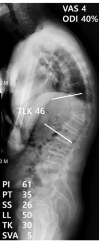
Fig. 2. Example of a post-osteoporotic vertebral compression fracture thoracolumbar kyphosis patient with sagittal balance. The patient was 84 years old and had thoracolumbar kyphosis due to a previous T12 osteopo- rotic compression fracture. The relevant parameters in this patient were:
PT: 35°, SS: 26°, TLK: 46°, SVA: 5 cm, VAS: 4, ODI: 40%. TLK: thoracolum- bar kyphosis, SS: sacral slope, PT: pelvic tilt, SVA: sagittal vertical axis, VAS: visual analogue scale, ODI: Oswestry Disability Index.
Journal of Korean Society of Spine Surgery
www.krspine.org
79 The Effect of Sagittal Spino-pelvic Alignment on Thoracolumbar Kyphosis in Osteoporotic Patients
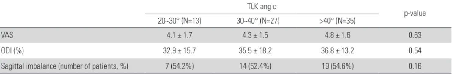
세 군(20~30
°
, 30~40°
, >40°
)으로 분류하여 임상 지표를 비교 하였을 때 통계적으로 유의한 차이를 보이지 않았다. 또한 흉요 추부 후만각에 따른 시상면 불균형 발생빈도에서도 통계적으 로 유의한 차이를 보이지 않았다(Table 2). 전체 환자를 대상으 로 SVA 5 cm 기준을 따라 시상면 균형 군과 시상면 불균형 군 으로 나누었을 때 나이, 성비, 체질량 지수 및 골밀도 T점수에 는 통계적으로 유의한 차이가 없었고, 최종 추시에서 시상면 불 균형이 있는 환자 군이 시상면상 균형 환자 군에 비해 임상적 평균값도 두 군 사이에 유의한 차이를 보이지 않았다(Table 1).최종 추시에서 전체 환자를 대상으로 흉요추부 후만각에 따라
Fig. 4. Example of a post-osteoporotic vertebral compression fracture thoracolumbar kyphosis patient with imbalance. (A) At baseline, the pa- tient was 84 years old and had thoracolumbar kyphosis due to a previous L1 compression fracture. She had the following values of the relevant parameters. PT: 25°, SS:15°, TLK: 28°, SVA: 6 cm, VAS: 4, ODI: 28%. (B) After 2 years of conservative treatment, the values changed to: PT: 31°, SS: 9°, TLK: 34°, SVA: 10 cm. Her clinical outcomes had deteriorated to a VAS of 7 and an ODI of 42%. TLK: thoracolumbar kyphosis, SS: sacral slope, PT: pelvic tilt, SVA: sagittal vertical axis, VAS: visual analogue scale, ODI: Oswestry Disability Index.
Fig. 3. Example of a senile thoracolumbar kyphosis patient with sagittal balance. She was 77 years old, with the following values of the relevant parameters. PT: 36°, SS: 20°, TLK: 50°, SVA: 2 cm, VAS: 3, ODI: 36%.
TLK:thoracolumbar kyphosis, SS: sacral slope, PT: pelvic tilt, SVA: sagittal vertical axis, VAS: visual analogue scale, ODI: Oswestry Disability Index.
Table 1. Average values of clinical measurements in post-osteoporotic vertebral compression fracture thoracolumbar kyphosis group (F group) and senile thoracolumbar kyphosis group (S group) at last follow-up.
F group (N=50) S group (N=25) p-value
VAS 4.7 ± 2.4 4.2 ± 2.1 0.75
ODI (%) 36.2 ± 20.2 35.6 ± 18.7 0.23
VAS: visual analogue scale, ODI: Oswestry disability index.
Table 2. Average values of clinical measurements and incidence of sagittal imbalance according to thoracolumbar kyphosis angle at last follow-up.
TLK angle
p-value 20~30° (N=13) 30~40° (N=27) >40° (N=35)
VAS 4.1 ± 1.7 4.3 ± 1.5 4.8 ± 1.6 0.63
ODI (%) 32.9 ± 15.7 35.5 ± 18.2 36.8 ± 13.2 0.54
Sagittal imbalance (number of patients, %) 7 (54.2%) 14 (52.4%) 19 (54.6%) 0.16
VAS: visual analogue scale, ODI: Oswestry disability index, TLK: thoracolumbar kyphosis.
A B
지표인 VAS 및 ODI 모두 유의하게 높게 나타났다(Table 3).
3. 방사선학적 결과
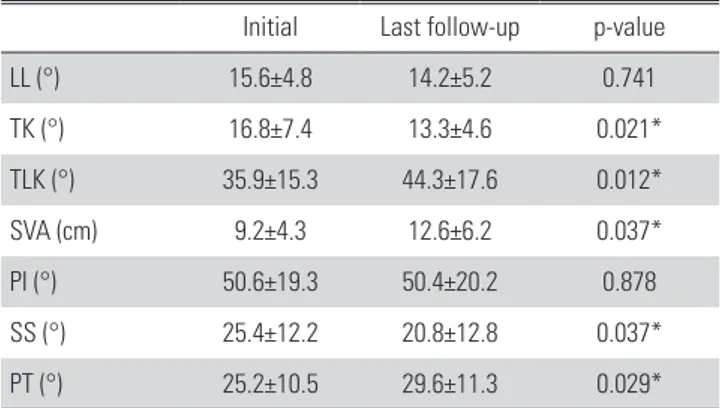
최종 추시에 측정한 모든 방사선학적 지표에서 두 군 사이에 유의한 차이를 보이지 않았다(Table 4). 시상면상 불균형이 있 는 환자 군(Fig. 4)에서는 흉추 후만각, 흉요추 후만각, 시상 수 직축 및 골반 경사각이 초기 진단 시에 비해 최종 추시에서 유 의하게 증가되었으며 천추 경사는 유의하게 감소되었다(Table 5).
고찰
고령화 사회가 되면서 퇴행성 성인 척추 변형으로 내원하는 환자들이 늘어나고 있다. 이러한 성인 척추 변형은 크게 관상
면, 시상면 불균형으로 나눌 수 있으며 이 중 시상면 불균형은 임상 증상과 밀접한 관련이 있어 삶의 질을 향상시키기 위해 최 근 수술적 치료가 빈번히 시행되고 있다.16,17) 또한, 골다공증의 유병률이 증가하며 골다공증성 흉요추부 척추체 압박 골절로 인한 흉요추부 후만 변형도 증가하고 있다. 이러한 시상면상 불 균형을 동반한 흉요추부 후만 변형은 통증 및 일상생활의 불편 을 일으킬 뿐 아니라 척추 신전근력 저하와 연관성이 있어 노인 환자에서 낙상 및 척추 골절의 발생을 증가시키는 중요한 요인 이다.10) 일반적인 흉요추부 후만증의 치료는 보존적 치료에서 부터 수술적 치료까지 다양하나 골다공증성 척추체 압박골절 후 발생한 흉요추부 후만증 및 노인성 흉요추부 후만증에 대한 치료 방법에 대해서는 특히 논란의 여지가 있다. 외상성 흉요추 부 후만증에 대하여 Bedbrook 등18) 은 흉요추부 Cobb’s angle 이 40도가 넘는 경우에도 후만각과 환자의 통증과 연관성 이 없다고 하였으나, Gertzbein 등19) 은 Cobb’s angle이 30 도가 넘는 경우에 통증의 위험도가 증가하며, 수술적 치료 가 필요하다고 하였고 Munting E 등9) 또한 Cobb’s angle 이 20도가 넘는 경우 수술적 치료가 필요하다고 하였다.
Chang 등12)은 보존적 치료에도 심각한 신체적 장애와 신경 학적 증상이 동반되는 경우 수술적인 치료가 요구된다고 하 였다. 이처럼 흉요추부 후만증 환자에서 후만각과 환자의 통 증 및 장애 정도에 관한 연구는 아직까지 명확히 정립되지 않아 치료방침을 정하는데 논란의 여지가 있다.
이번 연구에서 흉요추부 후만증을 제 10흉추의 상위 골단판 과 제 2요추의 하위 골단판 사이의 Cobb’s angle이 20도를 넘 는 경우로 정의하였다. 일반적으로 대부분의 흉요추부 손상 이 생역학적인 취약성으로 인해 제 11흉추에서 제 2요추 사이 분절에서 발생하는 것으로 알려져 있다.19,20) 본 연구에서는 골 Table 4. Average values of spinopelvic parameters in post-osteoporotic
vertebral compression fracture thoracolumbar kyphosis (F group) and se- nile thoracolumbar kyphosis (S group) at last follow-up.
F group (N=50) S group (N=25) p-value
LL (°) 20.2±9.4 18.7±8.2 0.84
TK (°) 19.5±10.4 17.8±9.6 0.58
TLK (°) 38.8±19.7 37.6±18.4 0.07
SVA (cm) 10.1±4.5 9.3±3.7 0.12
PI (°) 51.4±22.3 49.7±21.6 0.41
SS (°) 23.7±13.4 24.4±12.9 0.48
PT (°) 27.7±11.6 25.3±10.8 0.18
LL: lumbar lordosis angle, TK: thoracic kyphosis angle, TLK: thoracolumbar kyphosis angle(T10-L2 Cobb’s angle), SVA: sagittal vertical axis, PI: pelvic incidence, SS: sacral slope, PT: pelvic tilt.
Table 5. Average values of radiologic parameters in patient with sagittal imbalance.
Initial Last follow-up p-value
LL (°) 15.6±4.8 14.2±5.2 0.741
TK (°) 16.8±7.4 13.3±4.6 0.021*
TLK (°) 35.9±15.3 44.3±17.6 0.012*
SVA (cm) 9.2±4.3 12.6±6.2 0.037*
PI (°) 50.6±19.3 50.4±20.2 0.878
SS (°) 25.4±12.2 20.8±12.8 0.037*
PT (°) 25.2±10.5 29.6±11.3 0.029*
LL: lumbar lordosis angle, TK: thoracic kyphosis angle, TLK: thoracolumbar kyphosis angle, SVA: sagittal vertical axis, PI: pelvic incidence, SS: sacral slope, PT: pelvic tilt, *: Statistically significant.
Table 3. Average values of clinical measurements in sagittal balance groups and sagittal imbalance groups at last follow-up.
Sagittal balance
(N=32) Sagittal imbalance
(N=43) p-value
Age (yr) 71.3±5.2 74.7±8.1 0.09
Gender (M:F) 1:3.1 1:3.2 0.98
BMI (kg/m2) 23.3±3.4 22.7±4.2 0.07
BMD (T-score) -3.0±0.6 -3.1±0.8 0.23
VAS 3.6±1.7 5.2±2.4 0.02*
ODI (%) 31.5±15.7 39.4±19.2 0.01*
BMI: body mass index, BMD: bone marrow density, VAS: visual analogue scale, ODI: Oswestry disability index, *: Statistically significant.
Journal of Korean Society of Spine Surgery
www.krspine.org
81 The Effect of Sagittal Spino-pelvic Alignment on Thoracolumbar Kyphosis in Osteoporotic Patients
절 취약 부위를 충분히 포함할 수 있도록 제 10흉추에서 제 2요 추 사이를 범위로 하여 흉요추부 후만각을 측정하였다. 또한, 선행 연구들에서 평균 60세 이상의 정상 노인군에서의 흉요추 부 후만각이 평균 5~10도 정도로 보고 되었으며, 제 11흉추 상 위 골단판과 제 2요추 하위 골단판 사이의 각도가 15도가 넘는 경우 골다공증성 흉요추부 척추체 압박 골절과 감별이 필요함 이 보고되었고21), 외상 후 흉요추부 후만증에서 국소 후만각이 20도를 넘는 경우 수술적인 적응증으로 제시하였던 것에서 착 안하였다.9,21-23)
본 연구에서 골다공증성 흉요추부 척추 압박 골절의 유무와 후만각의 정도는 흉요추부 후만증 환자의 임상 증상과 연관성 이 없음을 알 수 있었다. 방사선상 지표 역시 척추 압박 골절의 유무와는 관련이 없었다. 최소 2년 이상의 추시 관찰기간 동안 보존적 치료는 전체 흉요추부 후만증 환자에서 흉요추부 척추 체 압박골절 유무에 상관없이 임상 증상 및 방사선 지표를 유지 되는데 도움이 되었다. 하지만, 시상면상 불균형이 동반된 환자 의 경우에서는 보존적 치료만으로 임상적 증상의 악화를 막지 못했을 뿐 아니라 흉요추부 후만곡의 악화와 이차적인 척추 변 형의 진행이 관찰되었다. 이는 흉요추부 후만증 환자에서 후만 변형에 대한 불충분한 보상 기전이 작용하고 척추 신전근의 지 속적인 피로로 인해 환자의 통증 및 장애를 유발하게 되었기 때 문으로 생각된다. 골반 경사의 증가와 천추 경사의 감소는 이러 한 골반의 불충분한 보상 작용으로 생각 할 수 있으며 역동적 시상면상 균형을 얻기 위한 흉요추부 후만각 교정 및 고정적인 치료가 필요할 수도 있다. 따라서 흉요추부 후만증 환자에서 보 존적 치료를 시행하였을 때 그 결과가 흉요추부 후만각이 아닌 시상면상 균형 여부에 따라 차이가 있었기 때문에 치료 계획을 세우는데 있어 시상면상 균형 여부를 반드시 고려해야 할 것으 로 생각된다. 시상면 불균형과 삶의 질의 연관성은 널리 알려져 있지만 이번 연구를 통해 국소적인 흉요추부 후만각보다 전신 적인 시상면 불균형이 중요함을 다시 확인할 수 있었다.
흉요추부 후만각도가 크면 시상면상 불균형이 더 많이 발생 할 것으로 예상하였으나 결과적으로 큰 연관성이 없었다. 나이 가 들면서 시상면 중심축이 전방으로 이동하는 경향이 있기 때 문에 시상면 불균형에 있어 흉요추부 후만각 외에 영향을 주는 인자로 작용했을 가능성을 생각해 볼 수 있다. 전체 환자군 중 시상면상 불균형을 보인 환자군이 절반 이상을 차지하였는데 이는 전체 환자군이 65세 이상의 고령으로 시상면 중심축이 전 방으로 전이되었을 것이며 평균적인 흉요추부 후만각이 20도 이상으로 기립 전신 척추 사진 촬영 시에 발생 할 수 있는 측정 의 오차 또한 영향을 주었을 수 있다고 생각된다.
본 연구의 제한점으로는 첫째, 표본집단의 수가 적어 모집단 전체의 성격을 대표 하기에는 한계가 있었다. 둘째, 연구의 대
상에서 수술적 치료를 시행 받았던 환자는 제외되어 비교적 증 상이 경미한 환자들 대상으로 하였기 때문에 선택 편향(selec-selec- tion bias)이 발생할 수 있다는 점이다. 셋째, 시상면 균형 여부 를 분류할 때 단순히 시상면 수직축만을 기준으로 하여 동적 시 상면 불균형(dynamic sagittal imbalance), 숨겨진 시상면 불균 형(hidden sagittal imbalance) 등의 요소를 세심히 고려하지 못 하였다. 넷째, 기립 전신 척추 사진 촬영 시 일부 환자에서 통증 으로 인해 고관절, 슬관절의 적절한 신전 자세를 유지하지 못해 척추-골반 지표의 오차가 있을 수 있다는 점이다. 마지막으로, 후향적 연구로서의 한계점이 있다고 할 수 있다. 아직까지 흉요 추부 후만증 환자들에 대한 치료에 대한 정확한 결론은 없으며 수술적 치료에 대한 적응증 역시 논란이 있다. 또한 해당 질환 의 자연 경과에 대해서도 잘 알려져 있지 않기 때문에 이를 포 함한 다기관 전향적 연구가 추후에 필요할 것으로 사료된다. 흉 요추부 후만증 환자의 치료에 있어서 병인 및 후만각 자체 보다 는 시상면 균형 여부를 고려한 치료적 접근이 필요할 것으로 판 단된다.
결론
흉요추부 후만증 환자에서 골절 또는 퇴행성 변화의 발생 원 인 및 흉요추부 후만 각도는 임상 증상과의 연관성이 없었던 반 면에 시상면 균형의 유무는 임상 증상과의 연관성을 보였고 방 사선학적 지표의 시간에 따른 변화와 연관됨을 보였다. 따라서 흉요추부 후만증 환자의 보존적 치료에 따른 결과에 시상면 불 균형이 좋지 않은 영향을 미치는 것을 알 수 있었고 치료 방침 을 정하는데 있어 시상면 균형 여부를 반드시 고려해야 할 것으 로 생각된다.
REFERENCES
1. Khan BI, Yost MT, Badkoobehi H, et al. Prevalence of sco- liosis and thoracolumbar kyphosis in patients with achon- droplasia. Spine Deform. 2016 Mar;4(2):145-8. DOI:
10.1016/j.jspd.2015.08.003.
2. Li S, Li Z, Hua W, Wang K, Li S, et al. Clinical outcome and surgical strategies for late post-traumatic kyphosis after failed thoracolumbar fracture operation: case re- port and literature review. Medicine (Baltimore). 2017 Dec;96(49):e8770. DOI: 10.1097/MD.0000000000008770 3. Tsirikos A, McMaster M. Infantile developmental tho-
racolumbar kyphosis with segmental subluxation of the spine. J Bone Joint Surg Br. 2010 Mar;92(3):430-5. DOI:
10.1302/0301-620X.92B3.23075.
4. Rajasekaran S. Kyphotic deformity in spinal tuberculosis and its management. International orthopaedics. Int Orthop.
2012 Feb;36(2):359-65. DOI: 10.1007/s00264-011- 1469-2.
5. Lee C-S, Lee C-K, Kim Y-T, et al. Dynamic sagittal im- balance of the spine in degenerative flat back: significance of pelvic tilt in surgical treatment. Spine (Phila Pa 1976).
2001 Sep 15;26(18):2029-35. DOI: 10.1097/00007632- 200109150-0017.
6. Lee CS, Chung SS, Chung KH, et al. Significance of pelvic incidence in the development of abnormal sagittal align- ment. Journal of the Korean Orthopaedic Association.
2006;41(2):274-80. DOI: 10.4055/jkoa.2006.41.2.274.
7. Zeng Y, Qu X, Chen Z, et al. Posterior corrective surgery for moderate to severe focal kyphosis in the thoracolumbar spine: 57 cases with minimum 3 years follow-up. Eur Spine J. 2017 Jul;26(7):1833-41. DOI: 10.1007/s00586-016- 4875-8. Epub 2016 Dec 28.
8. Cecchinato R, Berjano P, Damilano M, et al. Spinal oste- otomies to treat post-traumatic thoracolumbar deformity.
Eur J Orthop Surg Traumatol. 2014 Jul;24 Suppl 1:S31-7.
DOI: 10.1007/s00590-014-1464-6.
9. Munting E. Surgical treatment of post-traumatic kypho- sis in the thoracolumbar spine: indications and technical aspects. Eur Spine J. 2010 Mar;19 Suppl 1:S69-73. DOI:
10.1007/s00586-009-1117-3.
10. Finocchiaro FM, Nena U, Scalzo VL, et al. Treatment of kyphotic deformities in adults: our experience. Eur Spine J. 2012 May;21 Suppl 1:S100-7. DOI: 10.1007/s00586- 012-2224-0.
11. Kim BG, Dan JM, Shin DE. Treatment of thoracolum- bar fracture. Asian Spine J. 2015 Feb;9(1):133-46. DOI:
10.4184/asj.2015.9.1.133.
12. Chang KW, Chen YY, Lin CC, et al. Apical lordosating osteotomy and minimal segment fixation for the treat- ment of thoracic or thoracolumbar osteoporotic kyphosis.
Spine (Phila Pa 1976). 2005 Jul 15;30(14):1674-81. DOI:
10.1097/01.brs.0000170450.77554.bc.
13. Dang NR, Moreau MJ, Hill DL, et al. Intra-observer reproducibility and interobserver reliability of the radio- graphic parameters in the Spinal Deformity Study Group’
s AIS Radiographic Measurement Manual. Spine (Phila
Pa 1976). 2005 May 1;30(9):1064-9. DOI: 10.1097/01.
brs.0000160840.51621.6b.
14. Kim MS, Chung SW, Hwang C, et al. A radiographic analysis of sagittal spinal alignment for the standardization of standing lateral position. Journal of the Korean Ortho- paedic Association. 2005;40(7):861-7. DOI: 10.4055/
jkoa.2005.40.7.861.
15. Jackson RP, McManus AC. Radiographic analysis of sagittal plane alignment and balance in standing volun- teers and patients with low back pain matched for age, sex, and size. A prospective controlled clinical study.
Spine (Phila Pa 1976). 1994 Jul 15;19(14):1611-8. DOI:
10.1097/00007632-199407001-00010.
16. Glassman SD, Bridwell K, Dimar JR, et al. The impact of positive sagittal balance in adult spinal deformity. Spine (Phila Pa 1976). 2005 Sep 15;30(18):2024-9. DOI: 10.1097/01.
brs.0000179086.30449.96.
17. Schwab F, Ungar B, Blondel B, et al. Scoliosis Research Society—Schwab adult spinal deformity classifica- tion: a validation study. Spine (Phila Pa 1976). 2012 May 20;37(12):1077-82. DOI: 10.1097/BRS.0b013e31823e15e2.
18. Bedbrook GM. Treatment of thoracolumbar dislo- cation and fractures with paraplegia. Clin Orthop Relat Res. 2012 May 20;37(12):1077-82. DOI: 10.1097/
BRS.0b013e31823e15e2.
19. Gertzbein SD. Scoliosis Research Society. Multicenter spine fracture study. Spine (Phila Pa 1976). 1992 May;17(5):528- 40. DOI: 10.1097/00007632-199205000-00010.
20. DeWald RL. Burst fractures of the thoracic and lumbar spine. Clin Orthop Relat Res. 1984 Oct;(189):150-61.
21. Wei Y, Tian W, Zhang G, et al. Thoracolumbar kyphosis is associated with compressive vertebral fracture in postmeno- pausal women. Osteoporos Int. 2017 Jun;28(6):1925- 1929. DOI: 10.1007/s00198-017-3971-x.
22. Kim YB, Kim YJ, Ahn YJ, et al. A comparative analysis of sagittal spinopelvic alignment between young and old men without localized disc degeneration. Eur Spine J. 2014 Jul;23(7):1400-6. DOI: 10.1007/s00586-014-3236-8.
23. Kang KB, Kim YJ, Muzaffar N, et al. Changes of sagittal spinopelvic parameters in normal Koreans with age over 50. Asian Spine J. 2010 Dec;4(2):96-101. DOI: 10.4184/
asj.2010.4.2.96.
© Copyright 2019 Korean Society of Spine Surgery
Journal of Korean Society of Spine Surgery. www.krspine.org. pISSN 2093-4378 eISSN 2093-4386
This is an Open Access article distributed under the terms of the Creative Commons Attribution Non-Commercial License (http://creativecommons.org/licenses/by-nc/4.0/) which permits unrestricted non-commercial use, distribution, and reproduction in any medium, provided the original work is properly cited.
83
J Korean Soc Spine Surg. 2019 Sep;26(3):76-83. https://doi.org/10.4184/jkss.2019.26.3.83
Original Article
골다공증이 동반된 흉요추부 후만증 환자에서 시상면상 척추-골반 정렬이 임상 증상에 미치는 영향
김환정 • 송대건 • 이재원 • 장선호 • 박건영 • 김용호 • 마상범 • 최원식 을지대학교 의과대학 정형외과학교실
연구 계획: 후향적 연구
목적: 시상면상 척추-골반 정렬이 골다공증이 동반된 흉요추부 후만증(T10-L2 Cobb’s angle>20°) 환자의 임상 증상에 미치는 영향을 알아보고자 하였 다.
선행 연구문헌의 요약: 아직까지 외상 또는 선행 척추질환 병력 없이 퇴행성 변화에 의해 발생하는 흉요추부 후만증의 임상 증상 및 방사선학적 특징에 대한 연구는 거의 없는 실정이다. 또한 흉요추부 후만증 환자에서 후만 변형의 각도에 따라 임상 증상이 영향을 받는지, 혹은 어떤 치료방법을 선택해야 하는지에 대해서는 논란이 있다.
대상 및 방법: 2005년 5월부터 2016년 5월까지 본원에서 흉요추부 후만증(T10-L2 Cobb’s angle>20°)으로 진단되어 보존적 치료를 받았던 골다공증이 동반된 65세 이상의 환자 75명을 대상으로 하였다. 신경학적 증상이 있거나 불안정 골절 또는 선행 척추 질환의 병력이 있는 경우 연구에서 제외하였다.
골다공증성 척추체 압박 골절로 인한 흉요추부 후만증 환자 50명 및 노인성 흉요추부 후만증 환자 25명에 대해서 임상 증상 및 방사선학적 지표를 비교 분석하였다. 또한 대상자를 후만 각도 정도, sagittal balance 여부에 따라서도 나누어 비교 분석하였다. Visual analogue score (VAS), Oswestry disability index (ODI)를 이용해 평가하였다.
결과: 골다공증성 척추체 압박골절 이후 발생한 흉요추부 후만증 환자군과 노인성 흉요추부 후만증 환자군간 VAS, ODI 및 방사선학적 지표는 유의한 차 이가 없었다. 전체 환자를 흉요추부 후만각(20~30°, 30~40°, >40°)에 따라 분류한 후 임상 증상 및 방사선학적 지표를 비교하였을 때 역시 유의한 차이 가 없었다. 하지만, 전체 환자를 시상면 균형(SVA <5 cm, >5 cm) 여부로 분류하였을 때 시상면 불균형이 발생한 환자들의 VAS, ODI가 유의하게 증가되 어 있었다. 또한 처음 진단 시 시상면 불균형이 존재한 환자에서 최종 추시에서 VAS, ODI가 통계적으로 유의하게 증가 되었다.
결론: 흉요추부 후만 변형의 원인 및 후만각의 정도보다 시상면 균형의 유무가 환자의 통증 및 장애 정도에 영향을 주는 중요한 인자라고 생각된다. 따라 서, 골다공증이 동반된 흉요추부 후만 변형에서 시상면 불균형을 반드시 고려하여 치료 방법을 선택해야 할 것이다.
색인 단어: 흉요추부 후만증, 골다공증, 시상면상 척추-골반 지표, 시상면 불균형 약칭 제목: 시상면상 척추-골반 정렬이 골다공증이 동반된 흉요추부 후만증에 미치는 영향
접수일: 2019년 8월 6일 수정일: 2019년 8월 7일 게재확정일: 2019년 9월 3일 교신저자: 송대건
대전시 서구 둔산동 1306 을지대학교 의과대학 정형외과학교실
TEL: 042-611-3280 FAX: 042-611-3283 E-mail: jaystar4u@naver.com