Hypereosinophilic syndrome is a spectrum of diseases characterized by prominent peripheral eosinophilic leukocytosis without an identifiable cause (1, 2).
Eosinophilic infiltration occurs in various systems and organs such as the nervous system, heart, hematopoietic system, liver, spleen, bowel and lung.
The diagnostic criteria of idiopathic hypereosinophilic syndrome includes peripheral eosinophilia of more than 1500 eosinophils/mm3 for more than six months, ab- sence of parasitic, allergic or other causes of eosinophilia and evidence of organ involvement (3). Several reports have described the sonographic and computed tomogra- phy (CT) imaging findings in patients with hepatic in-
volvement (4, 5); however, portal, splenic and mesen- teric thrombosis in hypereosinophilic syndrome has been rarely reported (6). We report a case of portal, splenic and mesenteric thrombosis in a 33-year-old man with hypereosinophilic syndrome.
Case Report
A 33-year-old man was admitted for evaluation of dys- pnea, dizziness and abdominal distention. On a physical examination, a large amount of ascites was suggested.
Based on laboratory results, the WBC was 15,300/mm3 with 53% of eosinophils. The patient had no history of any allergic disease or ingestion of raw animal meat or liver. Stool, skin and ELISA tests for parasites such as Paragonimus westermani, Clonorchis sinensis, Sparganosis and Toxocara canis were negative. The patient under- went a high-resolution chest CT examination for evalua- tion of dyspnea and a multiphase abdominal CT exami-
Portal, Splenic and Mesenteric Thrombosis in Hypereosinophilic Syndrome: A Case Report1
Su Yeon Hwang, M.D., Kyung Mi Jang, M.D., Min Jeong Kim, M.D., Kwan Seop Lee, M.D., Sung Hye Koh, M.D., Eui Yong Jeon, M.D., Hyun Lee, M.D.,
Ju Hyun Choi, M.D., Mi Yeon Yie, M.D.
1Department of Radiology, Hallym University, College of Medicine Received February 18, 2009 ; Accepted March 19, 2009
Address reprint requests to : Kyung Mi Jang, M.D., Department of Radiology, Hallym University, College of Medicine, 896 Pyungchon- dong, Dongan-gu, Anyang-city, Kyungki-do 431-070, Korea
Tel. 82-31-380-3885 Fax. 82-31-380-3878 E-mail: jkm7290@hallym.or.kr
Idiopathic hypereosinophilic syndrome is a spectrum of diseases characterized by prominent peripheral eosinophilic leukocytosis without an identifiable cause. Several reports have described hepatic involvement as depicted on sonography and CT imag- ing in patients with hypereosinophilic syndrome. However, thrombosis of the portal, splenic and mesenteric veins in hypereosinophilic syndrome has been rarely reported.
We present here a case of portal, splenic and mesenteric thrombosis in a 33-year-old man with hypereosinophilic syndrome.
Index words :Thrombosis Portal vein Mesenteric veins Eosinophilia Liver
Tomography, X-ray computed
administration. A chest CT image showed multifocal patchy consolidations and ground-glass opacities with mainly peripheral distribution in both lungs and bilater- al pleural effusion (Fig. 1A). An unenhanced abdominal CT image showed the presence of a hyperdense throm- bus in the portal vein and the splenic vein (Fig. 1B, C). A contrast-enhanced abdominal CT image obtained during
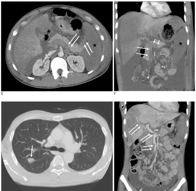
After the initial CT examinations, an abdominal CT scan was performed three days later for evaluation of newly developed acute severe left upper abdominal pain. A contrast-enhanced abdominal CT image obtained during the portal venous phase showed progression of throm- bosis in the splenic vein, acute splenic infarction and newly developed thrombosis in the superior mesenteric
A B
C D
Fig. 1. Imaging findings are presented for a 33-year-old man with hypereosinophilic syndrome combined with venous thrombosis in the portal, splenic and superior mesenteric veins.
A. An axial chest CT image with the lung window setting shows two areas of patchy consolidations (arrows), surrounded by zones of ground glass attenuation in the right upper lobe and a small amount of bilateral pleural effusion (asterisks).
B. An axial unenhanced CT image shows the presence of a hyperdense thrombus in the right and left portal veins (arrows).
C. A CT image obtained just inferiorly shows the hyperdense thrombus in the main portal vein and splenic vein (arrows).
D. An axial contrast-enhanced CT image obtained during the portal venous phase shows diffuse low attenuation of the enlarged liv- er with heterogeneity, splenomegaly and ascites, in addition to portal vein thrombosis (asterisks).
vein (Fig. 1E, F). Under the clinical impression of hyper- eosinophilic syndrome, the patient underwent systemic corticosteroid therapy for one month. As determined from follow-up laboratory results, the WBC was 7,400/mm3 with 0.7% of eosinophils. Follow-up chest and abdominal CT examinations were performed after the corticosteroid therapy. A chest CT image demon- strated improvement of the multifocal patchy areas of
consolidations and ground-glass opacities, except for a small consolidation in the right upper lung (Fig. 1G). On an abdominal CT image, portal, splenic and mesenteric thrombosis disappeared except for the presence of small thrombi in the main portal vein, the right portal vein and superior mesenteric vein (Fig. 1H). With the find- ings of hypereosinophilia in the peripheral blood, evi- dence of organ involvement and the response to cor-
E F
G H
Fig. 1. E. A follow-up CT image obtained three days after the initial CT examinations demonstrates the progression of the splenic venous thrombus (arrows) and acute splenic infarction (asterisk).
F. On a coronal CT image obtained during the portal venous phase, superior mesenteric vein thrombosis (long arrows) is demon- strated beside the splenic vein thrombosis (short arrows) and splenic infarction (asterisk). The CT image also shows heterogeneous low attenuation of the liver and a large amount of ascites.
G. A follow-up CT image obtained four weeks after the initial steroid therapy reveals disappearance of patchy consolidations and ground glass attenuation, except for a small focal consolidation (arrow) in the right upper lobe.
H. On a coronal CT image, thrombi in the portal, splenic and mesenteric veins have disappeared, except for small thrombi (arrows) in the right portal vein and superior mesenteric vein. The CT image also shows improvement of heterogeneous low attenuation of the liver and ascites.
Discussion
Hepatic involvement occurs in 40-90% of patients with hypereosinophilic syndrome, and usually presents with hepatomegaly and abnormal liver function (1, 2).
Previous studies about the CT findings of hepatic in- volvement in hypereosinophilic syndrome have demon- strated the presence of small low attenuation lesions that are scattered throughout the liver, especially in ar- eas adjacent to the portal veins, narrowing of the intra- hepatic portal veins and lobar or segmental low-attenu- ated lesions (4, 5). However, portal, splenic and mesen- teric thrombosis in hypereosinophilic syndrome has rarely been reported (6). The most frequent causes of portal vein thrombosis are a myeloproliferative disor- der, liver cirrhosis with portal hypertension, deficiency of natural anticoagulant proteins, gene mutations and hepatocellular carcinoma (7). Hypercoagulable syn- drome can lead to portomesenteric and splenic vein thrombosis and can be complicated with acute or suba- cute intestinal angina (8). Thus, early diagnosis and treatment of portomesenteric and splenic vein thrombo- sis in hypercoagulable syndrome is important to avoid complications such as intestinal angina (8). One of the causes of hypercoagulability is hypereosinophilic syn- drome (6, 9, 10).
Hypereosinophilic syndrome has been associated with various thrombotic manifestations such as deep venous thrombosis, cerebral venous thrombosis, hepatic veno- occlusive disease or the presence of intracardiac throm- bi (9, 10). Activated eosinophils and granular proteins in- cluding eosinophil cationin protein and major basic pro- tein are thought to modify coagulation and fibrinolysis in eosinophilia, resulting in thrombosis (6, 9, 10). Thus, venous thrombosis with hypereosinophilic syndrome
In summary, we experienced a case of rapidly pro- gressing portal, splenic and mesenteric thrombosis in a patient with hypereosinophilic syndrome. The splenic infarction was complicated during disease progression.
When portal, splenic and mesenteric thrombosis is asso- ciated with eosinophilia and pulmonary consolidation, hypereosinophilic syndrome should be considered in the differential diagnosis to avoid complications due to rapid progression of venous thrombosis.
References
1. Hardy WR, Anderson RE. The hypereosinophilic syndrome. Ann Intern Med 1968;68:1220-1229
2. Fauci AS, Harley JB, Roberts WC, Ferrans VJ, Gralnick HR, Bjornson BH. The idiopathic hypereosinophilic syndrome: clinical, pathophysiologic, and therapeutic considerations. Ann Intern Med 1982;97:78-92
3. Chusid MJ, Dale DC, West BC, Wolff SM. The hypereosinophilic syndrome: analysis of fourteen cases with review of the literature.
Medicine 1975;54:1-27
4. Lim JH, Lee WJ, Lee DH, Nam KJ. Hypereosinophilic syndrome:
CT findings in patients with hepatic lobar or segmental involve- ment. Korean J Radiol 2000;1:98-103
5. Kim GB, Kwon JH, Kang DS. Hypereosinophilic syndrome: imag- ing findings in patients with hepatic involvement. AJR Am J Roentgenol 1993;161:577-580
6. Kikuchi K, Minami K, Miyakawa H, Ishibashi M. Portal vein thrombosis in hypereosinophilic syndrome. Am J Gastroenterol 2002;97:1274-1275
7. Hidajat N, Stobbe H, Griesshaber V, Schroder RJ, Felix R. Portal vein thrombosis: etiology, diagnostic strategy, therapy and man- agement. Vasa 2005;34:81-92
8. Inagaki H, Sakakibara O, Miyaike H, Eimoto T, Yura J. Mesenteric venous thrombosis in familial free protein S deficiency. Am J Gastroenterol 1993;88:134-138
9. Kanno H, Ouchi N, Sato M, Wada T, Sawai T. Hypereosinophilia with systemic thrombophlebitis. Hum Pathol 2005;36:585-589 10. Kocaturk H, Yilmaz M. Idiopathic hypereosinophilic syndrome as-
sociated with multiple intracardiac thrombi. Echocardiography 2005;22:675-676
대한영상의학회지 2009;61:45-49
호산구증가증 환자에서 나타난 간문맥과 비장정맥 및 장간막정맥 혈전증: 증례 보고1
1한림대학교 의과대학 영상의학교실
황수연∙장경미∙김민정∙이관섭∙고성혜∙전의용∙이 현∙최주현∙이미연
특발성 호산구증가증은 밝혀진 원인 없이 말초혈액의 호산구가 증가하는 질환이다. 호산구증가증에서 다양한 장 기에 다량의 호산구들이 침착하여 다양한 영상 소견을 보일 수 있다. 주로 간을 침범한 환자에서 초음파 소견과 CT 소견에 대해 보고되었으나, 간문맥과 비장정맥 및 장간막정맥을 침범한 혈전증은 잘 알려져 있지 않다. 이에 본 저자 들은 특발성 호산구증가증에 동반된 간문맥과 비장정맥 및 장간막정맥을 침범한 혈전증에 대한 증례를 보고하고자 한다.