10.3988/jcn.2010.6.4.216 J Clin Neurol 2010;6:216-220
Introduction
Arteriovenous malformation (AVM) is a vascular conglomer- ate known as a nidus which has a main arterial feeder that is con- nected directly to the draining veins. The incidence of AVM is below 1 : 100,000 and accounts for about 1-2% of all strokes, and 3% of those in young patients.1 Although the association between AVMs and aneurysms is well known, an AVM in com- bination with moyamoya disease or progressive arterial occlu- sion has been reported only rarely.2
AVMs are generally thought to be congenital vascular anom- alies that arise as a result of the abnormal development of blood vessels during the early embryonic period.3 Conversely, moy- amoya disease is a progressive vascular disease that is defined as bilateral distal occlusion or stenosis of the internal carotid ar- tery (ICA) accompanied by a telangiectatic connection in the basal ganglia, the so-called ‘moyamoya vessels’.4 In particular, unilateral involvement is considered to be probable moyamo- ya disease. The etiology of vascular pathogenesis such as uni-
lateral stenosis and occlusion is unknown, although variable se- condary causes of arterial pathogenesis have been described, in- cluding atherosclerosis, brain tumor, and radiation.
We report herein a case of AVM with a feeding arterial occlu- sion and unilateral moyamoya disease coexisting in an acute st- roke patient.
Case Report
A 41-year-old man was admitted with sudden dysarthria and left facial palsy. He had no history of hypertension, diabetes, hyperlipidemia, or smoking. Brain computed tomography (CT) demonstrated slightly hyperdense cortical lesions in the right frontal lobe with calcified foci and adjacent low-density isch- emic lesions. CT angiography revealed occlusion of the right middle cerebral artery (MCA). T2-weighted magnetic resonance image (MRI) revealed the tightly packed vascular signals of flow voids without thrombus in the frontal lobe, and an adja- cent ischemic lesion was confirmed by diffusion-weighted MRI.
Arteriovenous Malformation with an Occlusive Feeding Artery Coexisting with Unilateral Moyamoya Disease
Seong Hwan Ahn, MD; In Seong Choo, MD; Jin Ho Kim, MD, PhD; Hoo Won Kim, MD
Department of Neurology, Chosun University School of Medicine, Gwangju, Korea
Received November 6, 2008 Revised September 23, 2009 Accepted September 23, 2009 Correspondence Hoo Won Kim, MD Department of Neurology, Chosun University School of Medicine, 588 Seoseok-dong,
Dong-gu, Gwangju 501-717, Korea Tel +82-62-220-3128
Fax +82-62-232-7587 E-mail [email protected]
BackgroundzzArteriovenous malformations (AVMs) with vascular abnormalities, including an- eurysms, have been reported frequently. However, the coexistence of AVM and unilateral moyamo- ya disease is rare. We report herein an AVM patient who presented with acute ischemic stroke with unilateral moyamoya disease and occlusion of the feeding artery.
Case ReportzzA-41-year old man was admitted with sudden dysarthria and facial palsy. Brain computed tomography and magnetic resonance imaging revealed an acute infarction adjacent to a large AVM in the right frontal lobe. Cerebral angiography revealed occlusions of the proximal right middle cerebral and proximal anterior cerebral arteries, which were the main feeders of the AVM.
Innumerable telangiectatic moyamoya-type vessels between branches of the anterior cerebral artery and dilated lenticulostriate arteries on the occluded middle cerebral artery were detected. However, a nidus of the AVM was still opacified through the distal right callosomarginal artery, which was supplied by the remaining anterior cerebral artery and leptomeningeal collaterals from the posterior cerebral artery.
ConclusionszzWhile AVM accompanied by unilateral moyamoya disease is rare, our case suggests an association between these two dissimilar vascular diseases. J Clin Neurol 2010;6:216-220 Key Wordszz arteriovenous malformations, moyamoya disease, ischemic stroke.
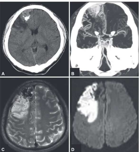
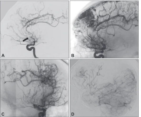
No evidence of previous hemorrhagic lesions was detected on either the CT or MRI (Fig. 1). Digital subtraction angiography revealed occlusion of the right proximal callosomarginal artery, a branch of anterior cerebral artery (ACA), and many small mo- yamoya-type telangiectatic connections between the mid cal- losomarginal and the pericallosal arteries. Occlusion of the right proximal MCA and dilated lenticulostriate arteries were also seen during the arterial phase. The right superior branches of the MCA and an AVM nidus were drained into the posterior cerebral artery-leptomeningeal collateral connection during the capillary-venous phase. However, no aneurysm was detected in the angiographic study (Fig. 2). Moreover, we failed to find any cardioembolic or atherothrombotic source in the electrocardio- gram, carotid Doppler, or transthoracic echocardiography. Lab- oratory testing revealed normal levels and activities of protein C, S antigen, antiphospholipid antibody, anticardiolipin anti- body, rheumatoid factor, fluorescent antinuclear antibody, lupus erythematosus cell, and homocysteine. Surgical interventions were not performed on the patient due to the mild symptoms and the presence of abundant collateral vessels. The patient was
discharged without major disability on the 8th day of hospital treatment.
Discussion
Our patient exhibited coexistence of two rare arterial lesions: 1) an AVM with proximal callosomarginal artery occlusion, a feed- er of the AVM, and 2) unilateral MCA occlusion with basal mo- yamoya vessels. The patient presented no evidence of athero- sclerosis, inflammation, radiation, or trauma. Acute cerebral le- sions in the adjacent AVM were the result of occlusion of the MCA. Abundant telangiectatic connections between the callo- somargianal and pericallosal arteries were revealed with angi- ography.
AVMs may be accompanied by dysplastic vascular changes including occlusion, stenosis, aneurysms, and ectasis. The rate of coexistence of aneurysm and AVMs in the same patient has been reported to be up to 58%. However, progressive feeding- artery occlusion of the AVM has been detected in less than 3%
of cases.2,5 The finding of an AVM accompanied by moyamo-
Fig. 1. Results of brain computed tomo- graphy (CT) and magnetic resonance im- aging (MRI). A: Pre-contrast brain CT scan showing hypodense lesions and a slightly hyperdense area with an irregu- lar calcified lesion in the right frontal cor- tex. B: Coronal CT angiography showing enlarged draining veins and proximal right middle cerebral arterial occlusion.
C: T2-weighted MRI, flow-void signals in the right frontal area. D: Diffusion-weight- ed MRI showing high signal intensity le- sion in the right anterior and middle ce- rebral arterial territories.
A
C
B
D
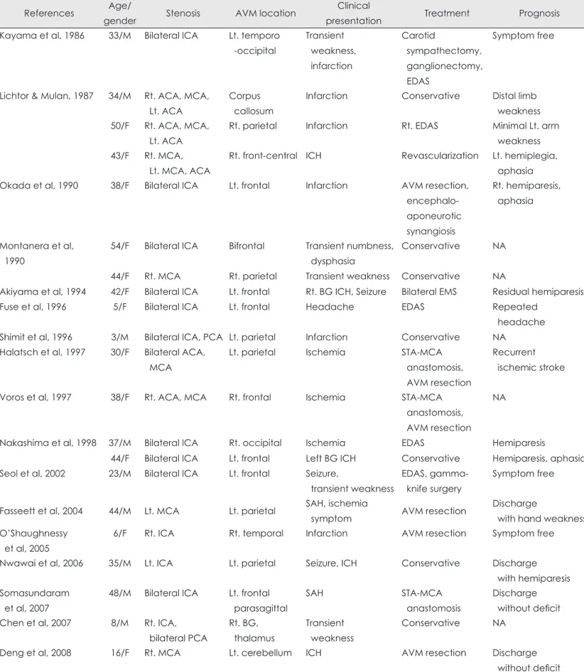
ya disease is even more rare. Only 21 cases have been report- ed in the English literature, as revealed by a scanning of the PubMed search and related articles (Table 1). The mean age of these patients was 32.1 years (range 3-54 years) with no gender predilection. Clinical presentation resulted in ischemic symp- toms or infarction in 13 patients, intracranial hemorrhage in 7, and headache in 1. The mean age of patients with hemorrhagic symptoms was greater than that of those with ischemic symp- toms (38.3 and 28.1 years, respectively). Most ischemic presen- tations seemed to be associated with moyamoya disease, be- cause ischemic stroke in AVM patients is rare feature.
It is unknown which vascular anomaly developed first: the AVM or moyamoya disease. Most reported cases show that AVMs located in the ipsilateral side of moyamoya vessels are associated with a moyamoya-type collateral.6-16 One hypothe- sis is that vascular occlusion drives from acquired dysplasia as a consequence of high blood flow from the AVM. The clinical evidence of high-flow-induced vascular change can be found in two case series of 19 patients with AVMs with progressive stenosis or occlusion of the feeding artery.5,17 Enam and Malik reported that six patients had a AVM with unilateral ICA occlu- sion with proximal ACA or MCA occlusions, and one had a ce- rebellar AVM with proximal occlusion of the vertebral artery.5 Mawad et al.17 reported nine AVM patients with moyamoya- type collaterals and three patients with isolated MCA or ACA occlusion without collaterals. In all cases, occlusive arterial le- sions were observed in the proximal artery that supplied the
AVM and were located unilaterally on the ipsilateral side of AVM. AVMs with feeding-arterial occlusion were accompa- nied with abundant angiectatic capillaries or collaterals resem- bling those found in moyamoya disease. The authors assumed that increased blood flow through an AVM causes turbulent flow at the carotid artery bifurcation, leading to focal intimal hyperplasia, with subsequent ICA stenosis and development of the moyamoya-type collaterals.
Others have suggested that the AVM most likely occurs sec- ondarily to the moyamoya disease. There are three case reports of an initial evaluation that failed to find AVM in a moyamoya patient, but where subsequent study discovered an AVM to be present.18-20 Those authors assumed that the ischemic environ- ment caused by moyamoya disease could influence the devel- opment of an AVM, since proximal large-arterial stenosis could be attributed to vasodilation in distal vessels, resulting in arte- rio-vascular shunts. Increased levels of basic fibroblast growth factor, which is a known angiogenic factor found in the endo- thelium or smooth muscle cells, have been detected in the ce- rebrospinal fluid of moyamoya disease patients.21 These factors may lead to opening and distension of anastomotic channels from the moyamoya vessles to the cortical veins concurrently with hyperangiogenic environments, in combination with the local angiogenic stimulation of previously injured neural tissue.
However, such AVMs were small (less than 3 cm), and were fill- ed with contrast dye during the late rather than the early arteri- al phase, which differs from the case in typical AVMs.
Fig. 2. Digital subtraction angiography.
A: Lateral early arterial phase of the in- ternal carotid artery shows the calloso- marginal artery, which is occluded proxi- mally (arrow), reappeareing in the distal part through abundant telangiectatic connections from the pericallosal artery.
B: A nidus with dilated cortical draining veins was detected in the late arterial phase. C: Occlusion of the proximal mid- dle cerebral artery with prominent moy- amoya vessels evident in the right ante- rior oblique view. D: Distal branches of the middle cerebral artery are opacified from anterotemporal and prieto-occipital branches of the right posterior cerebral artery in the capillary phase of lateral ver- tebral angiography.
A
C
B
D
Another hypothesis is that the coexistence of AVMs and mo- yamoya disease is coincidental finding. In three cases, moyam- oya vascular changes were found in locations unrelated to the
AVM.22-24 However, this differs from the cases mentioned above wherein the AVMs were supplied by normal feeding arteries, not by moyamoya collateral vessels.
Table 1. Summary of cases reported with an arteriovenous malformation (AVM) accompanied by moyamoya disease References Age/
gender Stenosis AVM location Clinical
presentation Treatment Prognosis
Kayama et al, 1986 33/M Bilateral ICA Lt. temporo -occipital
Transient weakness, infarction
Carotid
sympathectomy, ganglionectomy, EDAS
Symptom free
Lichtor & Mulan, 1987 34/M Rt. ACA, MCA, Lt. ACA
Corpus callosum
Infarction Conservative Distal limb weakness 50/F Rt. ACA, MCA,
Lt. ACA
Rt. parietal Infarction Rt. EDAS Minimal Lt. arm weakness 43/F Rt. MCA,
Lt. MCA, ACA
Rt. front-central ICH Revascularization Lt. hemiplegia, aphasia Okada et al, 1990 38/F Bilateral ICA Lt. frontal Infarction AVM resection,
encephalo- aponeurotic synangiosis
Rt. hemiparesis, aphasia
Montanera et al, 1990
54/F Bilateral ICA Bifrontal Transient numbness, dysphasia
Conservative NA
44/F Rt. MCA Rt. parietal Transient weakness Conservative NA
Akiyama et al, 1994 42/F Bilateral ICA Lt. frontal Rt. BG ICH, Seizure Bilateral EMS Residual hemiparesis
Fuse et al, 1996 5/F Bilateral ICA Lt. frontal Headache EDAS Repeated
headache Shimit et al, 1996 3/M Bilateral ICA, PCA Lt. parietal Infarction Conservative NA Halatsch et al, 1997 30/F Bilateral ACA,
MCA
Lt. parietal Ischemia STA-MCA
anastomosis, AVM resection
Recurrent ischemic stroke
Voros et al, 1997 38/F Rt. ACA, MCA Rt. frontal Ischemia STA-MCA anastomosis, AVM resection
NA
Nakashima et al, 1998 37/M Bilateral ICA Rt. occipital Ischemia EDAS Hemiparesis 44/F Bilateral ICA Lt. frontal Left BG ICH Conservative Hemiparesis, aphasia Seol et al, 2002 23/M Bilateral ICA Lt. frontal Seizure,
transient weakness
EDAS, gamma- knife surgery
Symptom free
Fasseett et al, 2004 44/M Lt. MCA Lt. parietal SAH, ischemia
symptom AVM resection Discharge
with hand weakness O’Shaughnessy
et al, 2005
6/F Rt. ICA Rt. temporal Infarction AVM resection Symptom free
Nwawai et al, 2006 35/M Lt. ICA Lt. parietal Seizure, ICH Conservative Discharge with hemiparesis Somasundaram
et al, 2007
48/M Bilateral ICA Lt. frontal parasagittal
SAH STA-MCA
anastomosis
Discharge without deficit Chen et al, 2007 8/M Rt. ICA,
bilateral PCA
Rt. BG, thalamus
Transient weakness
Conservative NA
Deng et al, 2008 16/F Rt. MCA Lt. cerebellum ICH AVM resection Discharge
without deficit M: male, F: female, Rt: right, Lt: left, ICA: internal carotid artery, ACA: anterior cerebral artery, MCA: middle cerebral artery, PCA: pos- terior cerebral artery,VA: vertebral artery, BG: basal ganglia, ICH: intracranial hemorrhage, EDAS: encephaloduroarteriosynangiosis, STA: superficial temporal artery, EMS: encephalomyosynagiosis, NA: not available.
Unilateral moyamoya disease, which involves occlusion or stenosis of the ICA or proximal MCA with moyamoya vessels, is atypical and is especially rare in adult. Some patients with unilateral moyamoya disease progress to the bilateral form.
However, the low level of basic fibroblast growth factor in the CSF and lack of familial occurrence in unilateral moyamoya disease supported a different pathomechanism that is different from typical bilateral moyamoya disease.25 Clinically, increas- ed levels of atherogenic factors or high regional blood flow are reported in unilateral moyamoya disease.26
In our case, occlusion of the AVM feeder was detected in the right proximal ACA. During the injection of contrast dye into the vertebral artery, delayed contrast filling of right MCA from the leptomeningeal arteries supports the suggestion that the MCA was also a main feeder of the AVM. Well-developed bas- al and leptomeningeal collaterals were the result of slow pro- gressive vascular pathology.27 Therefore, high flow caused by the AVM may influence the development of unilateral moy- amoya disease, and acute ischemic stroke is thought to be the re- sult of progressive vascular occlusive disease. Although the si- multaneous presence of two vascular diseases that have dif- ferent characteristics is extremely rare, further study is ne- cessary to improve our understanding of the association be- tween AVMs and moyamoya disease.
Conflicts of Interest
The authors have no financial conflicts of interest.
REFERENCES
1. Al-Shahi R, Warlow C. A systematic review of the frequency and prog- nosis of arteriovenous malformations of the brain in adults. Brain 2001;
124:1900-1926.
2. Meisel HJ, Mansmann U, Alvarez H, Rodesch G, Brock M, Lasjaunias P. Cerebral arteriovenous malformations and associated aneurysms: an- alysis of 305 cases from a series of 662 patients. Neurosurgery 2000;46:
793-800.
3. Mullan S, Mojtahedi S, Johnson DL, Macdonald RL. Embryological basis of some aspects of cerebral vascular fistulas and malformations. J Neurosurg 1996;85:1-8.
4. Fukui M. Guidelines for the diagnosis and treatment of spontaneous oc- clusion of the circle of Willis (‘moyamoya’ disease). Research Commit- tee on Spontaneous Occlusion of the Circle of Willis (Moyamoya Dis- ease) of the Ministry of Health and Welfare, Japan. Clin Neurol Neuro- surg 1997;99 Suppl 2:S238-S240.
5. Enam SA, Malik GM. Association of cerebral arteriovenous malfor- mations and spontaneous occlusion of major feeding arteries: clinical and therapeutic implications. Neurosurgery 1999;45:1105-1111.
6. Kayama T, Suzuki S, Sakurai Y, Nagayama T, Ogawa A, Yoshimoto T.
A case of moyamoya disease accompanied by an arteriovenous malfor- mation. Neurosurgery 1986;18:465-468.
7. Lichtor T, Mullan S. Arteriovenous malformation in moyamoya syn-
drome. Report of three cases. J Neurosurg 1987;67:603-608.
8. Akiyama K, Minakawa T, Tsuji Y, Isayama K. Arteriovenous malfor- mation associated with moyamoya disease: case report. Surg Neurol 1994;
41:468-471.
9. Halatsch ME, Rustenbeck HH, Jansen J. Progression of arteriovenous malformation in moyamoya syndrome. Acta Neurochir (Wien) 1997;
139:82-85.
10. Vörös E, Kiss M, Hankó J, Nagy E. Moyamoya with arterial anoma- lies: relevance to pathogenesis. Neuroradiology 1997;39:852-856.
11. Seol HJ, Kim DG, Oh CW, Han DH. Radiosurgical treatment of a ce- rebral arteriovenous malformation in a patient with moyamoya disease:
case report. Neurosurgery 2002;51:478-481.
12. Fassett DR, Schloesser PE, Couldwell WT. Hemorrhage from moyam- oya-like vessels associated with a cerebral arteriovenous malformation.
Case report. J Neurosurg 2004;101:869-871.
13. Nawawi O, Sinnasamy M, Ramli N. Unilateral moyamoya disease with co-existing arteriovenous malformation. Br J Radiol 2006;79:e12-e15.
14. Somasundaram S, Thamburaj K, Burathoki S, Gupta AK. Moyamoya disease with cerebral arteriovenous malformation presenting as prima- ry subarachnoid hemorrhage. J Neuroimaging 2007;17:251-254.
15. Chen Z, Zhu G, Feng H, Lin J, Wu N. Giant arteriovenous malforma- tion associated with unilateral moyamoya disease in a child: case report.
Surg Neurol 2007;67:89-92.
16. Montanera W, Marotta TR, terBrugge KG, Lasjaunias P, Willinsky R, Wallace MC. Cerebral arteriovenous malformations associated with mo- yamoya phenomenon. Am J Neuroradiol 1990;11:1153-1156.
17. Mawad ME, Hilal SK, Michelsen WJ, Stein B, Ganti SR. Occlusive vascular disease associated with cerebral arteriovenous malformations.
Radiology 1984;153:401-408.
18. Fuse T, Takagi T, Fukushima T, Hashimoto N, Yamada K. Arteriove- nous malformation associated with moyamoya disease. Childs Nerv Syst 1996;12:404-408.
19. Schmit BP, Burrows PE, Kuban K, Goumnerova L, Scott RM. Ac- quired cerebral arteriovenous malformation in a child with moyamoya disease. Case report. J Neurosurg 1996;84:677-680.
20. O’Shaughnessy BA, DiPatri AJ Jr, Parkinson RJ, Batjer HH. Devel- opment of a de novo cerebral arteriovenous malformation in a child with sickle cell disease and moyamoya arteriopathy. Case report. J Neu- rosurg 2005;102:238-243.
21. Yoshimoto T, Houkin K, Takahashi A, Abe H. Angiogenic factors in moyamoya disease. Stroke 1996;27:2160-2165.
22. Nakashima T, Nakayama N, Furuichi M, Kokuzawa J, Murakawa T, Sakai N. Arteriovenous malformation in association with moyamoya disease. Report of two cases. Neurosurg Focus 1998;5:e6.
23. Deng ZH, Wang S, Li Z, Zhao JZ. Unilateral moyamoya disease associ- ated with cerebellar arteriovenous malformation: one case report. Chin Med J (Engl) 2008;121:1145-1147.
24. Okada T, Kida Y, Kinomoto T, Sakurai T, Kobayashi T. Arteriovenous malformation associated with moyamoya disease--case report. Neurol Med Chir (Tokyo) 1990;30:945-948.
25. Houkin K, Abe H, Yoshimoto T, Takahashi A. Is “unilateral” moyam- oya disease different from moyamoya disease? J Neurosurg 1996;85:
772-776.
26. Ogata T, Yasaka M, Inoue T, Yasumori K, Ibayashi S, Iida M, et al. The clinical features of adult unilateral moyamoya disease: does it have the same clinical characteristics as typical moyamoya disease? Cerebrovasc Dis 2008;26:244-249.
27. Liebeskind DS. Collateral circulation. Stroke 2003;34:2279-2284.