pISSN: 2234-8646 eISSN: 2234-8840 https://doi.org/10.5223/pghn.2018.21.4.361
Pediatr Gastroenterol Hepatol Nutr 2018 October 21(4):361-364
PGHN
Case Report
PEDIATRIC GASTROENTEROLOGY, HEPATOLOGY & NUTRITION
Accessory Hepatic Lobe: A Rare Cause of Prehepatic Portal Hypertension in a Child
Elif Sağ, Ayşegül Cansu*, Mustafa İmamoğlu
†, and Murat Çakır
Departments of Pediatric Gastroenterology, Hepatology and Nutrition, *Radiology, and †Pediatric Surgery, Faculty of Medicine, Karadeniz Technical University, Trabzon, Turkey
Accessory hepatic lobe is noted as and considered a rare disease in children. It can manifest with various symptoms and complications depending on the location, volume, type and position of the disease as presented on a child. The patient presented as a 14-month-old girl who was seen with a notable hepatosplenomegaly and portal hypertension.
A diagnosis was made after taking an extensive medical history, observation and radiological examinations. The formal diagnosis was a prehepatic portal hypertension associated with accessory hepatic lobe.
Key Words: Accessory hepatic lobe, Child, Portal hypertension
Received:October 2, 2017, Revised:December 19, 2017, Accepted:December 22, 2017
Corresponding author: Elif Sağ, Department of Pediatric Gastroenterology, Hepatology and Nutrition, Faculty of Medicine, Karadeniz Technical Universty, 61080, Trabzon, Turkey. Tel: +90-0546-580-50-86, Fax: +90-462-3250518, E-mail: drturkmen61@ gmail.com
Copyright ⓒ 2018 by The Korean Society of Pediatric Gastroenterology, Hepatology and Nutrition
This is an openaccess article distributed under the terms of the Creative Commons Attribution NonCommercial License (http://creativecommons.org/licenses/by-nc/4.0/) which permits unrestricted noncommercial use, distribution, and reproduction in any medium, provided the original work is properly cited.
INTRODUCTION
Accessory hepatic lobe (AHL) is a rare congenital anomaly found in children, which is known to have resulted from an error in the formation of the endo- dermal caudal foregut during the embryogenetic process of a developing fetus, as noted most com- monly in the third gestational week of a pregnancy.
It may be completely separate, pedunculated or ses- sileor in characteristic, or else appear in the form of a lobe attached to normal hepatic tissue [1]. It is classi- fied as type 1, a large lobe attached to the subphrenic or perihepatic region of the liver (>31 g); type 2, a lobe arising from around the right hepatic lobe or a wide base on the surface (11-30 g); and type 3, an en-
tirely separate lobe frequently seen in the pelvis and the thorax or else in punctate form attached to the surface of the gallbladder or liver (<10 g) [1-3].
AHLs are generally asymptomatic conditions and are most often detected during surgery or autopsies. But it can be seen that sometimes these conditions can be presented with a torsion, hemorrhage, palpable mass in the upper right quadrant, acute or can be ac- companied with chronic abdominal pain, and vari- ous symptoms associated with the compression and location of the AHL (such as dyspnea, chest pain, vomiting and solid organ obstruction) [4]. Here in, we reported a child with AHL that is associated with a prehepatic portal hypertension (PHT).
362 Vol. 21, No. 4, October 2018 Pediatr Gastroenterol Hepatol Nutr
Fig. 2. Upper gastrointestinal endoscopy revealed three strands grade 2 esophageal varices without red spots.
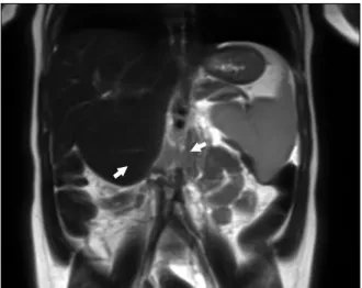
Fig. 1. T2-weighted coronal image demonstrates splenomegaly and 56×55 mm mass with same intensity as liver (accessory hepatic lobe, arrows) extending inferiorly from the segment 5-6 level, with separate portal and hepatic veins and compressing the right upper pole of the kidney.
CASE REPORT
In this case, we noted a 14-month-old girl who presented with a preliminary diagnosis of hepatos- plenomegaly. Upon review of her medical history, there were no pathological findings other than that a hepatosplenomegaly was present at the physical examination. Subsequently at the laboratory exami- nation, hepatic enzymes, alpha-fetoprotein, alpha-1 antitrypsin, liver autoantibodies and coagulation pa- rameters were normal, while the thrombocytopenia was observed at complete blood count (82×109/L).
The portal vein Doppler ultrasonography revealed an incidence of hepatosplenomegaly and AHL, which is in the same echogenicity of the liver and approx- imately 5.5 cm in size extending to the right upper pole of the kidney, as noted with separate portal and hepatic veins. The main portal vein diameter was measured at 15 mm (normal lower limit <13 mm), and the flow rate is 30 cm/second (normal range 13-23 cm/second) [5]. Upon review of the magnetic resonance imaging of the liver, it was revealed that the AHL is 56×55 mm size extending inferiorly from the segment 5-6 level, with a separate portal and hepatic veins, and was noted to be compressing the
right upper pole of the kidney on T2-weighted coro- nal image (Fig. 1). The endoscopic examination re- vealed grade 2 esophagial varices with three strands, and a notation that the varices were more evident when the patient lay on the left side during the endoscopy. The observed red spots were not visible during the review (Fig. 2). The patient was started on a program of propranolol (1 g/kg/day) and followed with an endoscopic examination with a six months interval. During the follow-up, the patient under- went endoscopy and the grade of varices were noted as stable upon follow up. The noted thrombocyte lev- els were 104×109/L. The surgical treatment was planned in the case of a progression of varices grade or with the incidence of patient variceal bleeding.
The researchers have obtained a written consent form from the family in this case study.
DISCUSSION
The incidence of an AHL is a rare condition that generally develops as a result of embryonic hetero- plasia in a patient. This rare condition is also thought to develop secondary to trauma or surgery in a patient. The condition may be associated with in- heritance of an autosomal recessively transmitted gene. It has been hypothesized that AHL forms as a
www.pghn.org 363
Elif Sağ, et al:Accessory Hepatic Lobe in a Child
result of the embryonic liver curving outward during the intrauterine development stage of a patient’s de- velopment, or of tunica muscularis recti develop- ment together with increased intra-abdominal pres- sure and liver growth [6].
The AHL is generally asymptomatic, and symp- toms may vary depending on the localization and complication of the condition. Torsion is the most common and severe complication, but it may also present in a patient with acute/chronic abdominal pain, nausea, vomiting, chest pain, constipation, a palpable mass in the abdomen, swelling, dyspnea if located in the thorax and hemorrhage, and torsion or rupture if complications develop [7].
The complications of AHL may vary depending on the patient’s age and location of the lobe. In infants it is generally associated with congenital acrompha- lus and biliary atresia. The incidence of peduncu- lated AHL is more risky because of complications, such as torsion, rupture or infraction. In addition, it may cause symptoms due to mechanical pressure placed on other organs or vascular structures, as in our case. A case of Riedel’s lobe causing obstruction by compressing the stomach was reported in the literature. In that case, the patient received a physi- cian performed cholecystectomy and fixing of the Riedel’ lobe to the abdominal wall, with a simple su- ture, in an effort to manage the condition [8].
Additionally, a case of PHT developing due to ex- ternal pressure caused by ectopic hepatic tissue was reported in an 8-year-old boy [9]. In another case, AHL was reported associated with vaginal varices and vaginal bleeding in a 25-year-old adult woman, while advanced tests revealed PHT and portal bilio- pathy developed due to extrinsic compression and occlusion of portal vein by the AHL. The patient in that case had perisplenic, perigastric collaterals vari- ces and mild splenomegaly. Additionally noted was the cause of vaginal bleeding, which was seen as the incidence of dilate and tortuous vessels in the pelvis surrounding the uterus. The patient in that case was followed with symptomatic medical therapy and has not needed a surgical operation [10]. In our case, we believe that the PHT is related to both the com-
pression of portal vein externally by AHL and in- crease portal vein blood flow by the AHL’s own circulation. The patient is receiving medical treat- ment and controlled by intermittent endoscopy.
Surgical treatment is performed when the patients are symptomatic or have complications as torsion, hemorrhage. There is no needed to treat patients with a sessile AHL connected to normal liver tissue [11]. In our patient, the AHL was identified coincidentally. The patient had only hep- atosplenomegaly, and was considered stable on the follow up without any variceal bleeding.
In conclusion, AHL is considered a rare disease in children. It can manifest with various symptoms and complications depending on location, volume, type and position. There are very few reports in the liter- ature of PHT developing secondary to the incidence of AHL compressing the portal vein. We want to share our experience with this case that AHL might be in the different diagnoses of prehepatic PHT.
REFERENCES
1. Carrabetta S, Piombo A, Podestà R, Auriati L. Torsion and infarction of accessory liver lobe in young man.
Surgery 2009;145:448-9.
2. Tancredi A, Cuttitta A, de Martino DG, Scaramuzzi R.
Ectopic hepatic tissue misdiagnosed as a tumor of lung.
Updates Surg 2010;62:121-3.
3. Mehta V, Arora J, Manik P, Suri RK, Rath G. Clinico- anatomical aspects of accessory fissures obscuring the normal hepatic morphology. Clin Ter 2010;161:259-60.
4. Glenisson M, Salloum C, Lim C, Lacaze L, Malek A, Enriquez A, et al. Accessory liver lobes: anatomical de- scription and clinical implications. J Visc Surg 2014;
151:451-5.
5. Chavhan GB, Parra DA, Mann A, Navarro OM. Normal Doppler spectral waveforms of major pediatric vessels:
specific patterns. Radiographics 2008;28:691-706.
6. Pujari BD, Deodhare SG. Symptomatic accessory lobe of liver with a review of the literature. Postgrad Med J 1976;52:234-6.
7. Garba ES, Ameh EA. Isolated rupture of an accessory liver from blunt abdominal trauma in childhood.
Pediatr Surg Int 2002;18:62-3.
8. Akbulut S, Cakabay B, Sevinc MM, Basak F. Gastric outlet obstruction caused by Riedel's lobe of the liver:
364 Vol. 21, No. 4, October 2018 Pediatr Gastroenterol Hepatol Nutr
a diagnostic and therapeutic challenge for surgeons.
Hepatogastroenterology 2011;58:589-92.
9. Matley PJ, Rode H, Cywes S. Portal vein obstruction by ectopic liver tissue. J Pediatr Surg 1989;24:1163-4.
10. Chandramohan A, Pachuau EL, Eapen A. Accessory hepatic lobe: a rare cause of extra-hepatic portal vein
obstruction. Trop Gastroenterol 2014;35:190-3.
11. Wang C, Cheng L, Zhang Z, Xie T, Ding H, Deng Q, et al. Accessory lobes of the liver: A report of 3 cases and review of the literature. Intractable Rare Dis Res 2012;1:86-91.