Korean J Pediatr Infect Dis 2014;21:96-103
DOI: http://dx.doi.org/10.14776/kjpid.2014.21.2.96
Comparison of Group A, B and C Rotaviral Gastroenteritis among Children in Korea: Prevalence and Clinical Features
Kil-Seong Bae, M.D.*, Woo Ri Bae, M.D.*, Ji Hoon Kim, M.D.*, Joong Hyun Bin, M.D.*, Hyun Hee Kim, M.D.*, Hee Jin Lee, M.D.†, and Wonbae Lee, M.D.*
Department of Pediatrics*, Clinical Medical Research Institute†, The Catholic University of Korea, Bucheon St Mary’s Hospital, Bucheon, Korea
Purpose: The aim of this study is that the prevalence of rotavirus infection was evaluated by each group and clinical features of group A, B and C rotaviruses infections were described respectively to compare one with another.
Methods: Between January 2010 and December 2010, we enrolled a group of children below 10 years of age admitted for management of acute diarrhea at the Catholic University of Korea Bucheon St. Mary's Hospital. A total of 310 stool samples documented to be free of common bacterial pathogens were collected from children with diarrhea. The presence of group A, B or C rotavirus is indicated by amplification of DNA segments of the expected lengths after the first and second PCRs
Results: In a total of 310 stool specimens, 40 (12.9%) specimens were positive for rotaviruses. These included 23 (7.4%) positive for group A, 5 (1.6%) for group B and 12 (3.9%) for group C rotaviruses. Group B rotavirus infected patients had significantly less diarrheas per day (group A: P =0.01, group C: P =0.01) and shorter duration of vomiting days (group A:
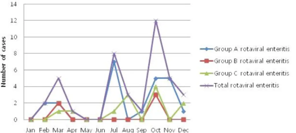
P =0.03, group C: P =0.03) than those with group A and C rotaviruses infection respectively. All the group B rotaviruses had been isolated in March and October. Group C rotavirus infections were prevalent during late summer and early winter and peaked in October.
Conclusion: These findings indicate that group B and C rotaviruses are notable causes or the contributing causes of diarrhea among infants and children in Korea.
Key Words : Rotavirus, Gastroenteritis, Prevalence
1)
Introduction
Rotavirus is one of the most important causes of acute gastroenteritis in mankind and animals world-
*The authors have no conflicts of interest relevant to this article to disclose.
Received : 15 August 2013, Revised : 17 February 2014 Accepted : 9 June 2014
Correspondence : Wonbae Lee, M.D.
Department of Pediatrics, the Catholic University of Korea, Bucheon St. Mary’s Hospital, South Korea
Tel : +82-32-340-7045, Fax : 82-32-340-2673 E-mail : [email protected]
widely1). Group A rotavirus has been considered as the major cause of severe diarrhea in mankind, whereas the clinical importance of group B and C rotaviruses gastroenteritis remains unclear2).
Occasionally called adult diarrhea rotavirus, group B rotavirus is distinct genetically and antigenically from group A and group C rotaviruses2). The pre- valence of group B rotavirus is not as high as that of group A rotavirus; however, serological findings indicate that group B rotaviruses are distributed so wide that most people could acquire antibodies to the virus early in life at least in prevalent areas3).
As few laboratories provide specific diagnoses of group B rotavirus, there is a scarcity of studies con- cerning group B rotavirus infections; therefore, the epidemiological patterns and clinical influence are poorly understood.
Averagely, the worldwide seroprevalence of group C rotavirus infection is nearly 33%, with a peak in the older age groups4-7). Despite a number of re- ported studies on sporadic cases of severe diarrhea or outbreaks of gastroenteritis caused by group C rotavirus in many countries, the prevalence of group C rotavirus and a clinical importance of the disease caused by this virus remain uncertain8-15).
The clinical features of group A rotavirus-infected patients have been described broadly16). Most studies have reported fever, a high frequency of vomiting and dehydration, which lasted for 1-9 days17). The majority of patients infected with group B rotavirus experienced vomiting and/or abdominal pain along with diarrhea, but fever was reported in only a few patients (4.5%)18). Also, other studies have reported that group B rotavirus was responsible for severe cholera-like diarrhea, with vomiting and severe de- hydration19). Group C rotavirus has been reported to be associated with relatively mild disease; fewer episodes of vomiting, less dehydration and fewer hospitalizations when compared to group A rotavirus infection in children9, 12).
However, above studies based their conclusions on very small sample sizes for analysis of clinical data. Moreover, very few studies had been executed to compare the clinical presentations and prevalence of patients infected with group A, group B and group C rotaviruses one another, which was not far suffi- cient to provide knowledge helpful to physicians treating patients infected with rotavirus. It spurred
us on trying this study even if this study still may mean preliminary. We analyzed 310 children with acute diarrhea for a year. Among them, the preva- lence of rotavirus infection was evaluated by each group and clinical features of group A, B and C ro- taviruses infections were described respectively to compare one with another.
Materials and Methods
1. Subjects
Between January 2010 and December 2010, we enrolled a cohort of children below 10 years of age admitted for treatment of acute diarrhea at the Catholic University of Korea Bucheon St. Mary's Hospital. A total of 310 stool samples documented to be free of common bacterial pathogens were collected from children with diarrhea.
2. DNA preparation by RT-PCR
The fresh or frozen stool used with in the first steps of the protocol. Stool samples are lysed in buffer ASL (QIAGEN GmbH, Hilden, Germany). DNA- damaging substances and PCR inhibitors present in stool sample are adsorbed to inhibit EX matrix. The inhibit EX matrix is pelleted by centrifugation and the DNA and RNA in the supernatant is purified on QIAamp mini spin columns (QIAGEN GmbH, Hilden, Germany).
Viral nucleic acid was extracted from 200 mL of the filtered supernatant, using a QIAamp DNA stool kit and QIAamp Viral RNA mini kit (QIAGEN GmbH, Hilden, Germany) according to the manufacturer’s instructions. Five microliters of the extracted nucleic acid was first incubated with 500 mM dNTPs and
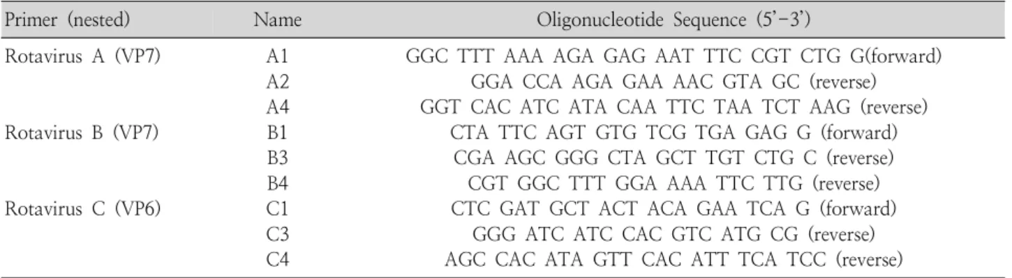
Table 1. Primers used for RT-PCR
Primer (nested) Name Oligonucleotide Sequence (5’-3’)
Rotavirus A (VP7) Rotavirus B (VP7) Rotavirus C (VP6)
A1 A2 A4 B1 B3 B4 C1 C3 C4
GGC TTT AAA AGA GAG AAT TTC CGT CTG G(forward) GGA CCA AGA GAA AAC GTA GC (reverse) GGT CAC ATC ATA CAA TTC TAA TCT AAG (reverse)
CTA TTC AGT GTG TCG TGA GAG G (forward) CGA AGC GGG CTA GCT TGT CTG C (reverse)
CGT GGC TTT GGA AAA TTC TTG (reverse) CTC GAT GCT ACT ACA GAA TCA G (forward)
GGG ATC ATC CAC GTC ATG CG (reverse) AGC CAC ATA GTT CAC ATT TCA TCC (reverse)
Fig. 1. PCR primers for detection of rotavirus groups A, B and C.
50 ng random primers at 65℃ for 5 min, followed by reverse transcription using 100 U SuperScript II reverse transcriptase (Invitrogen), 20 U RNase inhibitor, The reaction was incubated at 25℃ for 5 min, 50℃ for 60 min and heat inactivated at 70℃
for 15 min. Synthesized cDNA was used in PCR detection of the viruses.
3. DNA Amplication
The presence of group A, B or C rotavirus is in- dicated by amplification of DNA segments of the expected lengths after the first and second PCRs:
1062 and 257 bp for group A, 489 and 434 bp for group B, and 356 and 327 bp for group C rotavirus, respectively. For first PCR, primer pairs A1-A4, B1-B4 and C1-C4 (Table 1) were used separately in individual reverse transcription polymerase chain reaction (RT-PCR) assays of the three human ro- tavirus groups20). For second PCR, primer pairs A2- A4, B1-B3 and C1-C3 (Table 1, Fig. 1) were used in RT-PCR assays20).
4. Statistical analysis
Statistical analysis to compare clinical features among group A, B and C rotavirus-infected patients was performed using SPSS (SPSS Inc., version 18.0, Chicago, IL, USA) for Windows by chi-square test
or Fisher's exact test. A P value under 0.05 was considered statistically significant.
5. Results
40 (12.9%) cases were positive for rotaviruses in 310 stool specimens. These included 23 (57.5%
of the rotavirus infections) positive for group A, 5
Table 2. Comparison of Clinical Features of Patients Infected by Group A, B and C Rotaviruses Group A (N=23) Group B (N=5) Group C (N=12)
P-value
Median Range Median Range Median Range
Age (months) Hospitalization (days) Diarrhea duration (days) Diarrhea frequency (/day) Vomiting duration (days) Vomiting frequency (/day) Fever (days)
12 6 5 9 2 4 2
2-72 2-11 3-9 3-20 1-3 2-10 0-6
14 6 3 4 0 0 0
6-36 3-7 2-5 2-6 0-1 0-3 0-4
17 5 4 6 2 3 2
4-78 1-11 2-9 3-8 0-3 0-6 0-5
>0.05
>0.05
>0.05 0.01 0.03
>0.05
>0.05 Fig. 2. Monthly distribution of group A, B and C rotavirus-positive cases in Korea.
(12.5%) for group B rotavirus and 12 (30%) for group C rotavirus.
Epidemiologic and clinical data of patients infected with group A, B, and C rotaviruses were examined and compared with one another (Table 2). The age range of children excreting group A (2-72 months) and excreting group C rotaviruses (4-78 months) were higher than that of children infected with group B rotavirus (6-36 months), but the difference was not significant (P =0.12).
Although these three groups of patients had similar overall clinical features such as hospitalization, di- arrhea and fever days, vomiting per day, group B rotavirus patients had significantly less diarrheas per day than those with group A and C rotaviruses
infection (group A: P=0.01, group C: P=0.01). Pa- tients infected with group B rotavirus tend to have shorter duration of vomiting days than those infected with group A and C rotaviruses significantly (group A: P =0.03, group C: P =0.03). Amongst patients infected with group A rotavirus, only 2 patients showed severe dehydration and one patient suffered seizure during hospitalization.
Monthly distribution of group A, B and C rotavirus- positive cases was showed in Fig. 2. Group A rota- virus infections were prevalent during late winter, autumn, and summer, All the group B rotaviruses were isolated in spring and autumn. Any case of group B rotavirus infection was not detected in winter (January-February and November-December).
Group C rotavirus infections were prevalent during late summer and early winter, with most cases oc- curring between August and December.
Discussion
There are several reasons why very few studies have reported in detail on the clinical manifestations of group A, B and C rotavirus infections in terms of comparison. Most of all, distribution of group B ro- taviruses is restricted to one geographical area and as few as studies have investigated the epidemio- logy of these viruses.
China, India and Bangladesh, only three neigh- bouring countries, have reported group B rotavirus, with the current addition of Myanmar to the list. In India, during 2003-2004, group B rotaviruses con- stituted 2-4% of diarrhea patients attending Dhaka hospital in whom no common diarrhea pathogen was isolated. The present our study also detected a si- milar proportion (1.6%) of patients infected by this virus. However, a remarkably higher percentage (18.5%) of group B rotaviruses was isolated from Indian children during 2002-200418). In spite of lack of published references, it is apparent that group B rotavirus infection accounts for exraordinary per- centage of rotaviral gastroenteritis, which will re- quire further researches.
After finding the prevalence and clinical features of group B and C rotavirus-involved children, we compared them with those of group A rotavirus- involved children. Our study confirms that group B and C rotaviruses have been circulating at Bucheon St. Mary's Hospital and that they are responsible for a substantial proportion of pediatric diarrheal illness.
When compared with the clinical features of group A rotavirus infection, it was found that group B in- fection included most of the symptoms of group A infection. However, dehydration was milder in group B rotavirus-infected patients than in group A ro- tavirus-infected patients. While many investigators described previously that most of the group B- positive patients were older than 18 years, all the group B-positive patients in our study were younger than 4 years. According to 5 cases, it is highly im- plicative that group B rotavirus infections can cause less significant symptoms in infants and children. In restricted communities of Korea during the surveil- lance period, small-scale of group B rotavirus out- breaks might occur and go undetected, particularly if the severity of the disease is mild and hospitali- zation may not be required.
The frequency of group C rotavirus infections detected in this work (3.9%) was higher than in other studies carried out with pediatric population in Korea (0.7%), Turkey (0.8%), Japan (1.2%), Nigeria (1.8%), Argentina (2.8%) and Malawi (3.3
%)10-11, 21-24). However, the prevalence of the infec- tion was lower compared to Japan (10.2%), Spain (16%)12, 13). Group C rotavirus has also been found in co-infection with other enteric pathogens such as Vibrio cholerae, Shigella flexneri and group A rotavirus10, 12, 14). But, any mixed infections (Vibrio Cholerae, Shigella Flexneri) with group C rotavirus were not shown in this study.
The data presented above, together with other studies, demonstrate clearly that the selection of diagnostic methods can directly influence detection rates. With this very same collection, a 1% detection rate was reported previously using only one ELISA method for screening with nested PCR for confir-
mation. The incorporation of an additional ELISA method (CDC-ELISA), modifications in the set of primers for RT-PCR and nested PCR and the use of Southern blotting increased the estimate of group C rotavirus incidence to 3%25).
The median age of group C rotavirus detections in the current study were 17 months old. Previously, seroepidemiological investigations and detection data suggested a difference between age distribution in children with diarrhea caused by group A rotavirus, which typically infects children before 3 years of age, and group C rotavirus, which affects older chil- dren7, 25, 26). Although it is generally believed that group C rotavirus is more prevalent among older children (>4 years), detection of group C rotaviruses in both older and younger children is not surprising at all9-12, 15). However, the small number of samples in the present study did not allow us to draw solid conclusions about the prevalence in older children.
The sporadic nature of group C rotavirus infections along with the possibility of rapid dissemination among susceptible individuals in certain settings could be the cause of large outbreaks. It has been suggested that sporadic introduction from a hypo- thetical reservoir could explain at least in part the variability in detection rates observed in short stu- dies27). This study does not suggest that the group C rotavirus have currently a major epidemiological impact, even after the introduction of a group A rotavirus vaccine.
The fact that the group C rotavirus strains do not remain circulating in humans like group A rotavirus, may suggest that this group does not achieve the fitness required to become a successful human pathogen28). It is an alternative hypothesis that the group C rotavirus may cause a subclinical infection
in humans. Based on the study conducted in Hungary, the group C rotavirus were detected from raw se- wage samples, suggesting that the virus is in circu- lation; however, a significant increase in the number of sporadic cases or outbreaks was not observed29). It is noted that the group B and C rotavirus inci- dence and associated disease remain unclear once sensitive tests for its detection are not available to clinical laboratories. The diagnosis is difficult since most of the ELISA assays do not recognize the group B and C rotavirus specific VP7, VP6 antigen. The RT-PCR using group B and C rotavirus specific primer is a convenient option30). However, it has not been used widely due to the large costs for routine surveillance.
In conclusion, these findings suggest that group B and C rotaviruses are notable causes of diarrhea among infants and children in Korea. Until now, as mentioned above, group B rotaviruses have been confined regionally and for this reason little attention has been paid to them worldwide. The studies of group C rotavirus also were insufficient. Continuous monitoring of group B and C rotaviruses both in hospitals and in the community may be helpful to evaluate the true clinical importance of group B and C rotaviruses.
한 글 요 약
한국 소아에서의 로타 장염군의 비교: 유병율과 임상증상
배길성ㆍ배우리ㆍ김지훈ㆍ빈중현ㆍ김현희ㆍ이희진 이원배
가톨릭대학교 의과대학 소아과학교실
목적: 본 연구의 목표는 로타 바이러스 감염의 유병률을
로타 바이러스 A, B, C 군으로 나누어 평가하고 각 군의 임상 양상을 서로 상대적으로 비교함에 있다.
방법: 2010년 1월에서 2010년 12월 사이에, 부천 성 모병원에 급성 장염으로 입원한 10세 미만 환아를 대상으 로, 총 310개의 대변표본이 추출되었고, PCR을 통해 DNA 증폭과정을 거쳐, A, B, C 군의 로타 바이러스 군을 구분 하였다.
결과: 총 310개의 대변표본에서 40개(12.9%)에서 로 타 바이러스 양성이 확인되었으며, 이중 23개(7.4%)가 로타 바이러스 A군에 양성, 5개(1.6%)가 B군, 12개(3.9
%)가 C군에 양성 소견을 보였다. B군은 A군과 C군에 비 교하여 유의하게 경한 설사증상과 짧은 구토기간을 보였 다(group A: P =0.01, group C: P=0.01) 모든 B군 로 타 바이러스는 3월과 10월에 발견되었으며, C군은 늦은 여름과 초겨울에, 그 중에서 10월에 최고의 유병률을 보 였다.
결론: 국내에서 로타 바이러스 장염은 주로 로타 바이 러스 A군에 의하여 발생하였다. 그러나 일부에서는 로타 바이러스 C군과 B군이 원인으로 확인된바 향후 이에 대 한 지속적인 연구가 필요할 것으로 사료된다.
References
1) Kapikian AZ, Hoshino Y, Chanock RM. Rotaviruses.
In: Knipe D, Howley P, Griffin D, MArtin M, Lamb R, Roizman B, Straus S, eds. Fields virology. 4th ed.
Philadelphia: Lippincott Williams & Wilkins, 2001:
1787-822.
2) Saif LJ, Jiang B. Nongroup A rotaviruses of humans and animals. Curr Top Microbiol Immunol 1994;185:339-71.
3) Krishnan T, Sen A, Choudhury JS, Das S, Naik TN, Bhattacharya SK. Emergence of adult diarrhoea rotavirus in Calcutta, India. Lancet 1999;353:380-1.
4) James VL, Lambden PR, Caul EO, Cooke SJ, Clarke IN. Seroepidemiology of human group C rotavirus in the UK. J Med Virol 1997;52:86-91.
5) Riepenhoff-Talty M, Morse K, Wang CH, Shapiro C, Roberts J, Welter M, et al. Epidemiology of group C rotavirus infection in Western New York women of childbearing age. J Clin Microbiol 1997;35:486-8.
6) Steele AD, James VL. Seroepidemiology of human group C rotavirus in South Africa. J Clin Microbiol 1999;37:
4142-4.
7) Iturriza-Gomara M, Clarke I, Desselberger U, Brown D, Thomas D, Gray J. Seroepidemiology of group C rotavirus infection in England and Wales. Eur J Epidemiol 2004;19:589-95.
8) Maunula L, Svensson L, von Bonsdorff CH. A family outbreak of gastroenteritis caused by group C rotavirus.
Arch Virol 1992;124:269-78.
9) Jiang B, Dennehy PH, Spangenberger S, Gentsch JR, Glass RI. First detection of group C rotavirus in fecal specimens of children with diarrhea in the United States.
J Infect Dis 1995;172:45-50.
10) Cunliffe NA, Dove W, Jiang B, Thinwda Cert BD, Broad- head RL, Molyneux ME, et al. Detection of group C rotavirus in children with acute gastroenteritis in Blantyre, Malawi. Pediatr Infect Dis J 2001;20:1088-90.
11) Adah MI, Wade A, Oseto M, Kuzuya M, Taniguchi K.
Detection of human group C rotaviruses in Nigeria and sequence analysis of their genes encoding VP4, VP6, and VP7 proteins. J Med Virol 2002;66:269-75.
12) Sanchez-Fauquier A, Roman E, Colomina J, Wilhelmi I, Glass RI, Jiang B. First detection of group C rotavirus in children with acute diarrhea in Spain. Arch Virol 2003;
148:399-404.
13) Phan TG, Nishimura S, Okame M, Nguyen TA, Khamrin P, Okitsu S, et al. Virus diversity and an outbreak of group C rotavirus among infants and children with diarrhea in Maizuru city, Japan during 2002-2003. J Med Virol 2004;74:173-9.
14) Rahman M, Banik S, Faruque AS, Taniguchi K, Sack DA, Van Ranst M, et al. Detection and characterization of human group C rotaviruses in Bangladesh. J Clin Microbiol 2005;43:4460-5.
15) Steyer A, Poljsak-Prijatelj M, Bufon T, Sedmak M, Vidmar L, Mijovski JZ, et al. First detection of group C rotavirus in patients with gastroenteritis in Slovenia.
J Med Virol 2006;78:1250-5.
16) Estes M, Kapikian A, Knipe D, Howley P. Rotaviruses.
In: Knipe D, Howley P, Griffin D, Lamb R, Martin M, Roizman B, Straus S, editors. Fields virology. 5th ed.
Philadelphia: Lippincott Williams & Wilkins, 2007:
1917-74.
17) Rodriguez WJ, Kim HW, Arrobio JO, Brandt CD, Cha- nock RM, Kapikian AZ, et al. Clinical features of acute gastroenteritis associated with human reovirus-like agent in infants and young children. J Pediatr 1977;91:188-
93.
18) Rahman M, Hassan ZM, Zafrul H, Saiada F, Banik S, Faruque AS, et al. Sequence analysis and evolution of group B rotaviruses. Virus Res 2007;125:219-25.
19) Sanekata T, Ahmed MU, Kader A, Taniguchi K, Koba- yashi N. Human group B rotavirus infections cause severe diarrhea in children and adults in Bangladesh. J Clin Microbiol 2003;41:2187-90.
20) Gouvea V. PCR detection of rotavirus. In: Persing DH, Smith TF, Tenover FC, White TJ, editors. Diagnostic molecular microbiology: principles and applications.
Washington, DC: American Society for microbiology, 1993:383-88.
21) Moon S, Humphrey CD, Kim JS, Baek LJ, Song JW, Song KJ, et al. First detection of Group C rotavirus in children with acute gastroenteritis in South Korea. Clin Microbiol Infect 2011;17:244-7.
22) Mitui MT, Bozdayi G, Dalgic B, Bostanci I, Nishizono A, Ahmed K. Molecular characterization of a human group C rotavirus detected first in Turkey. Virus Genes 2009;39:157-64.
23) Castello AA, Arguelles MH, Villegas GA, Olthoff A, Glikmann G. Incidence and prevalence of human group C rotavirus infections in Argentina. J Med Virol 2002;
67:106-12.
24) Kuzuya M, Fujii R, Hamano M, Nishijima M, Ogura H. Detection and molecular characterization of human
group C rotaviruses in Okayama prefecture, Japan, bet- ween 1986 and 2005. J Med Virol 2007;79:1219-28.
25) Castello AA, Arguelles MH, Rota RP, Humphrey CD, Olthoff A, Gentsch JR, et al. Detection and characteri- zation of group C rotavirus in Buenos Aires, Argentina, 1997-2003. J Med Virol 2009;81:1109-16.
26) Kuzuya M, Fujii R, Hamano M, Ohata R, Ogura H, Yamada M. Seroepidemiology of human group C rota- virus in Japan based on a blocking enzyme-linked im- munosorbent assay. Clin Diagn Lab Immunol 2001;8:
161-5.
27) Banyai K, Jiang B, Bogdan A, Horvath B, Jakab F, Meleg E, et al. Prevalence and molecular characterization of human group C rotaviruses in Hungary. J Clin Virol 2006;37:317-22.
28) Luchs A, Morillo SG, de Oliveira CM, Timenetsky Mdo C. Monitoring of group C rotavirus in children with acute gastroenteritis in Brazil: an emergent epidemiological issue after rotavirus vaccine? J Med Virol 2011;83:1631-6.
29) Meleg E, Banyai K, Martella V, Jiang B, Kocsis B, Kisfali P, et al. Detection and quantification of group C rota- viruses in communal sewage. Appl Environ Microbiol 2008;74:3394-9.
30) Castello AA, Arguelles MH, Villegas GA, Lopez N, Ghi- ringhelli DP, Semorile L, et al. Characterization of human group C rotavirus in Argentina. J Med Virol 2000;62:
199-207.