ISSN 2234-3806 • eISSN 2234-3814
492 www.annlabmed.org https://doi.org/10.3343/alm.2018.38.5.492 Ann Lab Med 2018;38:492-494
https://doi.org/10.3343/alm.2018.38.5.492
Letter to the Editor
Diagnostic Genetics
PCM1-JAK2 Fusion in a Patient With Acute Myeloid Leukemia
Jong-Mi Lee, M.D.1,2, Jaewoong Lee, M.D.1,2, Eunhee Han, M.D.1,2, Myungshin Kim , M.D.1,2, Yonggoo Kim, M.D.1,2, Kyungja Han, M.D.1,2, and Hee-Je Kim, M.D.3
Department of Laboratory Medicine1 and Catholic Genetic Laboratory Center2, Seoul St. Mary’s Hospital, College of Medicine, The Catholic University of Korea, Seoul; Department of Hematology3, Catholic Blood and Marrow Transplantation Center, Seoul St. Mary’s Hospital, Leukemia Research Institute, College of Medicine, The Catholic University of Korea, Seoul, Korea
Dear Editor,
Over 34 myeloid neoplasm cases with a PCM1-JAK2 fusion have been reported worldwide since 1999, including one case reported in Korea [1, 2]. These cases share common features, such as splenomegaly, eosinophilia, myelofibrosis, and male predominance. Most cases have been diagnosed as myelopro- liferative neoplasm or myelodysplastic/myeloproliferative neo- plasm, in particular, as chronic eosinophilic leukemia and atypi- cal chronic myeloid leukemia. However, acute myeloid/lymphoid leukemia cases have also been reported [1]. Therefore, the 2016 WHO revision recognized the PCM1-JAK2 fusion gene as a provisional entity [3], joining the existing category of “myeloid and lymphoid neoplasms with eosinophilia and abnormalities of platelet-derived growth factor receptor α (PDGFRA), platelet-de- rived growth factor receptor β (PDGFRB), or fibroblast growth factor receptor 1 (FGFR1).”
A PCM1-JAK2 diagnosis can be made with or without eosino- philia, if the presence of the genetic rearrangement is proven.
However, identification of the genomic breakpoint in the PCM1- JAK2 fusion is quite difficult because it varies by case; as many as 14 different fusion transcripts from 15 patients have been re- ported [2, 4-6]. Moreover, the genetic lesions involved in the re-
arrangement of the two genes are widely distributed, from exon 23 to exon 36 in PCM1 and from exon 1 to exon 11 in JAK2 [5].
Therefore, it is important to report cases of newly confirmed breakage sites where chromosomal assays offer the only indica- tion of a PCM1-JAK2 fusion. We report a case of acute myeloid leukemia with a PCM1-JAK2 fusion that was not accompanied by eosinophilia. This study was approved by the institutional re- view board of Seoul St. Mary’s Hospital, Korea, and informed consent was obtained from the patient. A 51-year-old woman was diagnosed as having acute myeloid leukemia at a tertiary hospital in September 2009. At diagnosis, a complete blood count revealed 1.8×10⁹/L leukocytes, 66 g/L Hb, and 34×109/L platelets. A peripheral blood smear demonstrated a leukoeryth- roblastic reaction (nucleated red blood cells: 86/100 leukocytes;
left shifted maturation pattern). A bone marrow (BM) biopsy showed hypercellularity with increased erythroid precursors (64% of all nucleated cells) and blasts (47% of non-erythroid cells). The blasts were positive for CD13, CD33, CD117, human leukocyte antigen–antigen D related (HLA-DR), and myeloper- oxidase (MPO), and were weakly positive for CD64, CD56, and nuclear terminal deoxynucleotidyl transferase (TdT). A diagnosis of acute erythroid leukemia was made based on the 2008 WHO
Received: August 31, 2017
Revision received: December 17, 2017 Accepted: April 26, 2018
Corresponding author: Myungshin Kim https://orcid.org/0000-0001-8632-0168
Department of Laboratory Medicine, College of Medicine, Seoul St. Mary’s Hospital, The Catholic University of Korea, 222 Banpo-daero, Seocho-gu, Seoul 06591, Korea
Tel: +82-2-2258-1645, Fax: +82-2-2258-1719 E-mail: microkim@catholic.ac.kr
© Korean Society for Laboratory Medicine
This is an Open Access article distributed under the terms of the Creative Commons Attribution Non-Commercial License (http://creativecommons.org/licenses/by-nc/4.0) which permits unrestricted non-commercial use, distribution, and reproduction in any medium, provided the original work is properly cited.
1 / 1 CROSSMARK_logo_3_Test
2017-03-16 https://crossmark-cdn.crossref.org/widget/v2.0/logos/CROSSMARK_Color_square.svg
Lee J-M, et al.
AML with PCM1-JAK2 fusion
https://doi.org/10.3343/alm.2018.38.5.492 www.annlabmed.org 493
classification [7]. A cytogenetic examination could not be per- formed successfully because of inadequate specimens. The pa- tient experienced complete remission following induction che-
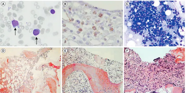
motherapy with an idarubicin and cytosine arabinoside regimen and subsequently received three courses of high dose consoli- dation therapy with cytosine arabinoside. In April 2011, the pa- Fig. 1. Bone marrow (BM) findings. (A) Myeloblasts showing less cytoplasm and distinct prominent nucleoli at initial diagnosis (April 2011) (Wright stain, ×400); (B) CD34-positive myeloblasts in the BM biopsy (November 2011) (immunohistochemistry stain, ×400); (C) in- creased immature cells in touch preparation (March 2012) (Wright stain, ×400); (D–F) serial BM biopsies showing fibrosis progression (April 2011, November 2011, and March 2012, respectively) (hematoxylin & eosin stain ×200).
A B C
D E F
Fig. 2. The t(8;9)(p21;p24) translocations and PCM1-JAK2 fusion gene. (A) Karyogram of BM showing 46,XX,t(8;9)(p21;p24), (B) Se- quence of the chimeric PCM1-JAK2 gene showing in-frame fusion between exon 30 of PCM1 and exon 9 of JAK2.
B PCM1 exon 30 JAK2 exon 9
A
8 9
Leu Gly Ser Ile Leu Gln Glu Ile Glu Leu Ser Ser
Lee J-M, et al.
AML with PCM1-JAK2 fusion
494 www.annlabmed.org https://doi.org/10.3343/alm.2018.38.5.492 tient re-developed pancytopenia, which lasted for three weeks;
she was transferred to Seoul St. Mary’s Hospital, and the BM bi- opsy confirmed increased myeloblasts (13%; Fig. 1A). The pa- tient received re-induction chemotherapy (FLANG regimen; 30 mg/m2/day fludarabine, 1 g/m2/day cytosine arabinoside, 10 mg/m2/day mitoxantrone, and 300 μg/day G-CSF for five days) twice because of persistent myeloblasts in the BM. After seven months, in November 2011, the BM revealed increased myelo- blasts (8%; Fig. 1B). The patient received an allogeneic periph- eral blood stem cell transplant from an HLA-matched unrelated donor. Unfortunately, her BM study revealed a relapse of acute myeloid leukemia at the 3-month follow-up post transplantation (myeloblasts 60%, Fig. 1C). In this case, well-known pheno- types of the PCM1-JAK2 fusion, such as splenomegaly and eo- sinophilia, were not observed; however, myelofibrosis, a mor- phological feature that matches myeloid neoplasms with PCM1- JAK2 was detected (Fig. 1D, 1E, and 1F). Chromosomal analy- ses of the specimen in November 2011, demonstrated abnor- malities in the short arm of chromosome 8 and 9 (Fig. 2A):
46,XX,t(1;14)(p36.1;q11.2),t(2;6)(q35;p21.1),t(8;9)(p21;p24) [10]/46,XX[3]. We amplified the PCM1-JAK2 fusion transcript from the BM specimen with the detected chromosomal rear- rangement by reverse-transcription PCR with primers designed using PRIMER 3 (available from: http://primer3.sourceforge.
net): PCM1 exon 28 forward (5´-GAGCGTATGAAGACTG-3´) and JAK2 exon 9 reverse (5´-GGCCATGACAGTTGCTTTGT-3´). Sanger sequencing also confirmed an in-frame fusion between PCM1 exon 30 and JAK2 exon 9 (Fig. 2B). Targeted next-generation sequencing, including 46 myeloid neoplasm-associated genes, identified a DNMT3A c.2644C>T, p.Arg882Cys mutation.
The PCM1-JAK2 fusion, together with the JAK2 V617F, MPL mutations, causes overexpression of the JAK2 pathway [5].
Therefore, a JAK2 inhibitor, ruxolitinib, could be used as a thera- peutic agent; in fact, several cases with positive treatment effi- cacy have been reported [8, 9]. Because eosinophilia and my- elofibrosis accompany 50–70% of myeloid neoplasms with PCM1-JAK2 [10], careful genetic evaluation is necessary for ad- equate diagnosis and the application of targeted therapy, even when a patient does not manifest well-known characteristics.
Authors’ Disclosures of Potential Conflicts of Interest
No potential conflicts of interest relevant to this article were re- ported.
Acknowledgments
This study was supported by a grant from the Korea Health Technology R&D Project, Ministry of Health and Welfare, Korea (A120175).
REFERENCES
1. Bain BJ and Ahmad S. Should myeloid and lymphoid neoplasms with PCM1-JAK2 and other rearrangements of JAK2 be recognized as spe- cific entities? Br J Haematol 2014;166:809-17.
2. Song I, Lee DH, Lee JH, Jang S, Huh JR, Seo EJ. A t(8;9)(p22;p24)/
PCM1-JAK2 translocation in a patient with myeloproliferative neoplasm and myeloid sarcoma: first report in Korea. Ann Lab Med 2016;36:79- 81.
3. Arber DA, Orazi A, Hasserjian R, Thiele J, Borowitz MJ, Le Beau MM, et al. The 2016 revision to the World Health Organization classification of myeloid neoplasms and acute leukemia. Blood 2016;127:2391-405.
4. Schwaab J, Knut M, Haferlach C, Metzgeroth G, Horny HP, Chase A, et al. Limited duration of complete remission on ruxolitinib in myeloid neo- plasms with PCM1-JAK2 and BCR-JAK2 fusion genes. Ann Hematol 2015;94:233-8.
5. Patterer V, Schnittger S, Kern W, Haferlach T, Haferlach C. Hematologic malignancies with PCM1-JAK2 gene fusion share characteristics with myeloid and lymphoid neoplasms with eosinophilia and abnormalities of PDGFRA, PDGFRB, and FGFR1. Ann Hematol 2013;92:759-69.
6. Reiter A, Walz C, Watmore A, Schoch C, Blau I, Schlegelberger B, et al.
The t(8;9)(p22;p24) is a recurrent abnormality in chronic and acute leukemia that fuses PCM1 to JAK2. Cancer Res 2005;65:2662-7.
7. Arber DA, Brunning RD, Orazi A. Acute myeloid leukaemia, not other- wise specified. In: Swerdlow SH, Campo E, et al. eds. WHO classifica- tion of tumours of haematopoietic and lymphoid tissues. 4th ed. Lyon:
International Agency for Research on Cancer (IARC), 2008:134-5.
8. Rumi E, Milosevic JD, Casetti I, Dambruoso I, Pietra D, Boveri E, et al.
Efficacy of ruxolitinib in chronic eosinophilic leukemia associated with a PCM1-JAK2 fusion gene. J Clin Oncol 2013;31:e269-71.
9. Lierman E, Selleslag D, Smits S, Billiet J, Vandenberghe P. Ruxolitinib inhibits transforming JAK2 fusion proteins in vitro and induces com- plete cytogenetic remission in t (8; 9)(p22; p24)/PCM1-JAK2–positive chronic eosinophilic leukemia. Blood 2012;120:1529-31.
10. Reiter A and Gotlib J. Myeloid neoplasms with eosinophilia. Blood 2017;129:704-14.