Received January 15, 2014; Accepted September 27, 2014 Correspondence to: Byung Sung Kim, MD, PhD
Department of Orthopedic Surgery, Soonchunhyang University Bucheon Hospital, Soonchunhyang University College of Medicine, 170 Jomaru-ro, Wonmi-gu, Bucheon 420-767, Korea
Tel: +82-32-621-5262, Fax: +82-32-621-5016 E-mail: kbsos@schmc.ac.kr
A locked the metacarpophalangeal joint (MPJ) of the in- dex finger occurs when the osseous prominence on the ra- dial condyle of the metacarpal head catches the accessory collateral ligament (ACL).1,2) When manual reduction fails,
Background: Locking of metacarpophalangeal joint (MPJ) of the index finger occurs when volar radial osteophytes of the meta- carpal head catch the accessory collateral ligament. We devised a ligament-preserving approach to quickly restore the MPJ motion while protecting the radial collateral ligament.
Methods: We retrospectively reviewed the results of nine patients treated for a locked MPJ of the index finger. In three patients, closed reduction was successful. In six cases, volar radial osteophytes were excised from the metacarpal head using a ligament- preserving technique through a longitudinal incision on the radial side. We analyzed osteophyte shape and height as demonstrated by X-ray and computed tomography (CT). Function was evaluated by examining the range of motion, recurrence, Disabilities of the Arm, Shoulder and Hand (DASH) score, and MPJ stability based on the key pinch strength. One male and eight female patients were followed for an average of 33 months (range, 12 to 65 months); the average age of patients was 41 years (range, 34 to 47 years). The average duration of locking of the MPJ was 23 days (range, 1 to 53 days).
Results: The sharp type of osteophytes was identified in six patients and the blunt type of osteophytes was indentified in three patients. The average height of radial osteophytes on the index finger metacarpal was 4.6 ± 0.4 mm in the axial CT image. At the final follow-up, the average extension limitation decreased from 26° (range, 10° to 45°) to 0°, and further flexion increased from 83° (range, 80° to 90°) to 86°. There was no recurrent locking after surgery. The DASH score improved from 24.3 to 7.2. Key pinch strength improved from 67.3% to 90.4%.
Conclusions: We obtained satisfactory outcomes in irreducible locking of the MPJ of the index finger by excising volar radial os- teophytes of the metacarpal head using a ligament-preserving approach.
Keywords: Metacarpophalangeal joint, Osteophyte, Index, Locking
operative treatment may be required.1,3) However, the ACL or collateral ligament (CL) may be injured if the conven- tional operative approach is used for resection of the osse- ous prominence. Stability of the MPJ in the index finger, which depends on the CL and the ACL, is especially im- portant for the key pinch function.1,2,4,5) Thus, preservation of the CL can improve postoperative rehabilitation follow- ing open reduction for a locked MPJ of the index finger.
A method to excise radial condyle osteophytes for treating MPJ locking has been reported.1,2,6) We believe that the CL preserving approach is feasible and effective for removal of radial condyle osteophytes and allows rapid
Copyright © 2015 by The Korean Orthopaedic Association
This is an Open Access article distributed under the terms of the Creative Commons Attribution Non-Commercial License (http://creativecommons.org/licenses/by-nc/3.0) which permits unrestricted non-commercial use, distribution, and reproduction in any medium, provided the original work is properly cited.
Clinics in Orthopedic Surgery • pISSN 2005-291X eISSN 2005-4408
recovery of the MPJ motion without instability or recur- rence. The purpose of this study is to analyze the outcomes after resection of osteophytes using an approach that does not interrupt the CL or the ACL in a locked MPJ of the index finger.
METHODS
Patient Selection
We retrospectively reviewed the results of nine patients who were treated for a locked MPJ of the index finger at our hospital between November 2004 and December 2011. The inclusion criteria for this study were: (1) passive extension limitation of greater than 20° without a history of trauma; (2) catching of the ACL on the osseous promi- nence of the radial condyle of the metacarpal head; and (3) follow-up of more than 12 months. The exclusion criteria were: (1) history of previous surgery or trauma; (2) locked MPJ in other fingers; and (3) flexion limitation of the MPJ.
One male and eight female patients were followed for an average of 33 months (range, 12 to 65 months) and their average age was 41 years (range, 34 to 47 years).
The average duration from the onset of joint locking to visit to our clinic was 23 days (range, 1 to 53 days). Two patients had several episodes of self-reduction. Four cases occurred in the right hand and 5 cases occurred in the left hand. The average extension limitation of the MPJ was 26°
(range, 10° to 45°) and further flexion was 83° (range, 80°
to 90°). Underlying diseases included chronic renal failure, lymphoma, and rheumatoid arthritis, each of which oc- curred in one case without any overlap.
The point of maximum tenderness was on the radial aspect of the MPJ. Initially, gentle manual reduction as de- scribed by Yagi et al.,3) which includes MPJ flexion, radial
deviation, and external rotation of the proximal phalanx followed by gradual extension, was attempted. When this method failed, we resorted to open reduction.
In three patients, closed reduction was successful.
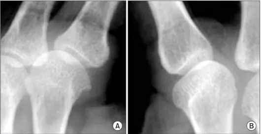
However, the remaining 6 patients required open reduc- tion. A plain radiographic examination, including antero- posterior, oblique, and lateral X-rays, was performed in all of the patients to define the metacarpal head morphology based on the example by Yagi et al.3) (Fig. 1).
Four patients, in whom the metacarpal head had a sharp edge, were examined by computed tomography (CT). Osteophyte height, defined as the distance between the base of the condyle and the most prominent radial Fig. 1. Radiographs of the metacarpal head. (A) A sharp edge (case 4). (B) A blunt edge (case 9).
A B
Fig. 2. Height of metacarpal head osteophytes of the index finger. The osteophyte height is defined as the distance between the base of the condyle and the most prominent radial margin of the osteophyte (arrow).
Metacarpal diameter is measured from a circle template. Template of metacarpal head (yellow circle).
margin of the osteophyte, was measured on the metacar- pal head and neck junction in the axial plane (Fig. 2). For evaluating the difference in the size of metacarpal bones between sexes and persons, we measured the diameter of the index finger metacarpal and the third metacarpal con- dyle radial osteophytes in the corresponding axial plane CT image. However, the difference in size was negligible.
In the six patients who underwent open reduction, function was evaluated by examining the range of motion, recurrences, Disabilities of the Arm, Shoulder and Hand (DASH) score, and MPJ stability using a key pinch gauge (B&L Engineering, Santa Fe Springs, CA, USA) (Fig. 3). If key pinch strength was more than 80% of that of the con- tralateral side, we considered that there was no instability of the MPJ. This study was approved by the Institutional Review Board of Soonchunhyang University Bucheon Hospital.
Surgical Procedure
The operation was performed under general anesthesia.
When the index MPJ was maximally flexed, a longitudinal
~2-cm incision was made on the radial aspect of the MPJ centered around the MPJ space. The radial part of the sag- ittal band was then split transversely along its fibers, and the MPJ capsule was opened along the dorsal border of the radial CL. Exposure of the metacarpal head confirmed that a volar radial osteophyte was impinging on the CL (Fig.
4). As the MPJ was flexed further, the proximal portion of the volar radial condyle osteophyte between the metacar- pal head and neck junction became more accessible (Fig.
5). After removing the osteophyte using a mini-osteotome, we checked whether the MPJ could be extended fully. If locking was not resolved completely, we excised more bone from the radial edge of the condyle while preserving as much articular cartilage as possible. Extension of the MPJ was re-examined, and upon resolution of locking, the Fig. 3. A 46-year-old female patient (case 6) with a locked metacarpophalangeal joint (MPJ) of the index finger. (A) Oblique radiograph shows a sharp edge of the index metacarpal radial condyle. Axial (B) and three-dimensional (C) computed tomography images show radial osteophyte of the index metacarpal head.
(D) Index MPJ extension was limited prior to surgery.
A B
C D
radial capsule and sagittal band were repaired sequentially.
Postoperative Management
A short arm splint was used to maintain extension of the MPJ for 1 week. Free and full motion of the MPJ was al- lowed thereafter.
RESULTS
All of the patients had tenderness on the volar radial side
of the metacarpal head, which resolved spontaneously af- ter the operation. At the final follow-up, the average exten- sion limitation of the MPJ decreased from 26° to 0°, and further flexion increased from a mean 83° to 86°.
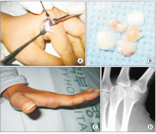
A plain radiograph showed that the osteophyte on the index metacarpal head had a sharp edge in six patients and a blunt edge in three patients. All six patients with sharp-edged metacarpal head osteophytes required open reduction, while all three locked fingers with blunt edged metacarpal head osteophytes were reduced manually. One Fig. 4. A 34-year-old female patient (case 2) with a locked metacarpophalangeal joint (MPJ) of the index finger. (A) Intra- operative photograph during ligament- preserving surgery on the flexed index MPJ with a radial osteophyte on the meta carpal head. (B) Osteophyte fragment was removed from the radial metacarpal head using a mini- osteotome. (C) Post operative photograph shows full ex tension of the MPJ. (D) Postoperative obli que radiograph does not show a visible sharp edge of the index metacarpal head.
A B
C D
Fig. 5. Diagram of our ligament-pre- serving surgical approach to remove condylar osteophytes. (A) When the metacarpophalangeal joint (MPJ) is flexed at 90°, the distal half of the radial osteophyte can be approached. (B) If the MPJ is hyperflexed beyond 90°, the more proximal portion of the radial osteophyte can be approached.
A B
six patients who underwent osteophyte excision, postop- erative oblique radiographs confirmed complete removal of the metacarpal head osteophytes (Fig. 4).
Following surgery, the DASH score decreased from 24.3 to 7.2 points and key pinch strength improved from 67.3% to 90.4% of that of the contralateral side. There were no complications such as recurrent locking and no MPJ instability after surgery.
DISCUSSION
Among our cohort of nine patients who underwent treat- ment for locked MPJ of the index finger, six patients required open reduction, for which we used a novel ligament-preserving approach. We obtained satisfactory results without recurrence or instability, allowing for rapid recovery of the MPJ motion.
We identified the radiological morphology of the metacarpal head that underlies failure of manual reduc- tion. Operative treatment for the locked MPJ was required in all of the patients with sharp metacarpal osteophytes.
Moreover in these patients, CT confirmed that the height of osteophytes on the index radial metacarpal head was twice that of osteophytes on the third metacarpal head.
MPJ locking usually occurs in the index finger.1,2) Although the reason for this preponderance is not clear, it is though that thumb-index finger pinching, which trans- mits repeated load to the index radial metacarpal head, may play a contributory role.
Many authors have suggested that the radial ACL is involved in locking caused by osseous prominences of the radial metacarpal.1,2) The radial ACL originates from the metacarpal fovea and inserts obliquely into the volar plate.
When the MPJ is flexed maximally, the ACL is relaxed and clears the proximal surface of the condyle. As the MPJ starts to extend, tension of the ACL over the osseous prominence of the volar metacarpal head increases, and the dorsal border of the ACL gets caught at the volar radial condyle edge, limiting MPJ extension.
The principle of reduction is to make space adjacent Table 1. Details of the Nine Reviewed Patients CaseSex
Age (yr)
Involved sideAssociated diseaseDuration from locking to surgery (day)
Treatment method
Follow-up (mo)Radiographic appearance at tip of condyle
Extension limitation PreoperativePostoperative 1Female38Right-25Open reduction12Sharp450 2Female34Left-50Open reduction65Sharp300 3Female43Right-22Open reduction60Sharp200 4Male43LeftChronic renal failure10Open reduction60Sharp300 5Female46RightLymphoma53Open reduction50Sharp200 6Female46Left-30Open reduction12Sharp200 7Female39Left-1Manual reduction12Blunt300 8Female47RightRheumatoid arthritis3Manual reduction12Blunt100 9Female39Left-19Manual reduction20Blunt200
to the metacarpal head by radial deviation and external rotation of the proximal phalanx to alleviate ligament ten- sion.3,7,8) However, reduction is not always successful and locking recurs occasionally.1) If an additional force, such as contraction of the lumbrical muscle, which passes through the radial side of the MPJ, is applied during reduction, it may interfere with resolution by reducing the space be- tween the ACL and metacarpal head. Compression of the radial aspect of the MPJ caused by tight lumbrical muscle is especially likely if the proximal phalangeal joint is flexed.
Surgeons should not apply excessive force during reduction maneuvers.3) Careless reduction maneuvers can cause fractures of the metacarpal condyle, especially under anesthesia because the surgeon cannot accurately perceive the resistance within the joint. Although the possibility of fractures depends on bone quality and careful technique, condylar fractures may eventually result in MPJ contrac- ture. In this study, fracture did not occur during manipu- lation.
In the study by Yagi et al.3) on 12 patients, the con- dyle tip appeared to be blunt in nine patients and sharp in three patients on X-ray. Radiographic examination did not show any association between manual reduction failure and either the sharp or blunt type. In our study, we ob- served a definite tendency for manual reduction failure in patients with sharp condyle tips, although statistical analy- sis was not possible due to the small number of patients.
Moreover, our cohort included a larger proportion of pa-
tients with sharp condyles than in the report by Yagi et al.3) This difference in the proportion of patients with sharp condyles explains why many of our patients required open reduction compared to those in the series by Yagi et al.3) Limitations
There were some limitations to this study. First, the study included a small number of patients and it had a retro- spective design, because locked MPJ is not very common.
Second, we excluded cases of locking due to causes other than radial metacarpal head osteophytes.9,10) The limited incisions made in our surgical approach might make it dif- ficult to identify other causes of MPJ locking. However, we did not find any causes other than volar radial osteophytes on the metacarpal head. Third, our study did not include any comparison group.
Our surgical approach to resolve MPJ locking of the index finger avoid injury to the CL and the ACL. We confirmed that this approach is feasible and effective for removal of osteophytes of the index radial metacarpal head and allows rapid recovery of MPJ motion without recurrence of locking.
CONFLICT OF INTEREST
No potential conflict of interest relevant to this article was reported.
REFERENCES
1. Inoue G, Miura T. Locked metacarpophalangeal joint of the finger. Orthop Rev. 1991;20(2):149-53.
2. Thomsen L, Roulot E, Barbato B, Dumontier C. Locked metacarpophalangeal joint of long fingers: classification, definition and treatment based on 15 cases and literature review. Chir Main. 2011;30(4):269-75.
3. Yagi M, Yamanaka K, Yoshida K, Sato N, Inoue A. Success- ful manual reduction of locked metacarpophalangeal joints in fingers. J Bone Joint Surg Am. 2000;82(3):366-71.
4. Langenskiold A. Habitual locking of a meta-carpo-pha- langeal joint by a collateral ligament, a rare cause of trigger finger. Acta Chir Scand. 1949;99(1):73-8.
5. Lee S, Yum JK, Kim JY. Locking of the metacarpophalan- geal joint of the thumb with the radial collateral ligament rupture after stress radiography. Arch Orthop Trauma Surg.
2010;130(2):237-9.
6. Kim JK, Chung MS, Baek GH. Locked metacarpophalange- al joint of the index finger: consideration about the surgical approach. J Hand Surg Eur Vol. 2009;34(2):278-80.
7. Guly HR, Azam MA. Locked finger treated by manipula- tion: a report of three cases. J Bone Joint Surg Br. 1982;
64(1):73-5.
8. Tajima K, Sato K, Sasaki T, Peimer CA. Vertical locking of the metacarpophalangeal joint in young adults. J Hand Surg Am. 2011;36(9):1482-5.
9. Aston JN. Locked middle finger. J Bone Joint Surg Br. 1960;
42(1):75-9.
10. Bloom MH, Bryan RS. Locked index finger caused by hy- perflexion and entrapment of sesamoid bone. J Bone Joint Surg Am. 1965;47(7):1383-5.