Enterobacter sakazakii : New emerging pathogen
Review on E. sakazakii and development of selective medium
오 세 욱 식품안전성연구본부
The first cases of neonatal meningitis believed to have been caused by Enterobacter sakazakii were reported in 1961. Prompted by several subsequent outbreaks of E. sakazakii infections in neonates and an increasing number of neonates in intensive care units being fed rehydrated powdered infant formula, considered to be a source of the pathogen, public health authorities and researchers are exploring ways to eliminate the bacterium or control its growth in dry infant formula, processing environments and formula preparation areas in hospitals. Reviewed here are advances in taxonomy and classification of E. sakazakii, methods of detecting, isolating and typing the bacterium, antibiotic resistance, clinical etilogy and pathogenicity. Outbreaks of E. sakazakii infections in neonates and adults are summarized. Reports on the presence of E. sakazakii in clinical settings, the environment and foods and food processing facilities are reviewed.
Ⅰ. 서론
1961년 Urmenyi와 Franklin은 1958년 영국 St.
Albans에서 발생하였던 두 가지 종류의 neonatal meningitis에 대하여 보고함으로서 Enterobacter sakazakii에 대한 최초의 보고자가 되었으며 이후 1965년 Joker 등도 Denmark에서 발병된 meningitis 에 대해 보고하였다. 보고된 yellow-pigmented Enterobacter cloacae는 1980년에 이르러서야 DNA relatedness, pigment production, biotyping, antibiotic susceptibility 등의 실험(Farmer et al., 1980; Izard et al., 1983)에 의해 Enterobacteriaceae
의 새로운 species로 분류되었으며 일본인 미생물 학자인 Riichi sakazakii의 이름을 따서 Enterobacter sakazakii로 명명되었다. Farmer 등(1977)과 Brenner 등(1977)이 E. sakazakii라는 이름을 최초로 사용 하였다고 알려져 있다. E. sakazakii로 명명되기 전까지 Urmenyi and Franklin bacillus, yellow coliform, yellow Enterobacter, pigmented cloacae 와 yellow-pigmented E. cloacae의 다섯 가지의 이름으로 명명되었다.
E. sakazakii infection은 주로 생후 3일부터 4년
까지의 신생아와 어린이에게 병을 유발하며 현재
까지 적어도 76건의 infection과 19건의 사망이 보 고되고 있다(Iversen and Forsythe, 2003). 또한 어른을 대상으로는 아홉 건 이상의 infection이 보 고되고 있다(Dennison and Morris, 2002; Emery and Weymouth, 1997; Hawkins et al. 1991;
Jimenez and Gimenez, 1982; Pribyl et al., 1985;
Lai, 2001). Infection은 세계적으로 8개국에서 보 고되고 있으며 또한 미국 내에서 9개주에서 발생 이 보고되고 있다.
최근의 E. sakazakii infection은 2004년 7월 뉴 질랜드에서 발생하여 미숙아가 사망하였으며, 동년 10월과 12월 사이에 프랑스에서 9명이 infection되 어, 4명이 발병하였으며 그 결과 2명이 사망하였다.
아직까지 E. sakazakii와 관련된 infection의 감 염원 및 경로는 밝혀져 있지 않지만, 역학적 조사 결과에 따르면 rehydrated infant milk formula(a non-sterile product), 분유를 탄 장소의 환경조건, 사용한 기구 등이 가장 유력한 오염경로라고 알려 져 있다(Bar-Oz et al., 2001; Biering et al., 1989;
Clark et al., 1990; Muytjens and Kollee, 1990;
Muytjens et al., 1983; Noriega et al., 1990;
Simmons et al., 1989; Van Acker et al., 1990).
E. sakazakii에 오염되어 있을 것으로 추정되는 조제분유에 대하여 2002년 미국에서 자발적인 recall(Wyeth nutrition)이 진행되었으며 2005년 1월 네덜란드에서도 전 세계를 대상으로 한 recall (Pregestimil)이 진행된 바 있다. 따라서 그 어느 때 보다도 조제분유에 대한 미생물 안전성 및 위 생 관리에 대한 관심이 고조되고 있는 상황이다.
Ⅱ. 본론
1. Taxonomy and biochemical characterization
E. sakazakii는 Enterobacteriaceae family에 속
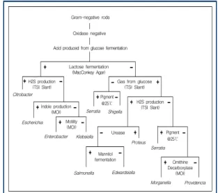
한다. 특징으로는 Gram-negative rod이며, glucose 를 발효하여 산을 생성하며 동시에 lactose를 분해 하여 산을 생산한다. 또한 hydrogen sulfide gas 및 indole을 생산하지 못하며 motility를 가지고 있 다(Coliform, Enterobacteriaceae spp.).
Enterobacter spp 중에서 D-sorbitol을 carbon source로 이용하지 못하는 것으로 알려져 왔으며 extracellular deoxyribonuclease를 생산한다고 알 려져 있다(Farmer et al., 1980). 그러나 최근 예외 적으로 D-sorbitol을 carbon source로 이용하는 균 주도 보고되고 있다(Heuvelink et al., 2001).
Gram-negative rods Oxidase negative Acid produced from glucose fermentation
Lactose fermentation (MacConkey Agar)
Gas from glucose (TSI Slant) H2S production
(TSI Slant) Citrobacter
Escherichia Enterobacter Indole production
(MOI)
Klebsiella Motility
(MOI)
Pigment
@25℃
Serratia Shigella
H2S production (TSI Slant)
Urease Proteus
Serratia Mannitol
fermentation Salmonella Edwardsiella
Pigment
@25℃
Morganella Providencia Omithine Decarboxylase
(MOI)
Fig. 1. Biochemical differentiation of Entero- bacteriaceae family
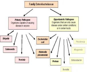
Enterobacteriaceae family는 항상 pathogen으로 활동할 수 있는 Shigella spp. Salmonella spp.
Yersinia spp. 등의 primary pathogen이 있으며
특정 환경조건이나 host에 따라서 pathogenisity를
나타내는 Proteus spp. Serratia spp. Morganella
spp.와 Enterobacter 등의 opportunistic pathogen
으로 구성되어 있으며 이러한 두 가지 성질을 동
시에 가지고 있는 Escherichia coli, Klebsiella
pneumoniae도 있다. 대부분의 Enterobacteriaceae
균주는 대장에 존재하는 normal microflora로 진 화되어 왔기 때문에 질병을 유발하지 않지만 plasmid, transposons와 phage 등에 의해 genetic information을 흡수하여서 pathogen이 되기도 한 다고 알려져 있다.
Fig. 2. Enterobacter sakazakii as an opportunistic pathogens
1980년대 이전에 yellow-pigmented E. cloacae 로 알려져 왔던 E. sakazakii는 DNA-DNA hybridization 실험 결과 non-pigmented strain과 비교하여 50% 이하의 homology를 나타내었기 때 문에 새로운 species로 분류되었다. 일반적으로 균 주를 분류할 때는 phenotypic characteristics, biochemical traits, serotyping, bacteriophage typing과 antibiotic resistance를 이용하며(Arbeit, 1995; Einstein, 1990, Nazarowec-White and Farber, 1999) 또한 실제적으로 phenotype test에 의하여 Enterobacter species를 분류하기도 하지 만 Enterobacter species 전체 분류에 적용하기 어 려우며 또한 그 결과가 그다지 효과적이지도 못한 것으로 알려져 있다(Gaston, 1988; Grattard et al., 1994; Nazarowec-White and Fraber, 1999;
Poilane et al., 1993). 따라서 대안으로서 유전자를 이용한 분류 방법이 개발되고 있다. Iverson 등 (2004)은 16S ribosomal DNA와 hsp60 sequencing 에 의해 상관관계를 조사한 결과, type strain의 16S rDNA sequence는 Cirobacter roseri와 97.8%
유사한 sequence를 가지고 있었으며 E. cloacae와 97.0% 유사한 것으로 나타났다고 하였으며 또한 최근 연구결과에 의하면 E. sakazakii는 hsp60 sequence에 의해 더욱 더 세분화 될 수 있는 2가 지의 특징적인 group으로 구분될 수 있으며 이러 한 group간의 biochemical profile이 서로 일치 하 지 않았다고 하여 phenotypic typing 보다 객관적 인 분류가 가능하다고 하였다.
따라서, phenotypic characterization 뿐만 아니라 PCR, randomly amplified polymorphic DNA(RAPD) PCR, pulsed-field gel electrophresis(PFGE), chromosomal DNA restriction analysis, ribotyping 과 plasmid typing(Grant and Kroll, 1993; Farber, 1996, Nazarowec-White and Farber, 1999)에 의 한 DNA와 RNA finger printing이 비약적으로 발 전되어 왔다. Nazarowec-White와 Farber(1999)는 EcoRI restriction endonuclease를 이용하여 18종 의 E. sakazakii isolate에 대하여 분석한 결과 10 종의 ribotype으로 분류 할 수 있었다고 하였으며 restriction endonuclease analysis(REA, Clark et al., 1990) 보다 더욱 더 효율적이었다고 하였다.
그러나 아직까지 가장 정확하게 분류에 이용할 수 있는 객관적인 finger printing 방법은 없는 것으 로 생각되며 AP-PCR, ribotyping, PFGE, plasmid typing 결과를 종합하여 molecular typing 하는 것 이 가장 효율적인 방법이라고 생각한다.
2. Isolation, identification and typing
U.S. Food and Drug Adminstration(FDA, 2002)
은 dehydrated powdered infant formula(Table 1)에
Step Time Temperature
√ Sterilize can lid margins and sample spoons N/A N/A
√ Dilute 100g, 10g, 1g of powdered infant formula with pre-warmed sterile distilled water at 1:10 ratio, mix and incubate
Overnight 36℃
√ Add 10 ml of each suspension to 90 ml of Enterobacteriaceae
enrichment broth and incubate Overnight 36℃
√ Mix suspensions and surface plate 0.1 ml on VRBG agar, streak on VRBG agar with a 10 μl incubating loop onto three quadrants for isolation and incubate
Overnight 36℃
√ Pick five presumptive-positive E. sakazakii colonies from both sets of VRBG plates and subculture by streaking onto TSA and incubate
48-72 h 25℃
√ Select yellow-pigmented colonies only and confirm per manufacture's instructions for the API 20E biochemical confirmation system
N/A N/A
√ Calculate the most probable number(MPN) after determining
the number of positive tubes at each dilution N/A N/A Table 1. Method for analyzing powdered infant formula for presumptive E. sakazakii
서 E. sakazakii를 분리, 계수할 수 있는 방법을 권고하였다. 이 방법은 Muytjens 등(1988)과 Nazarowec-White와 Farber(1997) 방법을 근간으 로 하고 있지만 FDA 방법은 powdered infant formula를 buffered peptone water에 reconstitute 하는 대신에 증류수를 사용하며 enrichment 후에 sample을 tryptic soy agar(TSA), violet red bile glucose(VRBG) agar에 transfer 하는 방법에서 차 이가 있다.
FDA가 권고하고 있는 방법은 크게 2가지의 단 점이 있다. 첫째, selective media로 사용되고 있는 VRBG의 selectivity가 낮아 검출을 목표로 하고 있는 E. sakazakii 이외의 많은 균들이 특징적인 purple colony를 형성하므로 분리를 방해할 수 있
다. 둘째, VRBG에서 E. sakazakii로 추정되는
colony를 선발하여 TSA에서 배양후 yellow
colony를 선발토록 권고하고 있지만, E. hermanii,
E. vulneris, L. acecarboxylata 등 Enterobacteriaceae
중에서 많은 균주가 yellow pigment를 생성하기
때문에 selectivity가 떨어진다고 할 수 있다. 따라
서 E. sakazakii를 selective하게 검출할 수 있는
배지 개발이 대한 연구가 진행되었다. Selective
배지를 개발하기 위해서는 먼저, 대상으로 하는
균주의 특성을 조사해야 한다. 대상균주만이 가지
고 있는 생화학적 특징이 있다면 이를 selection
marker로 이용하는 것이 가장 바람직하다고 할
수 있다. 특징적인 biochemical metabolites의 생산,
특징적인 enzyme의 생산, antibiotics resistance
등이 selection marker로 보편적으로 이용되고 있다.
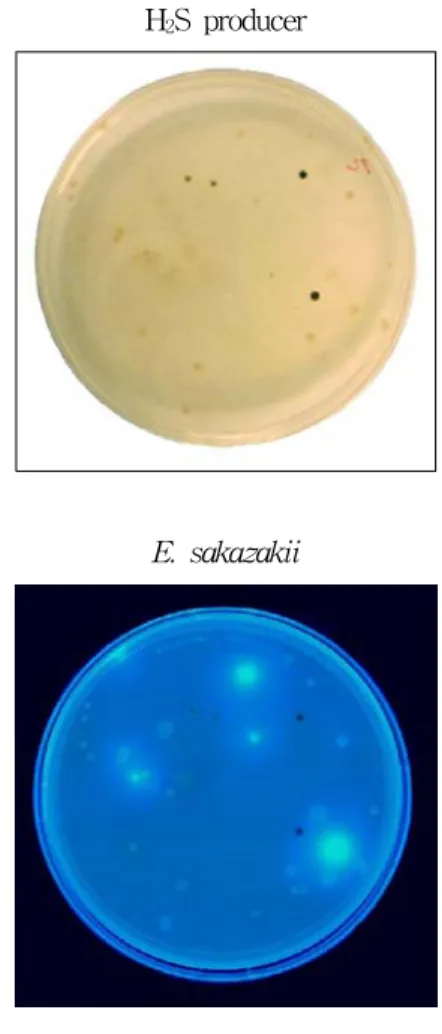
E. sakazakii는 Enterobacter spp. 중에서 유일 하게 α-glucosidase를 생산하기 때문에 이러한 특 징을 selection marker로 이용하여 differentiation media가 개발되었다(Oh and Kang, 2004; Iverson et al., 2004). Oh와 Kang은 E. sakazakii가 α -glucosidase를 생산하는 특징을 selection marker 로, hydrogen sulfide를 생산하지 못한다는 특징을 보조적인 marker로 이용하였다. 즉, Enterobacteria 속 균주의 성장이 가능한 배지조건(bile salt 존재) 에서 α-glucosidase를 생산하지만 hydrogen sulfide 를 생산하지 못하는 균주를 Enterobacter sakazakii 로 선발하도록 설계하였다. Alpha-glucosidase를 detect 하기 위하여 4-methylumbelliferyl-α-D-glucoside (α-MUG)를 기질로 사용하였으며 효소작용에 의하 여 분해, 분리된 4-methylumbelliferyl moiety의 형 광을 자외선(365 nm)을 이용하여 검출하였다. 상 업적으로 판매되고 있는 complex media인 TSA, VRBG agar, Tryptone bile agar중 basal medium 을 선정하였으며 이후 배지성분을 최적화 하여 E.
sakazakii에 의해 생산되는 α-glucosidase를 specific 하게 검출할 수 있도록(background noise 제거) 최적화 하였다. 또한, 보조 marker인 hydrogen sulfide를 생산하는 균주를 분별하기 위하여 sodium thiosulfate와 ferric citrate를 첨가하여 black colony (Salmonella spp. Edwardsiella spp. Proteus spp) 를 제외시켰다. Fig. 3에 나타난 바와 같이 α -glucosidase 활성에 의해 형광을 띠면서 동시에 black colony를 형성하지 않는 균을 E. sakazakii 로 설정하였다. 이후 API 20E identification kit와 oxidase test를 실시하여 confirmation 하였을 때 개발된 선택배지의 selectivity가 우수한 것으로 나 타났다.
H
2S producer
E. sakazakii
Fig. 3. OK medium developed for E. sakazakii isolation and differentiation. E. sakazakii appeared as blue fluorescent colonies and Hydrogen sulfide producer as black colonies.
Iverson 등(2004)도 α-glucosidase 활성을 검출
하는 동일한 원리를 이용하여 E. sakazakii 선택
배지(Druggan-Forsythe-Iverson agar, DFI)를 개
발하였으며 현재 Oxoid사를 통하여 상업화되어
판매되고 있다. 효소활성을 측정하기 위하여 사
용된 합성 chromogen은 5-bromo-4-chloro-
3-indolyl-α-D-glucopyranoside(XαGlc)으로 고가
이기 때문에 선택배지도 1 파운드(보통 1통)당
100만원 정도의 고가에 팔리고 있다. 이들은 95종 의 E. sakazakii 균주에 대한 검출실험 결과 FDA 에서 권고하고 있는 방법에 비하여 약 2일 정도의 시간이 단축되었다고 하였으며 또한 17 genera에 속하는 148 strain의 Enterobactriaceae 균주를 대 상으로 실험한 결과 DFI를 이용하였을 경우 19 strain이 false-positive 한 결과를 나타낸 반면 FDA 방법인 VRBG를 이용하는 방법은 31 strain 이 false-positive 하였다고 하여 FDA에서 권고하 고 있는 선택배지 보다 우수하였다고 하였다.
Presumptive-positive한 E. sakazakii의 confirmation 은 주로 API 20E, Enterotube II 등의 biochemical 특성을 판별하는 phenotypic 방법이 널리 사용되 고 있지만(Biering et al., 1989; Cottyn et al., 2001; Gassem, 1999; Kandhai et al., 2004;
Monroe and Tift, 1979; Mosso et al., 1994;
Muytjens et al., 1983; Nazarowec-White and Farber, 1999; No et al., 2002; Seo et al., 2003;
Simmons et al., 1989; Van Os et al., 1996; Willis and Robinson, 1988), 최근 API 20E biochemical test가 false-negative 및 false-positive한 결과를 나타내었다는 보고(Iverson et al., 2004)에 따라 추가적인 실험이 진행되기도 하였다. Seo 등(2003) 은 E. sakazakii를 API ZYM과 Vitek assay에 의 하여 confirmation 하였으며, Cottyn 등(2001)은 fatty acid methylester (FAME) analysis, API 20E kit, Biolog strips을 동시에 사용하여 정확성 을 높였다고 하였다.
Polymerase Chain Reaction(PCR)을 이용한 confirmation으로 Seo 등(2003) 은 real-time PCR assay 기술을 개발하였다. Primer는 partial macromolecular synthesis(MMS) gene을 증폭대상으로 하였으며 E. cloacae와 확실한 구분이 가능하였다고 하였다.
또한 1 ml 당 100 CFU 정도로 존재할 경우 분석 이 가능하다고 하였다. 따라서 보통 오염된 분유
의 경우 1,000g당 4 cell이 존재하는 것으로 가정 하고(Zink, 2003) enrichment broth에 reconstitute 비율을 1:10로 가정한 경우 약 15 generation이 경 과되면 100 CFU가 가능하게 되어 전체적으로 4-5 시간 만에 분석이 가능하다고 하였다.
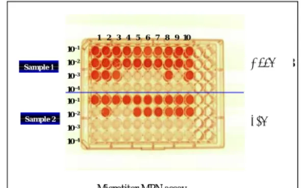
한편, Oh와 Kang 등(2005)은 분유 중에 존재하 는 E. sakazakii를 96 well microtiter plate를 이용 하여 빠르고 간편하게 detection 할 수 있는 miniaturized MPN 방법을 개발하였다. 이 방법은 differential and selective media로 보고된 OK medium(2004)의 특성을 이용한 것으로 α-glucosidase activity에 의한 형광발현을 endpoint determination 으로 활용하였으며 그 결과, 일반 고체배지를 사 용할 때 소요되는 시간(24시간 이상)에 비하여 10 시간 이내의 빠른 분석이 가능하였다고 하였다 (Fig. 4).
1 2 3 4 5 6 7 8 9 10 10 –1
10 –2 10 –3 10 –4 10 –1 10 –2 10 –3 10 –4 Sample 1
Sample 2
Microtiter MPN assay
1,678
⇒ 408
⇒ 1,678
408