Allergy Asthma Immunol Res. 2014 September;6(5):449-457.
http://dx.doi.org/10.4168/aair.2014.6.5.449 pISSN 2092-7355 • eISSN 2092-7363
INTRODUCTION
Nasal polyposis (NP) is a common chronic inflammatory dis- ease of the mucous membranes in the nose and paranasal si- nuses. NP is characterized by eosinophilic infiltration and tissue remodeling consisting of epithelial proliferation, goblet cell hy- perplasia, pseudocyst formation, basement membrane thicken- ing, focal fibrosis and edema.1 NP often accompanies chronic rhinosinusitis (CRS), and symptoms of NP include nasal con- gestion, loss of smell, and headache caused by secondary infec- tion.2 Although many studies for NP have been reported, its pathogenesis remains poorly understood and the treatment of NP treatment is limited.
Prostaglandin E2 (PGE2), a metabolite of arachidonic acid, has multiple physiological effects.3 These effects are mediated by 4 different E prostanoid (EP) receptors that belong to the G protein-coupled receptor family: EP1 increases Ca2+ channel gating via Gq, EP2 and EP4 increase the levels of cyclic adenos-
Prostaglandin E2 Induces IL-6 and IL-8 Production by the EP Receptors/Akt/NF-κB Pathways in Nasal Polyp-Derived Fibroblasts
Jung-Sun Cho,
1,2In-Hye Han,
1Hye Rim Lee,
1Heung-Man Lee
1,2,3*
1Brain Korea 21 Plus for Biomedical Science, Korea University College of Medicine, 2Institute for Medical Devices Clinical Trial Center, Guro Hospital, Korea University, Seoul, Korea
3Department of Otorhinolaryngology-Head and Neck Surgery, College of Medicine, Korea University, Seoul, Korea
ine monophosphate (cAMP) via Gs, whereas EP3 decreases cAMP via Gi.4 In previous studies, low production of PGE2 has been reported in NP and in both fibroblasts and epithelial cells.5 PGE2 suppresses eosinophilia-associated cellular re- sponses induced by staphylococcal enterotoxin, dominantly through an EP2–mediated pathway in NP.6 These observations suggest that PGE2 plays a role as an inflammatory mediator to induce inflammatory diseases, such as NP, allergic rhinitis, and bronchial asthma. However, the effect of PGE2 on the produc- tion of interleukin 6 (IL-6) and IL-8 in NP is unknown.
Many kinds of cells such as epithelial cell, T cell, mast and fi- Purpose: Interleukin 6 (IL-6) and IL-8 participate in the pathogenesis of chronic rhinosinusitis with nasal polyps, and their levels are increased by prostaglandin E2 (PGE2) in different cell types. The purposes of this study were to determine whether PGE2 has any effect on the increase in the lev- els of IL-6 and IL-8 in nasal polyp-derived fibroblasts (NPDFs) and subsequently investigate the possible mechanism of this effect. Methods: Differ- ent concentrations of PGE2 were used to stimulate NPDFs at different time intervals. NPDFs were treated with agonists and antagonists of E pros- tanoid (EP) receptors. To determine the signaling pathway for the expression of PGE2-induced IL-6 and IL-8, PGE2 was treated with Akt and NF-κB inhibitors in NPDFs. Reverse transcription-polymerase chain reaction for IL-6 and IL-8 mRNAs was performed. IL-6 and IL-8 levels were measured byenzyme-linked immunosorbent assay (ELISA). The activation of Akt and NF-κB was evaluated by western blot analysis. Results: PGE2 significant- ly increased the mRNA and protein expression levels of IL-6 and IL-8 in NPDFs. The EP2 and EP4 agonists and antagonists induced and inhibited IL-6 expression. However, the EP4 agonist and antagonist were only observed to induce and inhibit IL-8 expression level. The Akt and NF-κB inhibitors significantly blocked PGE2-induced expression of IL-6 and IL-8. Conclusions: PGE2 increases IL-6 expression via EP2 and EP4 receptors, and IL-8 expression via the EP4 receptor in NPDFs. It also activates the Akt and NF-κB signal pathways for the production of IL-6 and IL-8 in NPDFs. These results suggest that signaling pathway for IL-6 and IL-8 expression induced by PGE2 might be a useful therapeutic target for the treatment of nasal polyposis.
Key Words: Nasal polyps; prostaglandins E; interleukin-6; interleukin-8; E prostanoid receptor; Akt
This is an Open Access article distributed under the terms of the Creative Commons Attribution Non-Commercial License (http://creativecommons.org/licenses/by-nc/3.0/) which permits unrestricted non-commercial use, distribution, and reproduction in any medium, provided the original work is properly cited.
Correspondence to: Heung-Man Lee, MD, PhD, Department of
Otorhinolaryngology–Head and Neck Surgery, Guro Hospital, Korea University College of Medicine, 33-41 Gurodong-ro 28-gil, Guro-gu, Seoul 152-703, Korea.
Tel: +82-2-2626-3185; Fax: +82-2-868-0475; E-mail: lhman@korea.ac.kr Received: September 11, 2013; Revised: December 8, 2013;
Accepted: January 16, 2014
•There are no financial or other issues that might lead to conflict of interest.
broblast, are involved in the pathogenesis of NP. Among these cells, fibroblasts play a key role in the structural modification of the sinonasal mucosa. Fibroblasts differentiate into myofibro- blasts and produce large amounts of extracellular matrix mole- cules such as collagen and fibronectin.7 Recent studies have shown that fibroblasts are not just a structural modifier, but also important modulators of local inflammation.8 Fibroblasts ex- press many receptors for cytokines, growth factors, and hor- mones.9 Therefore, they play a role for mediator of immune function and have the capacity to release a variety of pro-in- flammatory mediators such as eotaxin, IL-6 and IL-8.10,11 Previ- ous studies revealed that IL-6 and IL-8 are increased by PGE2 via the EP4 receptor in different cell types.12,13 However, it is un- known whether PGE2 induces expression of IL-6 and IL-8 in nasal polyp-derived fibroblasts (NPDFs).
The purposes of this study were to determine whether PGE2 has any effect on the increase of IL-6 and IL-8 in NPDFs, and subsequently to investigate the possible mechanism underlying this effect.
MATERIALS AND METHODS Reagents
PGE2, purchased from Sigma (St. Louis, MO, USA) was dis- solved in dimethyl sulfoxide (Sigma) and then diluted with complete medium to concentrations suitable for use in this ex- periment. The receptor agonists and antagonists, obtained from Cayman Chemical (Ann Arbor, MI) were as follows: EP1 receptor and EP3 receptor agonist (Sulprostone, 10 nM), EP2- receptor agonist (Butaprost, 10 μM), EP4 receptor agonist (CAY10580, 10 μM), EP2 receptor antagonist (AH6809, 10 μM) and the EP4 receptor antagonist (AH23848, 10 μM). Akt inhibi- tor (LY294002, 10 μM) was purchased from Calbiochem (Biller- ica, MA, USA). NF-κB inhibitor (BAY-11, 1 μM) was bought from Sigma. NPDFs were previously exposed to PGE2 (20 μM) after pre-treatment for 1 hour with all agonists, antagonists and inhibitors.
Isolation and induction of NPDFs
Fibroblasts were cultured from 8 patients (4 women and 4 men; 32.3±5.2 years of age) who underwent endoscopic sinus surgery for CRS with NP at the Department of Otorhinolaryn- gology Head and Neck Surgery of the Korea University Medical Center. The study protocol was approved by the Institutional Review Board of the Korea University College of Medicine. NP- DFs were isolated from surgical tissues and purified according to our previous study.14 Cells used for the experiments were ob- tained from the fourth cell passage.
Reverse transcription-polymerase chain reaction (RT-PCR) NPDFs were stimulated with PGE2 in time (0-24 hours) and dose (0-20 μM, 12 hours) dependent manner. NPDFs were
stimulated with PGE2 (20 μM), with or without Sulprostone (10 nM), Butaprost (10 μM), CAY 10580 (10 μM), AH6809 (10 μM), AH23848 (10 μM), LY294002 (10 μM) and BAY-11 (1 μM) for 12 hours. Total RNA was isolated using Trizol reagent (Invitrogen, Carlsbad, CA) and 2 µg of the RNA were reverse-transcribed us- ing MMLV reverse transcriptase (Invitrogen). PCR was per- formed using the primer pairs targeting specific genes, as fol- lows: IL-6 (sense sequence, 5′-GCCTTCGGTCCAGTTGCC-3′;
anti-sense sequence, 5′-GCGCAGAATGAGATGAGTTGTCATG -3′; 566 bp), IL-8 (sense sequence, 5’-ATGACTTCCAAGCTGG CC-3′; anti-sense sequence, 5’-TCTTCAAAAA CTTCTCCACAA CCC-3′; 282 bp), GAPDH (sense sequence, 5′-GTGGATATTGTT GCCATCAATGACC-3′; anti-sense sequence, 5′-GCCCC AGCCT TCTTCATGGTGGT-3′; 271 bp). Amplification reactions were performed as follows: the initial denaturation step was per- formed at 94˚C for 5 minutes, followed by 30 cycles performed successively at 94˚C for 45 seconds, 55-65°C for 45 seconds, and 72˚C for 45 seconds. The final extension step was performed at 74˚C for 5 minutes. All these reactions were performed in a vol- ume of 20 μL and the products were electrophoresed on a 1.5%
agarose gel and visualized by staining with ethidium bromide.
Gel images were acquired using the Molecular Imager Chemi- Doc XRS + (Bio-Rad, Hercules, CA, USA).
Enzyme-linked immunosorbent assay (ELISA) of IL-6 and IL-8 NPDFs were stimulated with PGE2 for 48 hours in dose (0-20 μM)-dependent manner. NPDFs were stimulated with PGE2 (20 μM), with or without Sulprostone (10 nM), Butaprost (10 μM), CAY10580 (10 μM), AH6809 (10 μM), AH23848 (10 μM), LY294002 (10 μM) and BAY-11 (1 μM) for 48 hours. IL-6 and IL-8 production in the medium derived from NPDFs was deter- mined by ELISA (R&D Systems, Minneapolis, MN, USA). This assay was performed according to the manufacturer’s instruc- tions.
Western blot analysis
NPDFs were stimulated with PGE2 (20 μM), with or without LY294002 (10 μM) or BAY-11 (1 μM) for 1 hour. The fibroblasts were lysed in PRO-PREPTM protein extraction solution (iNtRON Biotechnology, Seongnam, Korea); proteins were separated by 10% sodium dodecyl sulfate–polyacrylamide gel electrophore- sis and transferred to polyvinylidene difluoride membranes (Millipore Inc., Billerica, MA, USA). These membranes were in- cubated with anti-rabbit polyclonal phosphorylated Akt, p50, and GAPDH (Santa Cruz, CA, USA). After incubation, the mem- branes were washed 3 times (5 minutes per wash) and treated with peroxidase-conjugated anti-rabbit IgG antibody (Vector Laboratories, Burlingame, CA, USA) for 1 hour. After washing, a substrate obtained from an enhanced chemiluminescence re- agent kit (Du Pont, Boston, MA, USA) was added to the mem- branes. The membranes were then exposed to X-ray films.
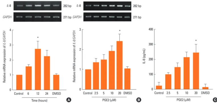
Fig. 1. Effect of PGE2 on IL-6 expression in NPDFs. (A) NPDFs were stimulated with PGE2 (20 μM) in a time-dependent manner. The expression level of IL-6 mRNA was examined using RT-PCR and quantified. (B, C) NPDFs were stimulated with PGE2 in a dose-dependent manner. The mRNA and protein expression levels of IL-6 were examined using RT-PCR for 12 hours (B) and ELISA for 48 hours (C). Values are the mean ± SEM of three independent samples. *P <0.05, †P <0.01 as com- pared to the mean IL-6 values of control cells.
566 bp
217 bp GAPDH
IL-6 566 bp
217 bp GAPDH
IL-6
Relative mRNA expression of IL-6/GAPDH
3
2
1
0
*
*
Control 6 12 24 DMSO
Time (hours) A
Relative mRNA expression of IL-6/GAPDH
3
2
1
0
† †
*
Control 2.5 5 10 20 DMSO
PGE2 (μM) B
IL-6 (pg/mL)
400
300
200
100
0
*
†
*
Control 2.5 5 10 20 DMSO
PGE2 (μM) C
Fig. 2. Effect of PGE2 on IL-8 expression in NPDFs. (A) NPDFs were stimulated with PGE2 (20 μM) in a time-dependent manner. The expression level of IL-8 mRNA was examined using RT-PCR and quantified. (B, C) NPDFs were stimulated with PGE2 in a dose-dependent manner. The mRNA and protein expression levels of IL-8 were examined using RT-PCR for 12 hours (B) and ELISA for 48 hours (C). *P <0.05 as compared to the mean IL-8 values of control cells.
282 bp
271 bp GAPDH
IL-8 282 bp
271 bp GAPDH
IL-8
Relative mRNA expression of IL-8/GAPDH
4
3
2
1
0
*
Control 6 12 24 DMSO
Time (hours) A
Relative mRNA expression of IL-8/GAPDH
3
2
1
0
*
Control 2.5 5 10 20 DMSO
PGE2 (μM) B
IL-8 (pg/mL)
400
300
200
100
0
*
Control 2.5 5 10 20 DMSO
PGE2 (μM) C
Statistical analysis
The statistical significance of the difference between the con- trol and experimental data was analyzed using Tukey’s test (GraphPad Prism, version 5; GraphPad Software, San Diego, CA, USA). A P value of <0.05 was considered statistically significant.
RESULTS
PGE2 induces IL-6 and IL-8 expressions in NPDFs
To determine the effect of PGE2 on IL-6 and IL-8 expressions in NPDFs, NPDFs were stimulated with PGE2 for 12 or 48
hours. PGE2 significantly increased IL-6 and IL-8 mRNA ex- pression levels in time-dependent (Fig. 1A and 2A) and dose- dependent (Fig. 1B and 2B). Also, PGE2 induced production of IL-6 and IL-8 in dose-dependent manner (Fig. 1C and 2C).
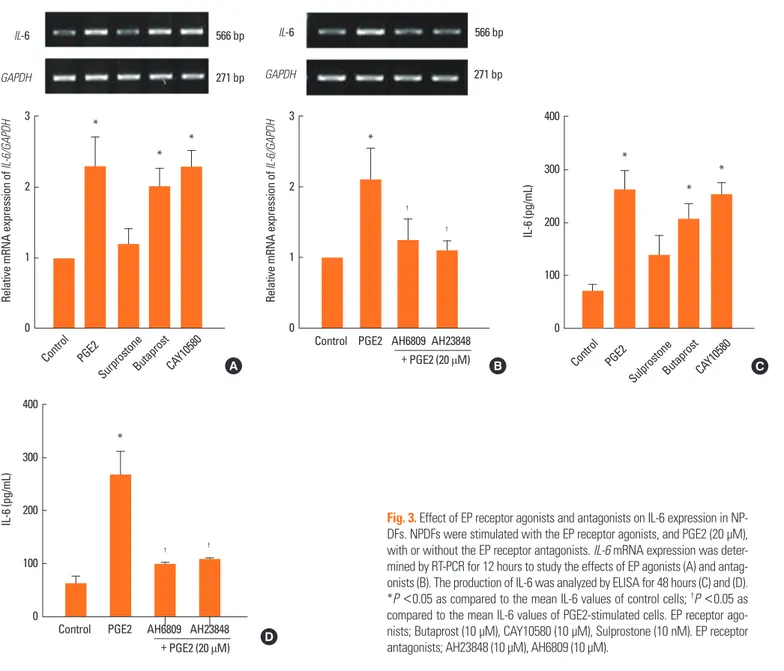
PGE2-induces IL-6 production increases with EP2 and EP4 in NPDFs
To identify the receptors that mediate the effect of PGE2-in duced IL-6 expression, we used agonists and antagonists spe- cific to the corresponding receptors. The specific EP2 agonist (Butaprost, 10 μM) and EP4 agonist (CAY10580, 10 μM) signifi- cantly induced IL-6 expression in NPDFs. However, the EP1and EP3 agonist (Sulprostone, 10 nM) did not induce IL-6 expres- sion in NPDFs (Fig. 3A and C). The increased expression of IL-6 was inhibited by both EP4 antagonist (AH23848, 10 μM) and EP2 antagonist (AH6809, 10 μM) (Fig. 3B and D).
PGE2-induces IL-8 production increases with only EP4 in NPDFs
To examine receptors that mediate PGE2-induced IL-8 ex- pression, we treated PGE2 with and without specific agonists and antagonists. The EP4 agonist (CAY10580, 10 μM) signifi- cantly induced IL-8 expression in NPDFs. However, EP1 adn EP3 agonist (Sulprostone, 10 nM) and EP2 agonist (Butaprost, 10 μM) did not increase IL-8 expression in NPDFs (Fig. 4A and C). IL-8 expression was inhibited by the EP4 antagonist alone (AH23848, 10 μM) (Fig. 4B and D).
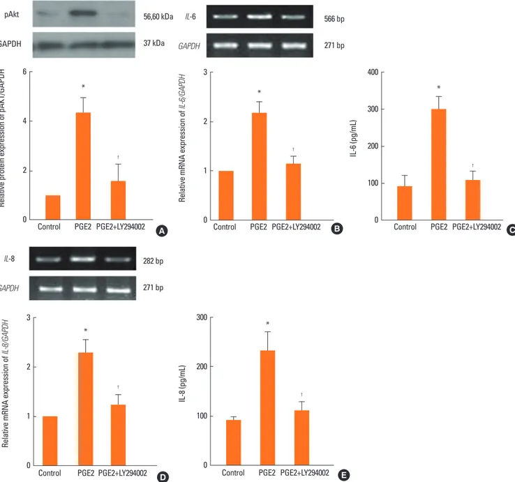
PGE2 increases IL-6 and IL-8 expression via the Akt pathway To determine the signal pathway for PGE2-induced IL-6 and IL-8 production in NPDFs, we evaluated the stimulation of Akt as a downstream marker of IL-6 and IL-8 signaling by western blot analysis and RT-PCR. In western blot analysis, PGE2-in-
Fig. 3. Effect of EP receptor agonists and antagonists on IL-6 expression in NP- DFs. NPDFs were stimulated with the EP receptor agonists, and PGE2 (20 μM), with or without the EP receptor antagonists. IL-6 mRNA expression was deter- mined by RT-PCR for 12 hours to study the effects of EP agonists (A) and antag- onists (B). The production of IL-6 was analyzed by ELISA for 48 hours (C) and (D).
*P <0.05 as compared to the mean IL-6 values of control cells; †P <0.05 as compared to the mean IL-6 values of PGE2-stimulated cells. EP receptor ago- nists; Butaprost (10 μM), CAY10580 (10 μM), Sulprostone (10 nM). EP receptor antagonists; AH23848 (10 μM), AH6809 (10 μM).
566 bp
271 bp GAPDH
IL-6
Relative mRNA expression of IL-6/GAPDH
3
2
1
0
Control PGE2
SurprostoneButaprost CAY10580
*
* *
A
IL-6 (pg/mL)
400
300
200
100
0
*
*
*
Control PGE2
SulprostoneButaprost CAY10580 C 566 bp
271 bp GAPDH
IL-6
Relative mRNA expression of IL-6/GAPDH
3
2
1
0
†
Control PGE2 AH6809 AH23848
†
*
+ PGE2 (20 μM) B
IL-6 (pg/mL)
400
300
200
100
0
†
Control PGE2 AH6809 AH23848
†
*
+ PGE2 (20 μM) D
Fig. 4. Effect of EP receptor agonists and antagonists on IL-8 expression in NPDFs. NPDFs were stimulated with the EP receptor agonist, and PGE2 (20 μM), with or without the EP receptor antagonist. IL-8 mRNA expression was determined by RT-PCR for 12 hours to study the effects of EP agonists (A) and antagonists (B). Pro- duction of IL-8 was analyzed by ELISA for 48 hours (C) and (D). *P <0.05 as compared to the mean IL-8 values of control cells; †P <0.05 as compared to the mean IL-8 values of PGE2-stimulated cells. EP receptor agonists; Butaprost (10 μM), CAY10580 (10 μM), Sulprostone (10 nM). EP receptor antagonists; AH23848 (10 μM), AH6809 (10 μM).
282 bp
271 bp GAPDH
IL-8
Relative mRNA expression of IL-8/GAPDH
4
3
2
1
0
Control PGE2
SurprostoneButaprostCAY10580
*
*
A
282 bp
271 bp GAPDH
IL-8
+ PGE2 (20μM)
Relative mRNA expression of IL-8/GAPDH
4
3
2
1
0 Control PGE2 AH6809 AH23848
*
*
†
B
+ PGE2 (20μM)
IL-8 (pg/mL)
400
300
200
100
0 Control PGE2 AH6809 AH23848
* *
†
Control PGE2 D
SulprostoneButaprostCAY10580
IL-8 (pg/mL)
300
200
100
0
*
*
C
duced stimulation of Akt decreased significantly when an Akt inhibitor (LY294002, 10 μM) was treated (Fig. 5A). Moreover, PGE2-induced expression levels of IL-6 and IL-8 were specifi- cally inhibited by the Akt inhibitor (Fig. 5B–E).
PGE2 increases IL-6 and IL-8 expression via the NF-κB transcription factor
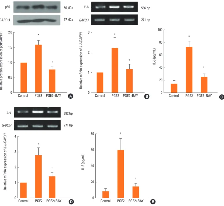
To determine whether the NF-κB transcription factor was in- volved in PGE2-induced IL-6 and IL-8 production in NPDFs, we treated PGE2 under two conditions: in the presence of the
NF-κB inhibitor (BAY-11, 1 μM) and in the absence of the inhib- itor. The western blot analysis showed that PGE2 increased the expression of p50 (subunit of NF-κB) and that p50 expression decreased in the inhibitor-treated NPDFs (Fig. 6A). The pro- duction of IL-6 and IL-8 was specifically inhibited by treatment with the NF-κB inhibitor (Fig. 6B and C).
DISCUSSION
In this study, we found that PGE2 significantly induced the ex-
Fig. 5. The role of Akt in the expression of IL-6 and IL-8 in NPDFs. NPDFs were stimulated with PGE2 (20 μM), with or without the Akt inhibitor (LY294002, 10 μM).
The activation of Akt was determined by western blot analysis (A). Akt inhibition of IL-6 expression was analyzed by RT-PCR for 12 hours and ELISA for 48 hours (B and C). Akt inhibition of IL-8 expression was analyzed by RT-PCR for 12 hours and ELISA for 48 hours (D and E). *P <0.05 as compared to the mean IL-6 or IL-8 values of control cells; †P <0.05 as compared to the mean IL-6 or IL-8 values of PGE2-stimulated cells.
56,60 kDa
37 kDa GAPDH
pAkt
Control PGE2 PGE2+LY294002
Relative protein expression of pAKT/GAPDH
6
4
2
0
*
†
A
566 bp
271 bp GAPDH
IL-6
Control PGE2 PGE2+LY294002 B
Relative mRNA expression of IL-6/GAPDH
3
2
1
0
*
†
Control PGE2 PGE2+LY294002
IL-8 (pg/mL)
300
200
100
0
*
†
E 282 bp
271 bp GAPDH
IL-8
Control PGE2 PGE2+LY294002
Relative mRNA expression of IL-8/GAPDH
3
2
1
0
*
†
D
Control PGE2 PGE2+LY294002 C
IL-6 (pg/mL)
400
300
200
100
0
*
†
pression of IL-6 and IL-8 in NPDFs. PGE2 increased IL-6 ex- pression via EP2 and EP4,however, increased IL-8 expression via EP4 alone. PGE2 activated the Akt and NF-κB signal path- ways for IL-6 and IL-8 expression.
The roles of cytokines in the development of NP are widely in- vestigated. IL-6 has a chemotactic effect on eosinophils and IL-8 affects both neutrophils and eosinophils, causing their mi- gration to the site of inflammation.15 According to recent stud- ies, increased levels of IL-6 and IL-8 may participate in the pri- mary pathogenesis of CRS and NP as well as in the recurrent
episodes.16-18 IL-6 is a multifunctional cytokine implicated in various inflammatory conditions including NP pathogenesis. It stimulates fibroblast proliferation, increases collagen deposi- tion, and decreases collagen breakdown. IL-8 releases other in- flammatory mediators such as histamine and leukotriene B4.19,20 IL-8 is a critical cytokine in the pathogenesis of CRS, and high levels of IL-8 have been detected within NP.21,22 PGE2-in- duced IL-6 and IL-8 release is mediated by EP4 receptor in co- lonic epithelial cells, pulmonary endothelial cells, and astro- cytes.12,13,23 In this study, PGE2 induced both IL-6 and IL-8 ex-
50 kDa
37 kDa GAPDH
p50
Control PGE2 PGE2+BAY
Relative protein expression of p50/GAPDH
2.0
1.5
1.0
0.5
0
*
†
A
566 bp
271 bp GAPDH
IL-6
Control PGE2 PGE2+BAY
Relative mRNA expression of IL-6/GAPDH
3
2
1
0
*
†
B
282 bp
271 bp GAPDH
IL-8
Relative mRNA expression of IL-8/GAPDH
4
3
2
1
0
*
†
Control PGE2 PGE2+BAY D
IL-8 (pg/mL)
80
60
40
20
0
*
†
Control PGE2 PGE2+BAY E
Control PGE2 PGE2+BAY
IL-6 (pg/mL)
100
80
60
40
20
0
*
†
C
Fig. 6. Role of NF-κB on the expression of IL-6 and IL-8 in NPDFs. NPDFs were stimulated with PGE2 (20 μM), with or without the NF-κB inhibitor (BAY-11, 1 μM).
Western blot analysis showed increased expression of NF-κB in case of when PGE2-treated NPDFs and decreased expression of NF-κB in case of BAY-11-treated NPDFs (A). Inhibition of IL-6 or IL-8 expression by BAY-11 treatment was analyzed by RT-PCR for 12 hours (B and D) and ELISA for 48 hours (C and E). *P <0.05 as compared to the mean IL-6 or IL-8 values of control cells; †P <0.05 as compared to the mean IL-6 or IL-8 values of PGE2-stimulated cells.
pression in NPDFs. PGE2-induced IL-6 expression was mediat- ed by EP2 and EP4 receptors. On the other hand, PGE2 induced IL-8 expression only by the EP4 receptor.
PGE2 are mediated via four different G-protein-coupled PGE receptors (e.g. EP1, EP2, EP3, and EP4), which are involved in the activation of phospholipase C (EP1) and activation (EP2, EP4) or inhibition (EP3) of adenyl cyclase.24 Recent studies showed EP4 agonist induced potent relaxations in asthma.25 PGE2 decreases proliferation of bronchial smooth muscle cell via EP2 and EP4 receptors in nonasthmatic eosinophilic bron- chitis.26 In our data, activation of EP2 and EP4 receptor induced
expression of IL-6 and activation of EP4 receptor stimulated ex- pression of IL-8 in NPDFs. However, sulprostone (EP1/3 ago- nist) did not induce both IL-6 and IL-8 expression in NPDFs.
These data suggest that PGE2 induced expression of IL-6 and IL-8 via EP2 and/or EP4 receptor(s) in NPDFs.
Activation of EP receptors can initiate kinase signaling by phosphatidylinositol 3-kinase (PI3K)/Akt pathways and then affects target gene transcription.27 A previous study has demon- strated that PGE2 induces IL-6 expression in human chondro- cytes via the PI3K/Akt-dependent pathway.28 Therefore, we evaluated Akt as a downstream molecule for PGE2-induced
IL-6 and IL-8 signaling. In western blot analysis, phosphoryla- tion of Akt significantly increased in PGE2-stimulated NPDFs.
Additionally, treatment with the Akt inhibitor (LY294002) spe- cifically inhibited the activation of Akt in PGE2-stimulated NP- DFs. In RT-PCR and ELISA data, the expression levels of IL-6 and IL-8 were also inhibited by treatment with LY294002. These findings show that PGE2-induced IL-6 and IL-8 production was mediated by the Akt pathway in NPDFs.
Activated Akt can phosphorylate IκB. The phosphorylated IκB frees from NF-κB, allowing translocation to the NF-κB nucleus.
The translocated NF-κB subsequently activates target genes.29,30 Activation of Akt regulates the binding of the NF-κB to the IL-6 promoter and mediates PGE2-induced IL-6 expression.28 We demonstrated that NF-κB is a transcription factor for PGE2-in- duced IL-6 and IL-8 signaling. PGE2 increased the expression lev- el of p50, a subunit of NF-κB, and p50 was shown to be inhibited by the NF-κB inhibitor (BAY-11) in western blot analysis. PGE2- induced IL-6 and IL-8 production was blocked when treated with the NF-κB inhibitor, as found by ELISA. These findings show that PGE2-induced IL-6 and IL-8 production is mediated by the Akt pathway in NPDFs. Our data reveal that PGE2 enhances IL-6 and IL-8 expression via the activation of NF-κB pathway in NPDFs.
In conclusion, we have shown that PGE2 increases the ex- pression of IL-6 and IL-8 in NPDFs. PGE2-induced expression of IL-6 and IL-8 is mediated by EP2 and/or EP4 receptor(s) and via Akt and/NF-κB downstream pathways in NPDFs. Our find- ings show the effect of PGE2 on the expression of IL-6 and IL-8 and the underlying pathway in NPDFs. These results suggest that signaling pathway of PGE2 induced-IL-6 and IL-8 expres- sion might provide a therapeutic target for the treatment of NP.
ACKNOWLEDGMENTS
This study was supported by a grant from the Korea Health- care Technology R&D Projects, Ministry for Health, Welfare &
Family Affairs, Republic of Korea (A090084) and a Korea Uni- versity Grant.
REFERENCES
1. Pawankar R. Nasal polyposis: an update: editorial review. Curr Opin Allergy Clin Immunol 2003;3:1-6.
2. Meltzer EO, Hamilos DL. Rhinosinusitis diagnosis and manage- ment for the clinician: a synopsis of recent consensus guidelines.
Mayo Clin Proc 2011;86:427-43.
3. Park JY, Pillinger MH, Abramson SB. Prostaglandin E2 synthesis and secretion: the role of PGE2 synthases. Clin Immunol 2006;119:
229-40.
4. Breyer RM, Bagdassarian CK, Myers SA, Breyer MD. Prostanoid re- ceptors: subtypes and signaling. Annu Rev Pharmacol Toxicol 2001;41:661-90.
5. Roca-Ferrer J, Pérez-Gonzalez M, Garcia-Garcia FJ, Pereda J, Pujols L, Alobid I, Mullol J, Picado C. Low prostaglandin E2 and cyclooxy-
genase expression in nasal mucosa fibroblasts of aspirin-intolerant asthmatics. Respirology 2013;18:711-7.
6. Okano M, Fujiwara T, Haruna T, Kariya S, Makihara S, Higaki T, Nishizaki K. Prostaglandin E(2) suppresses staphylococcal entero- toxin-induced eosinophilia-associated cellular responses domi- nantly through an E-prostanoid 2-mediated pathway in nasal pol- yps. J Allergy Clin Immunol 2009;123:868-74.e13.
7. Rinia AB, Kostamo K, Ebbens FA, van Drunen CM, Fokkens WJ.
Nasal polyposis: a cellular-based approach to answering ques- tions. Allergy 2007;62:348-58.
8. Xi X, McMillan DH, Lehmann GM, Sime PJ, Libby RT, Huxlin KR, Feldon SE, Phipps RP. Ocular fibroblast diversity: implications for inflammation and ocular wound healing. Invest Ophthalmol Vis Sci 2011;52:4859-65.
9. Kim EC, Zhu Y, Andersen V, Sciaky D, Cao HJ, Meekins H, Smith TJ, Lance P. Cytokine-mediated PGE2 expression in human colonic fi- broblasts. Am J Physiol 1998;275:C988-94.
10. Fray TR, Watson AL, Croft JM, Baker CD, Bailey J, Sirel N, Tobias A, Markwell PJ. A combination of aloe vera, curcumin, vitamin C, and taurine increases canine fibroblast migration and decreases tritiat- ed water diffusion across canine keratinocytes in vitro. J Nutr 2004;134:2117S-9S.
11. Ritchlin C. Fibroblast biology. Effector signals released by the syno- vial fibroblast in arthritis. Arthritis Res 2000;2:356-60.
12. Aso H, Ito S, Mori A, Morioka M, Suganuma N, Kondo M, Imaizu- mi K, Hasegawa Y. Prostaglandin E2 enhances interleukin-8 pro- duction via EP4 receptor in human pulmonary microvascular en- dothelial cells. Am J Physiol Lung Cell Mol Physiol 2012;302:L266- 73.
13. Fiebich BL, Schleicher S, Spleiss O, Czygan M, Hüll M. Mecha- nisms of prostaglandin E2-induced interleukin-6 release in astro- cytes: possible involvement of EP4-like receptors, p38 mitogen-ac- tivated protein kinase and protein kinase C. J Neurochem 2001;
79:950-8.
14. Cho JS, Moon YM, Park IH, Um JY, Moon JH, Park SJ, Lee SH, Kang HJ, Lee HM. Epigenetic regulation of myofibroblast differentiation and extracellular matrix production in nasal polyp-derived fibro- blasts. Clin Exp Allergy 2012;42:872-82.
15. Malerba M, Ricciardolo F, Radaeli A, Torregiani C, Ceriani L, Mori E, Bontempelli M, Tantucci C, Grassi V. Neutrophilic inflammation and IL-8 levels in induced sputum of alpha-1-antitrypsin PiMZ subjects. Thorax 2006;61:129-33.
16. Ghaffar O, Lavigne F, Kamil A, Renzi P, Hamid Q. Interleukin-6 ex- pression in chronic sinusitis: colocalization of gene transcripts to eosinophils, macrophages, T lymphocytes, and mast cells. Otolar- yngol Head Neck Surg 1998;118:504-11.
17. Danielsen A, Tynning T, Brokstad KA, Olofsson J, Davidsson A. In- terleukin 5, IL6, IL12, IFN-gamma, RANTES and Fractalkine in hu- man nasal polyps, turbinate mucosa and serum. Eur Arch Otorhi- nolaryngol 2006;263:282-9.
18. Ural A, Tezer MS, Yücel A, Atilla H, Ileri F. Interleukin-4, interleu- kin-8 and E-selectin levels in intranasal polyposis patients with and without allergy: a comparative study. J Int Med Res 2006;34:
520-4.
19. Rhyoo C, Sanders SP, Leopold DA, Proud D. Sinus mucosal IL-8 gene expression in chronic rhinosinusitis. J Allergy Clin Immunol 1999;103:395-400.
20. Shun CT, Lin SK, Hong CY, Huang HM, Liu CM. Hypoxia induces cysteine-rich 61, vascular endothelial growth factor, and interleu-
kin-8 expressions in human nasal polyp fibroblasts: An implica- tion of neutrophils in the pathogenesis of nasal polyposis. Am J Rhinol Allergy 2011;25:15-8.
21. Takeuchi K, Yuta A, Sakakura Y. Interleukin-8 gene expression in chronic sinusitis. Am J Otolaryngol 1995;16:98-102.
22. Allen JS, Eisma R, Leonard G, Lafreniere D, Kreutzer D. Interleu- kin-8 expression in human nasal polyps. Otolaryngol Head Neck Surg 1997;117:535-41.
23. Dey I, Giembycz MA, Chadee K. Prostaglandin E(2) couples through EP(4) prostanoid receptors to induce IL-8 production in human co- lonic epithelial cell lines. Br J Pharmacol 2009;156:475-85.
24. Mohan S, Ahmad AS, Glushakov AV, Chambers C, Doré S. Putative role of prostaglandin receptor in intracerebral hemorrhage. Front Neurol 2012;3:145.
25. Benyahia C, Gomez I, Kanyinda L, Boukais K, Danel C, Leséche G, Longrois D, Norel X. PGE(2) receptor (EP(4)) agonists: potent dila-
tors of human bronchi and future asthma therapy? Pulm Pharma- col Ther 2012;25:115-8.
26. Sastre B, del Pozo V. Role of PGE2 in asthma and nonasthmatic eo- sinophilic bronchitis. Mediators Inflamm 2012;2012:645383.
27. Bos CL, Richel DJ, Ritsema T, Peppelenbosch MP, Versteeg HH.
Prostanoids and prostanoid receptors in signal transduction. Int J Biochem Cell Biol 2004;36:1187-205.
28. Wang P, Zhu F, Konstantopoulos K. Prostaglandin E2 induces in- terleukin-6 expression in human chondrocytes via cAMP/protein kinase A- and phosphatidylinositol 3-kinase-dependent NF-kap- paB activation. Am J Physiol Cell Physiol 2010;298:C1445-56.
29. Han SS, Yun H, Son DJ, Tompkins VS, Peng L, Chung ST, Kim JS, Park ES, Janz S. NF-kappaB/STAT3/PI3K signaling crosstalk in iMyc E mu B lymphoma. Mol Cancer 2010;9:97.
30. Hanada T, Yoshimura A. Regulation of cytokine signaling and in- flammation. Cytokine Growth Factor Rev 2002;13:413-21.