endodontic pathologies, contribute to the loss of alveolar bone and subsequently complicate restorative treatments such as dental implants and dentures4. Many studies have demon- strated the potential merits of socket preservation following tooth extraction. To date, various materials and techniques have been used to preserve bone after tooth extraction5. For instance, the use of dense hydroxyapatite (HA) and bovine bone mineral integrated in collagen matrix have been report- ed after tooth extraction6,7.
Extracted teeth can be used as grafting materials8, and den- tin is of particular interest for this purpose due to its unique chemical composition9. Dentin is an acellular, avascular, col- lagen-rich tissue matrix, compared to bone that is a cellular tissue with vessels. However, dentin and bone have similar components; they both consist of 10% body fluid, 20% or- ganic materials, and 70% minerals (mainly HA) and contain bone morphogenetic proteins (BMPs), insulin-like growth factor (IGF)-II, and transforming growth factor (TGF)-β.
Cementum, a bone-like material surrounding tooth roots, also
I. Introduction
The preservation of the alveolus for jawbone health has been widely discussed in the scientific literature. In the ma- jority of cases, when a tooth is extracted, the surrounding alveolar bone undergoes a series of resorptive processes1. Studies have shown that natural healing post-extraction is associated with a significant loss in vertical and horizontal bone2,3. Moreover, a variety of factors, including trauma and
Shahriar Ahmadpour
Department of Anatomical Sciences, Medicine Faculty, North Khorasan University of Medical Sciences, Bojnurd 9414953536, Iran
TEL: +98-9157682132 FAX: +98-5842296764 E-mail: [email protected]
ORCID: http://orcid.org/0000-0002-7744-3358
This is an open-access article distributed under the terms of the Creative Commons Attribution Non-Commercial License (http://creativecommons.org/licenses/by-nc/4.0/), which permits unrestricted non-commercial use, distribution, and reproduction in any medium, provided the original work is properly cited.
CC
Effects of fresh mineralized dentin and cementum on socket healing:
a preliminary study in dogs
Mahdi Kadkhodazadeh1, Majid Ghasemianpour2, Negar Soltanian3, Gholam Reza Sultanian4, Shahriar Ahmadpour5, Reza Amid1
1Dental Research Center, Department of Periodontics, Dental School, Shahid Beheshti University of Medical Sciences,
2Endodontic Research Center, Shahid Beheshti University of Medical Sciences, 3Dental School, International Branch of Shahid Beheshti University of Medical Sciences, 4Iran Veterinary Organization, Tehran, 5Department of Anatomical Sciences, Medicine Faculty,
North Khorasan University of Medical Sciences, Bojnurd, Iran
Abstract(J Korean Assoc Oral Maxillofac Surg 2015;41:119-123)
Objectives: Dentin is composed of many minerals and growth factors. Based on this composition, we studied its effect as a possible regenerative ma- terial for alveolar healing.
Materials and Methods: This study was conducted using four 2.5-year-old mongrel dogs (male; weight, 25 to 30 kg). The third mandibular premo- lars were carefully mobilized with a dental elevator and then removed using forceps. The crown portions of the extracted teeth were removed with cut- ters, and the root portions of the remaining teeth were collectively trimmed as closely as possible to 350 to 500 μm. Dentin and cementum (DC) chips harvested from the extracted teeth were soaked in blood and packed into the fresh sockets (autograft). Biopsies were performed at the ends of day 14 and day 56 following implantation. Data were expressed as mean±standard deviation and compared with t-test results.
Results: The ratio of SA(bone) to total area of each probe was determined and was 170±16 μm2 for the control group and 71±14 μm2 for the DC group, a significant difference (P<0.05).
Conclusion: DC particulate grafts offered no improvement in bone regeneration in alveolar extraction sockets.
Key words: Dentin, Cementum, Tooth extraction, Healing
[paper submitted 2014. 12. 14 / revised 1st 2015. 2. 14, 2nd 2015. 3. 8 / accepted 2015. 3. 9]
Copyright Ⓒ 2015 The Korean Association of Oral and Maxillofacial Surgeons. All rights reserved.
was achieved via carprofen (50 mg/os/day, Rimadyls; Pfizer Santé Animale, Orsay, France) for 13 days. The dogs were placed on a soft diet throughout the entire observation period.
The sutures were removed under a brief period of general an- esthesia two weeks after the surgery.
Experiments performed and conducted in accordance with Regional Committee of Ethic complied with the regulations of the European Convention on Vertebrate Animals protec- tion (2005).
1. Collection and storage of the specimens
The biopsy procedure was performed at the ends of 14 and 56 days after implantation16. At that time, short-lasting general anesthesia was induced, and local infiltration anes- thesia was administered. A crestal incision was made, and the mucoperiosteal flap was elevated on both the buccal and lingual/palatal sides using a microsurgical periosteal eleva- tor (P-TROM; Hu-Friedy, Chicago, IL, USA) whenever the concomitant surgical procedure required a full-thickness flap.
When this was not the case (e.g., implant positioning with a flapless approach), the soft tissues overlying the extrac- tion socket were included. The tissue specimens were col- lected with a trephine bur (2 mm internal diameter and 8 mm length; Hu-Friedy) from the centers of the sockets. The depth of trephine bur insertion was related to the measurements previously made by the stent. Tissue was obtained from the center of each socket, with the insertion axis of the trephine kept parallel to the long axis of the adjacent tooth. The apical portion of the specimen was marked with a fine indelible pen.
The specimens were immediately fixed in 10% formalin.
2. Histological analysis
The specimens were rinsed thoroughly with water. After decalcification6, the tissue blocks were embedded in paraffin.
Several transverse sections with a diameter of 5 μm were cut through the center of each defect using a microtome (Jung, Frankfort, Germany). We used systematic random sampling to select sections with 20-μm intervals17. The samples were then stained with H&E and examined with an optical mi- croscope at 40× magnification (Nikon Eclipse E400; Nikon, Tokyo, Japan) that was linked via a digital camera (Nikon Fuji HC-300 ZI; Nikon) to a personal computer. For quanti- fication of the studied fields, a simple morphometric analysis was used; tissues of the specimens were divided into two compartments: newly formed bone and connective tissue.
contains TGF-β, IGF-I, and type I and type III collagen10-14. The osteoinductive potential of dentin was discovered in 196715. Since then, several lines of studies have shown that animal demineralized dentine matrix induces ectopic bone formation in subcutaneous and intramuscular pockets in ro- dents9,11. Regarding the biochemical properties of dentin, we sought to assess the regenerative properties of fresh dentin as a novel graft material for alveolar bone regeneration.
II. Materials and Methods
This study was conducted on four 2.5-year-old mongrel dogs (male; weight, 25 to 30 kg). These animals were housed in temperature-controlled rooms and lived under a standard 12-hour light/dark cycle. The protocol of this prospective, randomized controlled trial was approved by the ethics com- mittee of the Dental Research Center at Shahid Beheshti Uni- versity of Medical Sciences (Tehran, Iran). The animals were sedated with an intramuscular injection of 2% acepromazin (0.1 mg/kg; Ingelheim Vetmedica Inc., St. Joseph, MO, USA) and subsequently anesthetized with 10% ketamine hydro- chloride (25 mg/kg; Parke-Davis, Morris Plains, NJ, USA) administered intravenously and maintained during anesthe- sia; 2% lidocaine with 1:80,000 epinephrine (Darouphakhsh, Tehran, Iran) was injected to control any hemorrhaging. The third mandibular premolars on both sides were carefully mo- bilized with a dental elevator and then removed using forceps without the elevation of a muco-periosteal flap or compro- mising the marginal gingiva. The sockets were checked after extraction and thoroughly debrided with a curette to remove the periodontal ligament. After irrigation with saline solution, the depth of each socket was measured with a probe. The pulp tissues of the roots were extirpated with a hand instru- ment, and the root surfaces were cleaned with sterile gauze and a soft periodontal curette. The crown portions of the ex- tracted teeth were removed with cutters, and the root portions of the remaining teeth were collectively trimmed as closely as possible to 350 to 500 µm using a calibrated mesh filter.
In one quadrant, dentin and cementum (DC) chips harvested from the extracted teeth were soaked in blood and packed into the fresh sockets (autograft) so that they were completely filled with graft material. In the other quadrant, no bone sub- stitute was placed. The sockets were sutured (Seralene 4-0s;
Serag-Wiessner, Naila, Germany), and the animals were ob- served once a day for any clinical abnormality. Antimicrobial prophylaxis (cephalosporin 15 mg/kg, twice a day) was con- tinued for 48 hours after surgery. Postoperative pain control
reaction was noted. At the end of eight weeks in group DC, granular tissue with fibroblast cells and collagen fibers was the dominant histological feature, and sparse bone forma- tion was noted in the vicinity of the particles.(Fig. 3) On the control side after eight weeks, the histological examination showed collagen fiber deposition, a few scattered osteoblasts, bone formation in a centripetal pattern, and venules packed with red blood cells.(Fig. 4, 5)
1. Morphometry
The ratio of SA(bone) to total area of each probe was as- A stereological frame was superimposed on each random-
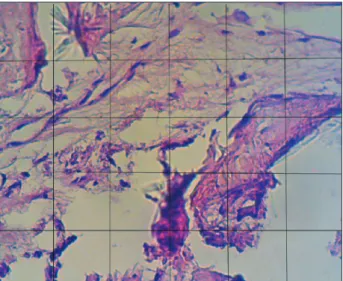
ized field.(Fig. 1) Based on the tissue contained within each randomized field, the boundary of each phase was defined on the image. And then the frame was superimposed on it; the hit points or intersections for each phase were counted and recorded. Using this method, the proportions of the histologi- cal phases (new bone and connective tissue) were obtained for each studied field. The surface area (SA) of each counting frame (150×120 µm) was 18,000 µm2. Finally, the number of hit of points was inserted into the following formula:
SA=I phase A/total number
where SA indicates the proportion of tissue, and I indicates the number of intersections for each phase. As mentioned earlier, the surface ratios for each section were calculated17.
3. Statistical analysis
All data are expressed as mean±standard deviation. The comparison was made by paired t-test, and P-values<0.05 were considered statistically significant. Calculations were performed using the SPSS Statistics version 17.0 software (SPSS Inc., Chicago, IL, USA).
III. Results
Histological examination of group DC at the end of the sec- ond week showed particles of unreacted ground dentin and some dilated vessels.(Fig. 2) On the control side, no specific
Fig. 1. A stereological counting frame was superimposed on each randomized field. This frame is composed of 30 squares (each square, 25 µm). ×40.
Mahdi Kadkhodazadeh et al: Effects of fresh mineralized dentin and cementum on socket healing: a preliminary study in dogs. J Korean Assoc Oral Maxillofac Surg 2015
*
Fig. 2. Dentin/cementum particle (asterisk) and a blood vessel (arrow) in an alveolar socket at the end of the second week. H&E staining, ×40.
Mahdi Kadkhodazadeh et al: Effects of fresh mineralized dentin and cementum on socket healing: a preliminary study in dogs. J Korean Assoc Oral Maxillofac Surg 2015
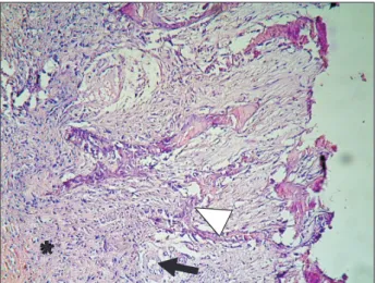
Fig. 3. New bone formation (arrowhead), particles of dentin/ce- mentum (arrow) in the periphery, and newly formed bone adjacent to the particles at the end of the eighth week. H&E staining, ×20.
Mahdi Kadkhodazadeh et al: Effects of fresh mineralized dentin and cementum on socket healing: a preliminary study in dogs. J Korean Assoc Oral Maxillofac Surg 2015
dentin and plaster repaired large jaw defects 57 months after surgery. They concluded that a dentin/plaster mixture was a useful biomaterial in the reconstructive surgery of jaw de- fects13. Register found that dentin allografts activated bone formation14, and Jeong et al.23 reported that autogenous teeth and bone could be used as good alternatives to autogenous bone. Collectively, the aforementioned studies have reported the potential of dentin as a grafting material. However, our results are not compatible with these studies. The discrepan- cies between our results and those of other studies may be explained by the wounds or defects, species, age, and method of material preparation. For instance, Nampo et al.22 used the extracted tooth with its pulp as a source of stem cells and neural crest cells. Devecioğlu et al.24 studied the effects of demineralized, freeze-dried DC on periodontal ligaments and fibroblasts. Their results implied that the applied materials had greater potential to form mineral-like nodules than did other applied materials. Gomes et al.25 studied the effects of demineralized dentin matrix on bone repair in diabetic rabbits and showed that dematerialized dentin was biocompatible in diabetic rabbits.
Given the results of these studies and our findings, fresh DC has little capability to induce significant bone formation in the alveolar socket. This result may be explained by the fact that such mineral acts as a barrier to chemotactic and morphogenic molecules of dentin, while acid initiates a de- mineralizing process that exposes osteoinductive molecules like BMPs. It seems that demineralization of biomaterials such as DC would be a key determinant factor in subsequent sessed and was 170±16 µm2 for the control group at the end
of eight weeks and 71±14 µm2 for the DC group, a significant difference (P<0.05).
IV. Discussion
This animal study was conducted to assess the healing of tooth extraction sockets using fresh DC. The results showed that the application of DC induced no effects on the healing process before two weeks. However, after eight weeks, gran- ular connective tissue was prominent in DC sockets. We used descriptive examination in conjunction with morphometric analysis to estimate the SA of newly formed bone. This method provides tangible and quantitative results from histo- logical sections and therefore enables the precise comparison of data18. The results of morphometric analysis revealed that bone formation proceeded at a lower rate in sockets treated with fresh DC. The present results are likely the first report on the use of fresh DC in reconstructive surgery. The application of biomaterials such as dentin has been studied extensively in the field of reconstructive surgery. Our data suggest that the route of preparation is a crucial step in the osteoinductiv- ity of biomaterials19. Studies have shown that demineralizing treatments of bone and dentin increase their osteoinductivity and decrease their antigenicity20,21. Nampo et al.22 studied the effect of iliac bone and teeth on new bone formation in the jawbones of rats. The results of their study showed that both types of materials could be used as suitable grafting materi- als. Kim et al.21 reported that a mixture of heat-pulverized
*
Fig. 4. Bone formed in a centripetal pattern. Bone spicules (arrow- head), connective tissue (asterisk) and a blood vessel (arrow) in the control group at the eighth week. H&E staining, ×20.
Mahdi Kadkhodazadeh et al: Effects of fresh mineralized dentin and cementum on socket healing: a preliminary study in dogs. J Korean Assoc Oral Maxillofac Surg 2015
Fig. 5. Red blood cells, venules (arrowhead), and connective tis- sue (asterisk) in the control group at the eighth week. H&E stain- ing, ×20.
Mahdi Kadkhodazadeh et al: Effects of fresh mineralized dentin and cementum on socket healing: a preliminary study in dogs. J Korean Assoc Oral Maxillofac Surg 2015
9. Huggins C, Wiseman S, Reddi AH. Transformation of fibroblasts by allogeneic and xenogeneic transplants of demineralized tooth and bone. J Exp Med 1970;132:1250-8.
10. Finkelman RD, Mohan S, Jennings JC, Taylor AK, Jepsen S, Bay- link DJ. Quantitation of growth factors IGF-I, SGF/IGF-II, and TGF-beta in human dentin. J Bone Miner Res 1990;5:717-23.
11. Bang G, Urist MR. Bone induction in excavation chambers in ma- trix of decalcified dentin. Arch Surg 1967;94:781-9.
12. Bessho K, Tagawa T, Murata M. Purification of rabbit bone mor- phogenetic protein derived from bone, dentin, and wound tissue after tooth extraction. J Oral Maxillofac Surg 1990;48:162-9.
13. Butler WT, Mikulski A, Urist MR, Bridges G, Uyeno S. Noncol- lagenous proteins of a rat dentin matrix possessing bone morpho- genetic activity. J Dent Res 1977;56:228-32.
14. Murata M, Kawai T, Kawakami T, Akazawa T, Tazaki J, Ito K, et al. Human acid-insoluble dentin with BMP2 accelerates bone induction in subcutaneous and intramuscular tissues. J Ceram Soc Jpn 2010;118:438-41.
15. Yeomans JD, Urist MR. Bone induction by decalcified dentine implanted into oral, osseous and muscle tissues. Arch Oral Biol 1967;12:999-1008.
16. Murata M. Bone engineering using human demineralized den- tin matrix and recombinant human BMP2. J Hard Tissue Biol 2005;14:80-1.
17. Reed MG, Howard CV, de Yanés GS. One-stop stereology: the estimation of 3D parameters using isotropic rulers. J Microsc 2010;239:54-65.
18. Wang X, Zauel RR, Rao DS, Fyhrie DP. Cancellous bone lamellae strongly affect microcrack propagation and apparent mechanical properties: separation of patients with osteoporotic fracture from normal controls using a 2D nonlinear finite element method (bio- mechanical stereology). Bone 2008;42:1184-92.
19. Lewandrowski KU, Tomford WW, Schomacker KT, Deutsch TF, Mankin HJ. Improved osteoinduction of cortical bone allografts:
a study of the effects of laser perforation and partial demineraliza- tion. J Orthop Res 1997;15:748-56.
20. Reddi AH. Bone matrix in the solid state: geometric influence on differentiation of fibroblasts. Adv Biol Med Phys 1974;15:1-18.
21. Kim YK, Kim SG, Byeon JH, Lee HJ, Um IU, Lim SC, et al.
Development of a novel bone grafting material using autog- enous teeth. Oral Surg Oral Med Oral Pathol Oral Radiol Endod 2010;109:496-503.
22. Nampo T, Watahiki J, Enomoto A, Taguchi T, Ono M, Nakano H, et al. A new method for alveolar bone repair using extracted teeth for the graft material. J Periodontol 2010;81:1264-72.
23. Jeong HR, Hwang JH, Lee JK. Effectiveness of autogenous tooth bone used as a graft material for regeneration of bone in miniature pig. J Korean Assoc Oral Maxillofac Surg 2011;37:375-9.
24. Devecioğlu D, Tözüm TF, Sengün D, Nohutcu RM. Biomaterials in periodontal regenerative surgery: effects of cryopreserved bone, commercially available coral, demineralized freeze-dried dentin, and cementum on periodontal ligament fibroblasts and osteoblasts.
J Biomater Appl 2004;19:107-20.
25. Gomes MF, Destro MF, Banzi EC, Vieira EM, Morosolli AR, Goulart Md. Optical density of bone repair after implantation of homogenous demineralized dentin matrix in diabetic rabbits. Braz Oral Res 2008;22:275-80.
26. Urist MR, Granstein R, Nogami H, Svenson L, Murphy R. Trans- membrane bone morphogenesis across multiple-walled diffusion chambers. New evidence for a diffusible bone morphogenetic property. Arch Surg 1977;112:612-9.
inductive/conductive potential of in vivo grafted materi- als19-21. Studies have documented that demineralization of dentin exposes the sequestrated BMPs in the insoluble col- lagen of the dentin matrix. Decalcified dentin has revealed better bone induction activity due to the activation of BMPs, which bind to collagen matrices through the demineralization process17-21,26. Recent data provide evidence that addition of BMP2 to demineralized dentin accelerates its osteoinductiv- ity16,26.
V. Conclusion
In conclusion, the strength of our pilot study is the evidence that fresh mineralized DC autografts have little to no effect on the induction of new bone in the alveolar socket milieu.
We suggest using ultrastructural and molecular approaches in similar studies in order to determine the underlying mecha- nisms.
Conflict of Interest
No potential conflict of interest relevant to this article was reported.
References
1. Irinakis T. Rationale for socket preservation after extraction of a single-rooted tooth when planning for future implant placement. J Can Dent Assoc 2006;72:917-22.
2. Botticelli D, Berglundh T, Lindhe J. Hard-tissue alterations follow- ing immediate implant placement in extraction sites. J Clin Peri- odontol 2004;31:820-8.
3. Tan WL, Wong TL, Wong MC, Lang NP. A systematic review of post-extractional alveolar hard and soft tissue dimensional changes in humans. Clin Oral Implants Res 2012;23(Suppl 5):1-21.
4. Dimova C. Socket preservation procedure after tooth extraction.
Key Engineering Materials 2014;587:325-30.
5. Vignoletti F, Matesanz P, Rodrigo D, Figuero E, Martin C, Sanz M.
Surgical protocols for ridge preservation after tooth extraction. A systematic review. Clin Oral Implants Res 2012;23(Suppl 5):22-38.
6. Sattayasanskul W, Brook IM, Lamb DJ. Dense hydroxyapatite root replica implantation: measurement of mandibular ridge preserva- tion. Int J Oral Maxillofac Implants 1988;3:203-7.
7. Jung RE, Siegenthaler DW, Hämmerle CH. Postextraction tissue management: a soft tissue punch technique. Int J Periodontics Re- storative Dent 2004;24:545-53.
8. Kim YK, Kim SG, Oh JS, Jin SC, Son JS, Kim SY, et al. Analysis of the inorganic component of autogenous tooth bone graft mate- rial. J Nanosci Nanotechnol 2011;11:7442-5.