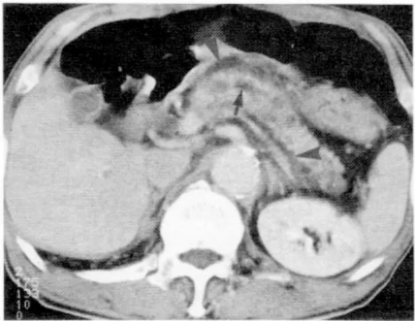
만성 췌장염 후에 발생한 췌장
전체 글
수치

관련 문서
Hepatitis B surface antigen loss in antiviral-treated patients with HBeAg(+) chronic hepatitis B (CHB) infection: observations from antiviral-naïve patients
만성췌장염에서 초음파내시경은 췌장 실질과 췌관의 변 화를 모두 평가할 수 있는 장점이 있으며 민감한 영상 소견 을 얻을 수 있어 만성 췌장염의 진단, 특히 조기 만성
Tenofovir alafenamide versus tenofovir disoproxil fumarate for the treatment of patients with HBeAg-negative chronic hepatitis B virus infection: a randomised, double-blind,
Patients with breast cancer; ovarian cancer; renal cell carcinoma; pancreatic neuroendocrine cancer; colorectal cancer; head and neck cancer; non-small cell lung
– Analyze unloading and cyclic loading behavior for both rheological models and for real materials, including cyclic stress-strain curves, irregular variation of strain with
Low intensity pulsed ultrasound used in this
newly formed periodontal ligament like tissue(PDL/NPDLT); green arrow, replacement resorption(RR); blue arrow, Cementum/ newly formed cementum-like tissue(C/NCeT).
relationship and and and and school school school school mark, mark, mark, mark, shows shows shows shows positive positive positive result positive result
