한수지 50(2), 219-221, 2017
219
Copyright © 2017 The Korean Society of Fisheries and Aquatic Science pISSN:0374-8111, eISSN:2287-8815 Korean J Fish Aquat Sci 50(2),219-221,2017
Note
서 론
다시마
(Saccharina japonica)
는갈조류에속하는한류성해조 류로서우리나라에서는동해안북부인원산만이북해역에서자연서식하는다년생해조류에속한다
.
다시마는만니톨(man-
nitol)
과글루타민산(glutamic acid)
이함유되어있어특유한맛 을나타내고,
또한요오드,
알긴산및라미나린을다량함유하 고있어건강식품으로각광을받고있는수산식물중하나이다(Jang, 2010).
국내다시마양식은1967
년일본북해도산다시 마어미엽체를도입하여인공양식의개발에성공하면서1974
년도에2,300
톤이생산되었고(Jang, 2010), 2004
년도에는약22,500
톤이생산되었다(KOSIS, 2015). 2005
년도에는전복양 식산업이활성화되면서다시마생산량이급격히늘어나(
전복 먹이로사용),
연간 생산량이 약100,000
톤으로증가하였고, 2014
년도에는 약370,000
톤으로증가하였다(KOSIS, 2015).
최근다시마생산량은우리나라해조류생산량의약
34%
를차 지하고있다(KOSIS, 2015).
다시마에서발생하는질병은원인에따라환경성질병과감염 성질병으로구분할수있다
.
환경성질병은조도불량,
담수유입
,
영양염의부족등으로인해발생하며,
감염성질병은세균,
진균,
착생동물등에의해발생한다(Park et al., 2009).
이중국 내다시마양식장에서는착생동물인히드라충과이끼벌레가다 시마엽체에부착하여상품가치를하락시키고있다(Park and Hwang, 2012; Oh et al., 2015).
다시마로부터분리한미생물또 는생물에대한병원성을조사하기위해서는수중에서사육중 인다시마를이용하거나배우체,
유엽등을사용해서감염실험 을실시해야한다.
이중배우체는실험실에서무균적으로관리 가가능하며,
연중저비용으로쉽게배양할수있는장점을가지 고있어유용하게사용될수있다. 2015
년다시마엽체로부터분 리한세균의병원성을조사하기위해다시마배우체를분양받아 관리하는과정에서,
전체배우체의약10%
에서진균의증식이 관찰되었다.
따라서,
본연구에서는다시마배우체에서발생하 는진균증에대해보고하고자한다.
재료 및 방법
2015
년전라남도해양수산과학원해조류연구센터로부터다 시마배우체를분양받아,
식물배양기에서관리[
온도, 15℃;
조다시마(Saccharina japonica) 배우체의 미동정 진균증
정하나·오명주·최성제 1 ·서정수 2 ·박명애 3 ·김위식*
전남대학교 수산생명의학과, 1전라남도 해양수산과학원, 2국립수산과학원 수산방역과, 3국립수산과학원 남동해수산연구소
Unidentified Mycosis of Kelp Saccharina japonica Gametophytes
Ha-Na Jeong, Myung-Joo Oh, Sung-Je Choi
1
, Jung-Soo Seo2
, Myoung-Ae Park3
and Wi-Sik Kim*Department of Aqualife Medicine, Chonnam National University, Yeosu 59626, Korea
1Algae Research Institute, Jeollanamdo Fisheries Research and Development Institute, Wando 59146, Korea
2Aquatic Life Disease Control Division, National Fisheries Research and Development Institute, Busan 46083, Korea
3Southeast Sea Fisheries Research Institute, National Institute of Fisheries Science, Tongyeong 53085, Korea
In 2015, white cottony tufts were observed on gametophytes of the kelp Saccharina japonica. Wet mount and histopa- thology examination revealed numerous fungal hyphae and mycelium around the gametophytes. The gametophytes surrounded by fungal hyphae were generally round and empty. A specific 610-bp fragment of the internal transcribed spacer (ITS)-5.8S rDNA-ITS gene of fungi was amplified by polymerase chain reaction and the nucleotide sequence showed 100% identity with those of Acremonium sclerotigenum, Acremonium sp. and Ascomycota sp. When fun- gus-infected gametophytes were mixed with healthy gametophytes, a high transmission rate (100%) resulted. This is the first report of mycosis of gametophytes in Korea.
Key words: Fungi, Gametophyte, Kelp, Saccharina japonica
This is an Open Access article distributed under the terms of the Creative Commons Attribution Non-Commercial Licens (http://creativecommons.org/licenses/by-nc/3.0/) which permits unrestricted non-commercial use, distribution, and reproduction in any medium, provided the original work is properly cited.
http://dx.doi.org/10.5657/KFAS.2017.0219 Korean J Fish Aquat Sci 50(2) 219-221, April 2017
Received 29 July 2016; Revised 19 October 2016; Accepted 23 March 2017
*Corresponding author: Tel: +82. 61. 659. 7177 Fax: +82. 61. 659. 7177
E-mail address: wisky@jnu.ac.kr
정하나
ㆍ
오명주ㆍ
최성제ㆍ
서정수ㆍ
박명애ㆍ
김위식220
도
, 1,000-1,500 lx;
광주기, 14:10 h (L:D)]
하였으며, 2
주간 격으로enriched seawater Saga-2 (ESS-2)
배지를교환하였다.
배우체의크기가지름
2.0 mm
이상으로성장하게되면멸균핀셋으로배우체를약
1/2
등분하여배양하였다.
위와같은방 법으로배우체를관리하던과정에서하얀솜털로뒤덮인배우 체가관찰되어본시료를채집하여실험에사용하였다.
채집된 시료로부터육안및현미경관찰을실시하였고,
병리조직학적 검사를위해시료를5%
중성포르말린액에고정한후상법에따라
70%
에서100%
까지단계별농도의에탄올수용액내에서탈수하고
,
자일렌으로투명화하여파라핀을침투시켜포매 후,
조직절편을만들어hematoxylin-eosin (H-E)
염색을실시 하여광학현미경으로관찰하였다.
배우체에부착된진균을동 정하기위해,
배우체로부터균사체를분리한후proteinase K
와phenol-chloroform
용액을사용하여DNA
를분리한후(Kim et al., 2005),
진균검출용primer
인internal transcribed spacer 5 (ITS5) (5'-GGAAGTAAAAGTCGTAACAAGG-3')
와ITS4 (5'-TCCTCCGCTTATTGATATGC-3')
를사용하여polymerase chain reaction (PCR)
을실시하였다(
타켓gene: ITS-5.8S rDNA- ITS
부위) (White et al., 1990). PCR
반응은30 mM KCl, 10 mM Tris-HCl (pH 9.0), 1.5 mM MgCl
2, 250 μM
의각dNTP, 1 U Taq DNA polymerase
가포함되어있는혼합물(PCR pre- mix, Bioneer, Korea)
에20 pmol
의각primer
와추출된DNA 1 μL (100 ng/ μL)
를첨가하여실시하였다.
반응조건은95℃
에서
5
분간pre-denature
시킨 후, 95℃
에서1
분간denature, 55℃
에서1
분간annealing, 72℃
에서1
분간extension
반응을30 cycles
을진행시키고, 72℃
에서5
분간post-extension
하였 다. PCR
증폭산물은1.5% agarose gel
을이용하여확인하였 고, gel purification kit (Bioneer, Korea)
를이용하여 정제후ABI PRISM dye terminator sequencing chemistry (Applied Biosystems, U.S.A.)
를사용하여염기서열을분석하였다.
분석 된염기서열은GenBank
의blast
분석을실시하여동정하였다.
감염실험은2
개의25 flask
에ESS-2
배지를30 mL
씩첨가한 후국립수산과학원해조류연구센터로부터분양받은건강한배 우체(
지름, 1.7-2 mm)
를5
개씩넣어주었다.
실험구에는진균이 부착되어있는배우체(1.7 mm)
를1
개넣어주었고,
대조구에는 건강한배우체(1.7 mm)
를1
개넣어준후15℃
에서1,500 lx
로7
일간배양하였다.
진균의부착여부는1
일간격으로현미경검 사를통해확인하였고,
건강한배우체의주위에균사가약20%
이상부착되었을경우양성으로판정하였다
.
감염실험에사용 한배우체는현미경검사를통해기생충과진균에대하여음성 인것을확인하였다.
결과 및 고찰
2015
년연구목적으로다시마배우체를관리하는과정에서전 체40
개중4
개의페트리디쉬에서(1
개의페트리디쉬: 3-4
개의배우체가들어있음
)
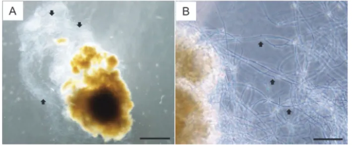
배우체에솜털모양의이물질이덮여있는 것이관찰되었다(Fig. 1A).
이물질로덮여있는배우체를현미 경으로관찰한결과,
가늘고긴균사들이배우체를뒤덮고있었 다(Fig. 1B).
진균이부착된배우체는약30
일간정상적인배우 체의색을유지하였으나,
시간이경과됨에따라배우체가하얗 게변화하면서폐사되었다.
균사로뒤덮인배우체를대상으로 병리조직학적검사를실시한결과,
배우체내부에는균사가관 찰되지않았으며,
배우체주위로다수의균사체와균사들이관 찰되었다(Fig. 2).
균사로뒤덮인배우체는정상배우체와달리 전반적으로둥그런형태를보이거나내부가비어있었다(Fig.
2).
이상의연구결과,
배우체에솜털모양으로뒤덮인이물질은 진균으로확인되었다.
또한진균은배우체내부로침입하지는 않지만진균주변의배우체들은형태가변형되거나내부가비 어있는것으로보아배우체는진균에의해영향을받는것으로 추정된다.
배우체에부착된진균을이용하여PCR
을실시한결 과,
약610 bp
의PCR
산물이확인되었다(data not shown). PCR
산물을사용하여염기서열을분석한결과(556 bp, primer
염기 서열미포함), Acremonium sclerotigenum, Ascomycota sp., Acremonium sp. (GeneBank Accession Number KU059865
의염기서열17-572 bp
와동일)
와100%
일치하는것으로확인 되었다.
배우체로부터분리한진균을사용하여감염실험을실A B
A B
m m
m
m m
0 20 40 60 80 100
0 1 2 3 4 5 6 7
Attachment rate (%)
Days after cohabitation Cohabitation with fungi-attached gametophyte and healthy gametophyte
Cohabitation with healthy gametophyte and healthy gametophyte
Fig. 1. Gametophyte attached to fungi (arrows) (A). Magnification of fungi (arrows) (B). Scale bar=500 μm (A), 50 μm (B).
A B
A B
m m
m
m m
0 20 40 60 80 100
0 1 2 3 4 5 6 7
Attachment rate (%)
Days after cohabitation Cohabitation with fungi-attached gametophyte and healthy gametophyte
Cohabitation with healthy gametophyte and healthy gametophyte
Fig. 2. Histopathology of gametophyte attached to fungi. Normal gametophyte (A) and fungi-affected gametophyte (B). Numerous fungal hyphae and mycelium (m) were observed around gameto- phyte and the fungi-affected gametophyte were generally rounded and empty (arrows). Scale bar=20 μm (A, B).
다시마 배우체의 미동정 진균증
221
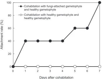
시한결과
,
건강한배우체(5
개)
와진균에부착된배우체(1
개)
를 혼합배양한실험구에서는1
일째부터건강한배우체의40%
에 서진균의부착이관찰되었고7
일째는모든배우체에서진균이 관찰되었다(
부착률, 100%) (Fig. 3).
대조구에서는진균의부착 이관찰되지않았다.
이상의연구결과,
배우체에부착된진균 은Acremonium sclerotigenum, Ascomycota sp., Acremoni- um sp.
와염기서열이100%
일치하므로Acremonium sp.
또는Ascomycota sp.
로추정되며,
빠른전파력을지닌것으로확인 되었다.
다시마에서분리되는진균으로는Penicillium sp., As- pergillus sp., Fusarium sp., Acremonium sp., Dendryphiella sp.
등이보고되어있다(Schatz, 1980; 1984; Zvereva, 1998).
이중
Penicillium sp., Aspergillus sp., Acremonium sp., Dend- ryphiella sp.
는다시마엽체에서분리되었으며, Fusarium sp.
는 다시마배우체에서분리되었다.
본연구에서는다시마배우체 에서처음으로Acremonium sp.
또는Ascomycota sp.
로추정 되는진균증을확인하였다.
진균의오염은배우체를관리하는 과정에서발생했기때문에사용한실험도구,
배지,
실내환경등 으로부터유입되었을것으로추정된다.
본진균은배우체에빠 르게전파되어증식하므로실내에서배우체를관리할경우,
본 진균에의한오염에주의가필요하다.
사 사
본연구는국립수산과학원수산과학연구사업수산생물방역 프로그램개발운영과제
(R20166069)
의지원으로수행된연구 입니다.
References
Jang GN. 2010. Seaweed and crustacean culture. 2nd ed, Samk- wang publishers, Seoul, Korea. 88-129.
Kim WS, Oh MJ, Jung SJ, Kim YJ and Kitamura SI. 2005.
Characterization of an iridovirus detected from cultured turbot Scophthalmus maximus in Korea. Dis Aquat Org 64, 175-180.
Korean statistical information service (KOSIS). 2015. Fishery pro- duction survey: Statistics by type of fishery and species[Internet].
Retrieved from http://kosis.kr/statisticsList/statisticsList_01List.
jsp on July 2016.
Oh MJ, Kim WS, Kim JO, Jeong HN and Kim JO. 2015. Study on management of seaweed (kelp) disease. Nation Fisheri Res Develop Instit. Busan, Korea.
Park CS and Hwang EK. 2012. Seasonality of epiphytic devel- opment of the hydroid Obelia geniculata on cultivated Sac-
caharina japonica (Laminariaceae, Phaeophyta) in Korea. J
Appl Phycol 24, 433-439. http://dx.doi.org/10.1007/s10811- 011-9755-3.Park SW, Lee JH and Kim YS. 2009. Seaweed disease. Biosci- ence, Seoul, Korea, 105-116.
Schatz S. 1980. Degradation of Laminaria saccharina by higher fungi: a preliminary report. Bot Mar 23, 617-622.
Schatz S. 1984. Degradation of Laminaria saccharina by sapro- bic fungi. Mycologia 76, 426-432.
White TJ, Bruns T, Lee S and Taylor J. 1990. Amplification and direct sequencing of fungal ribosomal RNA genes for phy- logenetics. In PCR Protocols: A Guide to Methods and Ap- plications. eds., Innis, M.A., Gelfand, D.H., Sninsky, J.J. and White, T.J., Academic Press, San Diego, U.S.A., 315-322.
Zvereva LV. 1998. Mycobiota of the cultivated brown alga Lam-
inaria japonica. Russ J Mar Biol 24, 19-23.
A B
A B
m m
m
m m
0 20 40 60 80 100
0 1 2 3 4 5 6 7
Attachment rate (%)
Days after cohabitation Cohabitation with fungi-attached gametophyte and healthy gametophyte
Cohabitation with healthy gametophyte and healthy gametophyte
Fig. 3. Attachment rate (%) of fungi in gametophyte cohabitated with fungi-attached gametophyte. Cohabitation with fungi-at- tached gametophyte and healthy gametophyte (●), cohabitation with healthy gametophyte and healthy gametophyte (○).