D ic l o f e n a c 투 여 후 시 간 경 과 에 따 른 장 손 상 과 장 내 세 균 전 위 의 변 화
김은정 • 김 정 욱 *'*
한 국 보 건 산 업 진 흥 원, * 중 앙 대학 교 의과 대학 내과 학 교 실 (Received May 22, 2008; Revised August 6, 2008)
T h e C h a n g e s in In te s t in a l D a m a g e a n d B a c te r ia l T r a n s lo c a tio n w it h T im e a fte r A d m in is t r a t io n o f D ic lo fe n a c
Eun Jeong Kim and Jeong Wook K im *'*
Korea Health Industry Developement Institute, Seoul 156-800, Korea
'^Department of Internal Medicine, Chungang University College of Medicine, Seoul 140-757, Korea
Abstract — Non-steroidal anti-inflammatory drug (NSAID)-induced gut damage and bacterial translocation (BT) have not been studies well, especially from the perspective of time after administration of NSAIDs. We therefore examined these changes in animals. The study was performed on 5 groups of rat; a control group (group A) and diclofenac groups (groups B, C, E, and F). Rats in the diclofenac groups were orally administered diclofenac sodium before intestinal permeability (IP) measurement (group B, 1 h before measurement; group C, 10 h before; group D, 22 h before; and group E, 52 h before).
The IF, stool pellet number, serum biochemical profile, enteric bacterial number, and BT in the mesenteric lymph nodes (MLNs), liver, spleen, kidney and heart were measured. The administration of diclofenac resulted in significantly increased
IP, caused intestinal protein loss, decreased stool pellet number, caused enteric bacterial overgrowth and increased BT in multiple organs in groups A, B, C, and D. IR intestinal protein loss, and the BT in the liver and the spleen in group E were decreased than those in group D. There were no differences in the other parameters between group D and E. In the recov
ery phase of the diclofenac-induced gut damage, enteric bacterial overgrowth and BT in the kidneys and the heart did not change while the BT in the reticuloendothelial systems such as in the MLNs and liver was decreased.
Keywords □ non-steroidal anti-inflammatory agents, adverse effects, protein-losing enteropathies, bacterial translocation
비 스테로이드소염제 (nonsteroidal anti-inflammatory drugs, NSAID)는 흔히 사용되고 있는 약물이나 부작용^ 약물 사용 에 제한을 받는 경우가 많다. 이 중 소화기계에서 발생하는 부작 용이 가장 중요하며 위장미나 샘창자 이외에도 소장이나 대장의 손상에 의하여 출혈이나 단백소실과 협착 등이 발생 한다.^'^^ 비 스테로이드소염제에 의한 장관손상의 기견은 아직 정확히 입증 되지 않았으나 장관장벽 기능이상에 의한 장투과성의 변화가 세 균이나 담즙 및 옴식물에서 유래된 작중 항원들의 침투를 유발 시켜 염증성 반응이 발생하는 것으로 설명되고 있다.^'^^
비스테로이드소염제에 의한 장관의 장투과성의 번화와 장내세 균의 파증식은 정상세균무리를 이루고 있는 세균 등이 장관 이 외의 곳으로 파급되는 현상인 장내세균견위률 유발한다. 장내세
"본 논문에 관한 문의는 저자께게로 (전화) 02-748-9941 (팩스) 02-790-2068 (E-mail) [email protected]
균견위의 주요 세가지 기전으로는 장투파성의 증가, 장내세균의 괴증식, 면역력의 약화가 제시되고 있다.®' 비스테로이드소염제에 의한 장내세균 전위는 비대상성 심부견 환자에서 심장 질환을 악 화 시 키 며 복 부 수술을 받는 환자에서 후 감염성 합병증인 장 내세균전위와 내득소혈증을 유발 한다.
비스테로이드소염 제는 실험동물에서 투여 후 장투과성 증가, 장관에서의 단백소실과 같은 장관손상의 소견이 관찰된다. 약물 투여 후 장투과성은 시간 경과에 따라 증가했다가 일정기간이 지 나면 다시 감 소 한 다 .^또 한 장관에서의 단백소실로 인하여 혈 청 알부민이 감소하였다가 시간의 경과에 따라 회복된다.^^* 그 러나 비스테로이드소염제 투여 후 장내세균전위의 변화와 그 기 견 중에 하나인 장내세균수의 번화쉬1 대해서는 아직 보고되지 않 았다.
이에 본 연구자는 비스테로이드소염제인 diclofenac의 투여 후 시간변화에 따른 장관손상의 번화와 장내세균수와 장내세균전위 의 변화률 알아보았다.
실험재료 및 방법
실험재료
7주령의 체중 140—160gm 정도의 수컷 Sprague-Dawley 백 서 35마리률 오리엔트 바이오사(Orient Bio Co., Ltd., Seoul, Korea)에서 공급받아 실험 견 7일간 적응시켰다. 백서 사육 기간 동안 1 2시간 간격으로 낮과 밤을 구별하고 사육 온도는 22±1°C 로 유지하였으며 고형사료인 Basal diet 5053(PMI Nutrition International, Inc., Richimond, California, USA)와 물을 자유롭 게 섬취하게 하였다. 백서들은 실험기간 동안 고형사료와 물을 자 게 섭취하게 하였으며 매일 물과 사료의 섭취량과 몸무게 의 번화률 측정하였다. 밑망을 설치하식 배설물이나 깔집 등 옴 식물이 아닌 것을 섬취하는 것을 방지하였다. 장투과성을 측정 하는 동안 환경변화에 따른 백서의 식이량과 옴용수의 섭취량이 감소하지 않게 위하여 대사케이지 (metabolic cage)안에 고정된 그뭇을 이 용 하 고 형 사 료 와 점성^ 공급하였다. 백서들은 실 험 시작 전 감염이나 기타 이상소견이 관찰되지 않았다. 백서둘 은 모두 5군으로 나누었으며 각 군 당 백서 7미리률 배정하였다.
A군은 대조군으로 하고 B, C, D, E군은 diclofenac 투여군으로 하였다.
Diclofenac에 의한 장손상 유발
장손상은 diclofenac sodium(Sigma Chemical Co., St. Louis, MO, USA)을 증류수 2 m/에 혼 함 뻐 ' 150 mg/kg의 용량으로 금 속 경구 투여관을 이용하여 일회 강제 경구투여 하쉬 유발하였 다. B군은 장투과성 죽정 시작 1시간 전에 diclofenac sodium을 투여하였고 C군은 10시간 전, D군은 22시간 전, E군은 52시간 견에 약물을 투여하였다.
장투과성과 배변수의 측정
5mg£l phenolsulfonphthalein(PSR Sigma Chemical Co., St. Louis, MO, USA)을 금속 경구 투여관을 이용'하식 백서 에 경구 투여 후 대소번을 분리하여 채취할 수 있는 대사케이지 (metabolic cage)에서 PSP 투여부터 20시간 동안 소번을 수집하 였다. 채집된 소변에서의 PSP 농도룰 구하기 위하여 10m/의 10% NaOH로 일칼리화 시킨 후 100 m/의 증류수를 이용하여 부 피룰 보정한 후에 559nm의 파장에서 분광광도계(Smart Spec 300, Biorad, Hercules, CA, USA)로 측정하였다.피 측정치률 계 산하식 복용한 PSP 중 회수된 앙을 백분율로 표시하였다. 또한 24시간 소변을 수집하는 동안 장통과시간의 측정을 위해 대사케 이지의 대번 회수통에 었는 대변의 수률 측정하였다.^^*
마취한 후에 무균조작으로 복부를 정중앙에서 절개하였다. 절개 직후 횡경막을 절개하고 심장에서 혈액을 채취하여 백서률 시켰으며 맹장으로부터 상방 20 cm의 회 정 서 장 내용물을 500 mg을 채취하였다. 장내세균전위 정도률 알아보기 위하여 무균조 작으로 장간막 림프절, 간, 비장, 콩괄, 심장을 절제하식 1 0 0 mg 씩 채취하였다. 맹장의 장내세균수률 측정하기 위하여 맹장을 1cm 정도 절개한 후 대번 500 mg을 채취하였다.
장내 호기성 세균수 및 장내세균전위에 대한 검사
회장과 맹장에서 획득한 장내용물 중 1 0 0 mg을 취하여 소득 된 생리식염수로 1 0배씩 연속적^ 희석한 후 장내 호기성균수 를 측정하기 위하여 혈액한천배지와 장내 그람 옴성균수롤 측정 하기 위하여 MacConkey 한천배지에 접종하여 배양기에서 37T 로 4&시간 동안 배양하였다. 배양 후 각 배지에서 자란 집락 수 률 관찰하여 CFU(colony-forming unit)/g으로 표시하였다. 장내 세균전위룰 알아보기 위하식 장간막 림프절, 간, 비장, 콩괄, 심
^ 100 m g# homogenizer(Heidolph-Elektro GmbH & Co., Schwabach, Germany)틀 이용하여 균질화 시킨 후 소득된 생리 식염수로 1 0배씩 연속적으로 희석하고 그람 옴성균수률 측정하 기 위하여 MacConkey 한천배지에 접종하여 배양기에서 37°C로 24시간 동안 배양하였다. 배양 후 각 배지에서 자란 집락 수률 관찰하여 CFU(colony-forming unit)/g으로 표시하였다.
혈액 검사
장관에서의 단백소실을 측정하기 위하식 백서에서 채취한 혈 액으로 혈청 총단백, 알부민을 측정하였다. 총단백 수치와 알부 민 수치의 번화가 약제에 의한 간 득 성 로 이차적으로 발생한 것 이 아님을 증명하기 위하여 알라닌아미노전이효소(alanine aminotransferase), 아스파르테 이 트아머 노견이 효소(aspartate aminotranferase), 총 빌리루번, 총콜레스테롤, 알칼리인산분해효 소 (alkaline phosphatase)률 자동화학 분석기 Advia 1650(Bayer Healthcare Co., Ltd, Tarrytown, NY, USA)로 동시에 죽정하여 비교하였다/ 효'
통계 처러
모든 수치는 평균±표준편차로 표시하고 각 군 간의 차이는 SPSS 11.5 프로그램 (SPSS Inc, Chicago, IL, USA)을 이용하여 각 실험군간 비교는 Mann-Whitney 검정법^ 설시하였다. p값 이 0.05 이하인 경우틀 유의성이 있는 것으로 관정하였다.
결 고 F
장투과성 검사틀 위한 24시간 소번 채취 후 백서는 에테르로
식이량과 체중 변화
백서의 1 0일 동안 체중번화, 섭취한 식이와 옴용수의 양은 정
j?<0.05 I 19.99'±6.55
p<0 .0 1
/;=0 .0 0 1 1 2 .6 9 ± 3 .5 3 8.29± 2.05
zr
2 .3 -l± 0 .5 8
/7=0.0()1
5.64± 1.65
/xO.Ol p<0.05
Group A Group B Group C Group D Group E Trial population
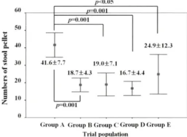
Fig. 2 - Numbers of stool pellet. Values are means ±SD. Group A, control group; Group B, group with measuring intestinal permeability 1 — 2 1 hours after diclofenac administration;
Group C, group with measuring intestinal permeability 10—30 hours after diclofenac administration: Group D, group with measuring intestinal permeability 22—42 hours after diclofenac administration; Group E, group with measuring intestinal permeability 52—72 hours after diclofenac administration.
Oi oup A Group B Group C Group D Group L Trial population
Fig. 1 - The Changes of intestinal permeability measured by 24 hour urinary excretion of PSP(phenolsulfonphthalein).
Values are means ±SD. Group A, control group: Group B, group with measuring intestinal permeability 1 — 2 1 hours after diclofenac administration; Group C, group with measuring intestinal permeability 10—30 hours after diclofenac administration; Group D, group with measuring intestinal permeability 22 —42 hours after diclofenac administration; Group E, group with measuring intestinal permeability 52—72 hours after diclofenac administration.
Table I - Body weight changes and amounts of food Intakes in the animals
Trial population Initial BW Body weight change (g/lOOg BW/day) Chow intakes (g/lOOg BW/day) Water intakes (g/lOOg BW/day)
Group A 152.1+8.5 5.10±0.62 13.72 ±3.30 29.63±9.96
Group B 151.3+8.0 5.99+0.88 14.19±1.23 35.01±1.49
Group C 154.1+2.9 5.78±0.23 13.58±L63 35.86+2.32
Group D 150.6+5.0 4.93 ±0.80 흐 12.71±0.73* 33.55±3.04
Group E 152.7+1.8 2.25±0.67* 우용*냐 11.50+ 0.73'"'' 22.39±1.78'^**
BW, body weight; Group A, control group; Group B, group with measuring intestinal permeability 1—21 hours after diclofenac administration; Group C, group with measuring intestinal permeability 10—30 hours after diclofenac administration; Group D, group with measuring intestinal permeability 22—42 hours after diclofenac administration; Group E, group with measuring intestinal permeability 52—72 hours after diclofenac administration.
*^=0.001 compared with group A
=0.001 and ^<0.05 compared with group B
7J=0 . 0 0 1 and '^/xO.OS compared with group C 갓=0.001 and **^<0.01 compared with group D
상대조군인 A군에 비해 B, C군에서 차어가 없었다. D군에서는 B군에 비해 체중번화와 섭취한 식이의 앙이 적었으며 섬취한 응
^ 의 양은 차이가 없었다. E군에서는 다른 군과 비교하여 체 중번화와 섭취한 식이와 응용수의 앙이 적었다(Table I). 실험 도 중 사■망하거나 기타 이유로 식이섭취량이 감소한 백서는 없었다.
장투과성과 배변수의 변화
장관장벽의 기능이상을 측정하기 위한 PSP를 이용한 장투과 성 검사에서는 정상대조군인 A군에 비해 B, C, D 군에서 diclofenac 투여 후 시간 경과에 따라 장투과성이 증가하였으나 E군에서는 감소하늬 B, C, D군보다 낮았다(Fig. 1). 배변수는 정
/^O.OOl
상대조군인 A군에 비해 감소되었으며 B, C, D, 않았다(Fig. 2).
diclofenac이 투여된 B, C, D, E군에서 E군 사이에 배번수의 처식는 관찰되지
p<0 .01
장관 내 세균수
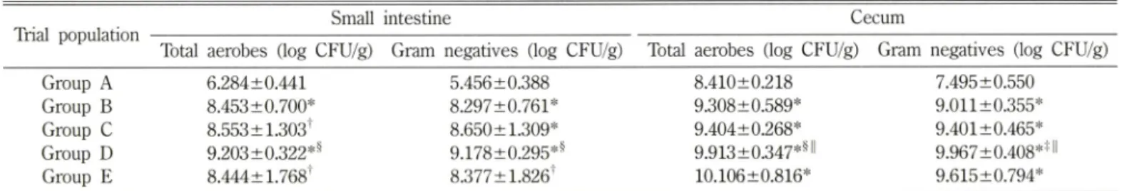
정상대조군인 A군보다 diclofenac이 투여된 B, C, D, E군에서 소장과 맹장에서 총호기성균주와 그람음성균주의 과증식이 관찰 되었다. D군에서 맹장에서의 총호기성 균주와 그람옴성균주는 B 군과 C군에 비해 균의 수가 증가하였다(Table II).
장내세균전위
장내세균전위률 관찰하기 위한 장간막 림프절, 간, 비장, 콩괄
»<0.05
p = O M ) l
2 4 .9 ± 1 2 .3
41.6± 7.7 19.0±7.1
18.7소4 3 ~ I ~ 16.7 + 4 .4
/7=0.001
0 0 0 03 2 1
001 o
o
o
o
o
4 3 2 1
껍
sddSJdqsn
조
Table II - Changes of the number of enteric bacterial numbers in the small intestine and colon
Trial population Small intestine Cecum
Total aerobes (log CFU/g) Gram negatives (log CFU/g) Total aerobes (log CFU/g) Gram negatives (log CFU/g)
Group A 6.284±0.441 5.456 ±0.388 8.410±0.218 7.495±0.550
Group B 8.453 ±0.700* 8.297±0.761* 9.308±0.589* 9.011±0.355*
Group C 8.553 ±1.303' 8.650± 1.309* 9.404 ±0.268* 9.401±0.465*
Group D 9.203±0.322*^ 9.178±0.295*^ 9.913±0.347*^>l 9.967±0.408* 해
Group E 8.444± 1.768' 8.377± 1.826' 10.106±0.816* 9.615±0.794*
Group k, control group; Group B, group with measuring intestinal permeability 1—21 hours after diclofenac administration; Group C, group with measuring intestinal permeability 10—30 hours after diclofenac administration; Group D, group with measuring intestinal permeability 22—42 hours after diclofenac administration: Group E, group with measuring intestinal permeability 52—72 hours after diclofenac administration.
*/j = 0.001 and '/) = 0.05 compared with group A 흐^ <0.01 and 보^ <0.05 compared with group B
용/) <0.05 compared with group C
Table III - Bacterial colony counts obtained from culture of the mesenteric lymph node, liver, spleen, kidney and heart
Trial population MLN (log CFU/g) Liver (log CFU/g) Spleen (log CFU/g) Kidney (log CFU/g) Heart (log CFU/g) Group A
Group B Group C Group D Group E
0 . 0 0 0 ±0 . 0 0 0
2.759±0.781*
3.062±0.860*
5.136± 1.427* II**
3.536+0.795*■니
0.468±0.810 2.264+1.255^
3.968±0.699=^^
4.387± 1.338*^
2.692±0.622=^
0.735 ±0.931 1.910± 1.011^
3.794±0.980*ll 4.378±1.00H 4.145±0.606*''
0.828± 1.068 1.462± 1.540 4.057±1.780■는합 3.965± 1.347*^
4.002±0.690*11
0.864± 1.125 1.329± 1.356 2.652± 1.883 3.129±0.870^^
3.498+0.909기 1 MLN, mesenteric lymph node; Group A, control group: Group B, group with measuring intestinal permeability 1—21 hours after diclofenac administration; Group C, group with measuring intestinal permeability 10—30 hours after diclofenac administration: Group D, group with measuring intestinal permeability 22—42 hours after diclofenac administration; Group E, group with measuring intestinal permeability 52—72 hours after diclofenac administration.
*/)=0.001, '^<0.01 and 궁^ <0.05 compared with group A 상/) = <0.01 and 하/) <0.05 compared with group B
**/)<0.01 compared with group C ' '/)<0.05 compared with group D
Table IV - Serum biochemical findings
Trial population TP (^d/) ALB (g/d/) ALT (lU//) AST (lU//) ALP (lU//) T-Bil (mg/d/) T-Chol (mg/d/) Group A 5.586±0.146 3.843 + 0.113 138.7±34.7 40.0 ±8.3 308.9±86.9 0.13 ±0.03 67.0±7.6 Group B 3.343 ±0.544* 2.214±0.334* 85.9±14.9' 28.9+2.9" 174.0±40.6^ 0.15 ±0.05 58.7±12.0 Group C 3.057±0.369* 2.071±0.275* 75.1+20.2' 25.9+3.9' 126.6±20.9* 하 0.10±0.06 66.9±18.3 Group D 3.129±0.355* 2.014±0.204* 84.1±41.7^ 23.4+6.1' 105.1±19.3*" 0.16±0.07 78.7±14.8 Group E 4.286±0.146* 힌**■미 ■ 2.800±0.129*"^**"" 84.0±20.3 우 25.1±5.2' 145.6 ±40.4 범 0.12 ±0.03 73.3±8.9
TF, total protein; ALB, albumin; ALT, alanine aminotransferase; AST, aspartate aminotranferase: ALR alkaline phosphatase; T-Bil, total bilirubin: T-chol, total cholesterol: Group A, control group; Group B, group with measuring intestinal permeability 1—21 hours after diclofenac administration: Group C, group with measuring intestinal permeability 10—30 hours after diclofenac administration: Group D, group with measuring intestinal permeability 22—42 hours after
permeability 52—72 hours after diclofenac administration,
*p=0.001, 'p<0.01 and ^/)<0.05 compared with group A
= 0.001 and ^<0.05 compared with group B
**/>=0.001 compared with group C
' '^=0.001and ■*■■는^ <0.05 compared with group D
diclofenac administration; Group E, group with measuring intestinal
및 심장에서의 그람옴성 세균수의 죽정결과 diclofenac 투여군에 서 정상대조군보다 장간막 림프절, 간, 비장, 콩괄 및 심장에서 그람옴성 세균수의 증가가 관찰되었다. C군에서는 B군보다 간, 비장, 콩괄에서 그람옴성균수가 증가하였다. D군에서는 B군에 비해 장간막림프절, 간, 비장, 콩팔, 심장에서 그람옴성균수가 증 가하였으며 , C군에 비해서는 장간막림프절에서 그람옴성균수가 증가하였다. E군에서는 D군보다 장간막림프절과 간에서 그람옴
성균의 수가 감소하였다(Table III).
혈액검사
정상대조군인 A군보다 diclofenac이 투여된 B, C, D, E군에서 혈청 총단백과 알부민이 감소하였다. 또한 알라닌아미노전이효 소, 아스파르테이트아미 노견이 효소, 알칼러 인산분해효소도 감소 하였으며 총빌리루빈과 총콜레스테롤수치는 처식가 없었다. E군
에서는 B, C, D군 보다 알라닌아머노전이효소, 아스파르테이트 아미노견이효소, 총빌리루번, 총콜레스테롤 수치의 번화 없이 총 단백파 알부민이 증가하였으며 D군에 비해 알칼리인산분해효소 도 증가하였다(Table IV).
고 찰
비스테로이드소염제의 부작용은 주로 소화성 궤양과 같은 상 부위장관 합병증에 대해서 많이 연구되었으나 하부장관의 손상 과 장내세균전위와 같은 이차적인 감염성 합병증에 대해서는 아 직 연구가 되지 않았다. 하지만 비스테로이드소염제의 하부장관 손상과 합병증은 상부위장관 합병증 보다 번도가 높으며, 장내 세균전위의 위험성은 상기 약물의 복 자 에 서 항상 존재하고 있 다. 장내세균전위는 장관 내에 정상세균무리로 존재하는 장내세 균이 다른 장기로 파급되는 현상을 말하며 특히 패혈증이나 다 발성 장기부견의 주요 원인인 호기성 그람음성균과 부산물인 내 득소가 주로 파급된다.®
비스테로이드소염제에 의한 장내세균견위는 비대상성 심 부견 환자에서 염증과 연관된 시토카인의 활성을 자국하쉬 심장기능 을 저하시킨다. 비대상성 심부전 환자는 심장기눙의 저하로 장 관부종이 동반되며 이로 인®M 장투과성의 증가와 장내세균전 위가 발생하는데 , 비스테로이드소염제는 추가적으로 장관손상과 장내세균전위를 증가시키므로 비대상성 심부견 환자에서는 허혈 성 심장질환이 동반되지 않은 경우에는 아스피린을 포함한 비스 테로이드소염제률 사용해서는 안 된다.®"® 복부 수술 전에 시용 한 비스테로이드소염제는 수술 후 내득소혈증과 장내세균견위률 유발하는데 수술 후 합병증^ 발생하는 각종 장기에서의 감염 성 합병증을 악화시킬 수 있다.®* 이외에 만성콩괄병, 비대상성 간경번증, 폐쇄성담도질환, 췌장염, 출혈성쇼크 등 많은 질환에 서 장내세균전위는 감염성 합병증을 유발하거나 악화시킬 수 있 는데 비 스테로이드소염제는 장관손상을 악화시켜 장내세 균전위 를 증가시킬 수 있다.
장내세균견위의 주요 유발 기전 중 가장 중요한 것은 장투과성 의 증가로 cyclooxygenase의 억제와 약물의 직접적인 점막세포손 상 등에 의해 발생된다.^' 다른 중요 유발 기전인 장내세균의 과 증식과 면역눙의 저하가 있더라도 장투과성의 증가가 없으면 장 내세균전위가 발생하지 않는다.® 비스테로이드소염제는 투여 후 장투과성이 증가하나 일정기간이 지나면 감소하고 장관손상과 연 관성이 있는 장관에서의 단백소실도 같은 양상을 보 인 다 . 그 러나 이와 같은 번화에 따른 장내세균전위와 또 하나의 중요유 발 인자인 장내세균수의 번화에 대해서는 아직 알려지지 않았다.
각종 장기에서의 장내세균전위의 경우 장관에서 유입되는 세 균 뿐 만 아니라 이미 장기에 유입된 세균이 증식하여 발생 할 수 있다. 그러나 그물내피계통(reticuloendothelial system)인 장
간막림프절, 간, 비장은 세균과 내득소를 제거하는 능력이 있지 만 콩괄과 심장은 그렇지 않다. 또한 장내세균전위에 중요한 유 발인자인 장내세균의 과증식은 약물에 의한 장관손상으로 발생 하는 장통과시간(intestinal transit time) 감소로 설명할 수 있으 나, 일정기간이 지난 후의 장내세균수의 변화와 장통과시간의 번 화에 대해서는 아직 연구되지 않았다.
이번 연구에서는 장투과성은 비스테로이드소염제 투여 후 시 간이 경과에 따라 증가하다가 일정 시간 경파 후에 감소하였다.
혈청 총단백과 알부민수치도 간기능의 연관된 수치와 식이량 번 화의 영향 없이 시간의 경과에 따라 감소하다가 장투과성의 증 가와 함께 증가하였다. 이는 비스테로이드소염제에 의해 발생한 장관에서의 단백소실이 장투과성의 번화처럼 일정 시간 경과 후 에 감소하였다는 것을 의미한다.
장내세균의 과증식은 비스테로이드소염제 투여 후 중가하였으 며 소장 원위부 보다 맹장에서 그 차의가 뚜렷하였다. 하지만 일 정 시간 경과 후 증가된 장투과성이 감소될 때 장내세균수의 변 화가 관찰되지 않았다. 장내세균의 과증식과 연관이 있는 장관 운동성을 나타내는 배번 수는 비스테로이드소염제 투여에 의해 감소하였으나 일정시간 경과 후 증가한 장투과성과 단백소실이 회복될 때 유의한 번화가 관찰되지 않았다. 이와 같이 장관운동 성이 회복되지 않는 기견에 대해서는 추가적인 연구가 월요하나 장내세균의 과증식의 지속과 연관성이 있다.
각 장기로의 장내세균견위는 비스테로이드소염제 투여에 의해 증가하였는데 장간막림프절, 간으로의 장내세균전위는 일정 시 간 경과 후에 장투과성의 번화와 같이 감소앙상을 보였다. 비장 로의 장내세균전위도 유사한 양상을 보였으나 통계학적으로는 의미가 없었으며 콩괄과 심장으로의 장내세균전위는 비스테로이 드소염제 투여에 의해 증가한 후 번화률 보이지 않았다. 이와 같 이 세균에 대한 면역기능이 있는 그물내괴계통인 장간막림프절, 간, 비장에서는 장내세균의 과증식이 번화가 지속되더라도 장투 과성이 감소하면 장내세균전위가 감소하나 그물내피계통이 아닌 콩괄과 심장에서 장내세균견위가 지속되었다.
이와 같은 현상의 이유 중에 하나로 포식세포, 쿠퍼세포, 조직 구 등 있어 항균작용이 있는 그물내피계통의 특성이 있다. 장내 세균전위에 의해 각 장기로 전파된 그람음성세균은 그물내피계 통에서는 일정시간이 경과하면 장투과성이 감소하여 장관에서의 그람옴성균 유입 감소와 함께 장기의 항균작용으로 인해 감소되 나, 그물내피계통이 아닌 장기에서는 장관에서의 세균유입이 감 소되더라도 장기 자체의 항균작용이 부족하여 이미 장기 안으로 유입된 세균의 증식으로 장내세균견위가 지속될 수 있다. 그러 나 이를 입증하기 위한 추가적인 연구가 필요하다.
또한 심장과 콩괄에서의 장내세균전위의 지속은 임상적으로 중요한 문제이다. 장내세균전위가 번번히 동반되는 비대상성 심 부전환자나 콩괄부 견 환자에서 일시적으로 비 스테로이드소염제를
사용 하였을 때 발생하는 이차적인 장내세균전위의 증가가 약제 중단이후에도 상당기간 동안 지속 될 수 있기 때문이다.®'^®
이번 실험에는 각 대상군에 대한 실험을 동시에 수행해야 하 는 실험진행 특성에 의한 제약 등^ 측정기간의 간격이 약간 일정하지 않고, 회복기에서의 측정이 한차례 밖에 이루어지지 않 아 회복기에서의 지속적인 번화률 관찰하지 못했다는 제한점이 있다. 그러나 비스테로이드소염제에 의한 장관손상미 회복되더 라도 장내세균의 과증식과 콩괄과 심장과 같은 주요 장기로의 장 내세균전위가 지속될 수 있다는 결과에는 영향을 주지 않았을 것 으로 생각된다.
결 론
백서 에서 비스테로이드소염 제는 장관손상을 유발하여 장투과 성의 증가와 장관에서의 단백소실이 발생하였으나 일정시간이 경과하면 회복되었다. 또한 비스테로이드소염제 투여에 의해 발 생한 장간막림프절과 간과 같은 그물내피 계통으로의 장내세균전 위도 감소하였다. 그러나 장통과시간의 감소, 장내세균의 과증식 과 콩팔과 심장으로의 장내세균견위는 약물 에 의해 발생 한 후 시간 경과에 따라 장관손상이 회복되더라도 변화가 없었다.
이와 같이 비스테로이드소염제 투여 후 발생한 콩괄과 심장과 같 은 중요 장기로의 장내세균전위는 약물 투여 후 시간 경과에 따 라 장투과성의 증가와 같은 장관손^0 ^ 1 개선되더라도 지속될 수 있으므로 상기 환자에서외 비스테로이드소염제 사용 시 주의가 필요 할 것으로 생각된다.
참고문헌
1) Allison, M. C., Howatson, A. G., Torrance, C. J., Lee, E D. and Russell, R. I .: Gastrointestinal damage associated with the use of non-steroidal anti-inflammatory drugs. N. Engl. /. Med. 327, 749 (1992).
2) Bjarnason, L, Zanelli, G., Smith, T., Prouse, R, Williams, E, Smethurst, R, Delacey, G., Gumpel, M. J. and Levi, A. J. : Nonsteroidal antiinflammatory drug-induced intestinal inflam
mation in humans. Gastroenterology 93, 480 (1987).
3) Reuter, B. K., Davies, N. M. and Wallace, J. L. : Nonsteroidal anti-inflammatory drug enteropathy in rats: role of per
meability, bacteria, and enterohepatic circulation. Gastroenterology
112, 109 (1997).
4) Davies, N. M., Saleh, J. Y. and Skjodt, N. M. : Detection and prevention of NSAID-induced enteropathy. J. Pharm. Pharm.
Sci. 3, 137 (2000).
5) Berg, R, D. : Bacterial translocation from the gastrointestinal tract. Adv. Exp. Med. Biol. 473, 11 (1999).
6) Page, J. and Henry, D. : Consumption of NSAIDs and the development of congestive heart failure in elderly patients: an underrecognized public health problem. Arch. Intern. Med.
160, 777 (2000).
7) Rauchhaus, M., Sharma, R. and Bolger, A ,: NSAIDs, intestinal cell integrity, and bacterial translocation in chronic heart failure. Arch. Intern. Med. 160, 3004 (2000).
8) Krack, A., Sharma, R., Figulla, H. R. and Anker, S. D. : The importance of the gastrointestinal system in the pathogenesis of heart failure. Eur, Heart. J. 26, 2368 (2005).
9) Brinkmann, A., Wolf, C. E, Berger, D., Kneitinger, E., Neumeister, B., Biichler, M., Radermacher, R, Seeling, W and Georgieff, M, : Perioperative endotoxemia and bacterial translocation during major abdominal surgery: evidence for the protective effect of endogenous prostacyclin? Crit Care. Med.
24, 1293 (1996).
10) Wright, M. R., Davies, N. M. and Jamali, E ; Toxicokinetics of indomethacin-induced intestinal permeability in the rat.
Pharmacol Res. 35, 499 (1997).
11) Atchison, C. R., West, A. B,, Balakumaran, A., Hargus, S. J., Pohl, L. R., Daiker, D. H., Aronson, J. E, Hoffmann, W. E.»
Shipp, B. K. and Treinen-Moslen M. ; Drug enterocyte adducts: possible causal factor for diclofenac enteropathy in rats. Gastroenterology 119, 1537 (2000).
12) Nakamura,]., Takada, S., Ohtsuka, N., Heya, T, Yamamoto, A., Kimura, T. and Sezaki, H. : An assessment of indomethacin- induced gastrointestinal mucosal damage in~vivo: enhancement of urinary recovery after oral administration of phenolsulfon- phthalein in rats. J. Pharm. Pharmacol. 35, 369 (1983).
13) Zittel, T. T, Lloyd, K. C., Rothenhofer, L, Wong, H., Walsh, J. H. and Raybould, H. E .: Calcitonin gene-related peptide and spinal afferents partly mediate postoperative colonic ileus in the rat. Surgery 123, 518 (1998).
14) Pardo, A., Bartoli, R., Lorenzo-Zuniga, V, Planas, R., Vinado, B., Riba, J., Cabre, E., Santos, J., Luque, T, Ausina, V and Gassull, M. A. : Effect of cisapride on intestinal bacterial overgrowth and bacterial translocation in cirrhosis. Hepatology
31, 858 (2000).
15) Kotanko, E, Carter, M. and Levin, N. W : Intestinal bacterial microflora-a potential source of chronic inflammation in patients with chronic kidney disease. Nephrol. Dial. Transplant.
21, 2057 (2006).