DOI 10.3339/jkspn.2008.12.2.227
1)
p
INTRODUCTION
Systemic lupus erythematosus(SLE) is an autoimmune disease characterized by an over- production of different autoantibodies and im- mune complex formation. Childhood-onset pa- tients more often have nephropathy, fever, and lymphadenopathy[1]. The higher frequency of renal disease at onset in childhood-onset pa- tients may contribute to the need for earlier aggressive treatment[2-4]. Nevertheless, it is
접수 : 2008년 8월 20일, 승인 : 2008년 9월 18일 책임저자 : 이재승, 서울시 서대문구 성산로 250
연세대학교 의과대학 소아과학교실 Tel : 02)2228-2054 Fax : 02)393-9118 E-mail : [email protected]
not easy to diagnose SLE when a patient pre- sents with only one or two criteria of SLE, which might also be overlapping symptoms of other diseases. Furthermore, an atypical pre- sentation of childhood SLE is common, which may lead to a delay in diagnosis[1, 5].
We report a case diagnosed as SLE during medical follow-up after urinary screening in a 16-year-old girl with suspected post-infec- tious glomerulonephritis.
CASE REPORT
A 16-year-old girl was referred to our hos- pital due to proteinuria and hematuria detected through mass urinary screening. The patient
Diagnosis of Systemic Lupus Erythematosus During Medical Follow-up After Urinary Screening
So Jin Yoon, M.D., Ji Eun Song, M.D., Jae Il Shin, M.D.
Il Cheon Jeong, M.D., Jae Seung Lee, M.D.
Hyo Sup Shim, M.D.* and Hyeon Joo Jeong M.D.*
The Institute of Kidney Disease, Department of Pediatrics, Pathology
*, Yonsei University College of Medicine, Severance Childrens Hospital, Seoul, Korea
= Abstract =
A 16-year-old girl presented with proteinuria and microscopic hematuria detected through mass urinary screening and was diagnosed as having suspected postinfectious glomerulo- nephritis by renal biopsy. However, heavy proteinuria did not respond to angiotensin con- verting enzyme inhibitor therapy. After 6 months, cervical lymphadenitis developed and a neck node biopsy showed subacute necrotizing lymphadenitis. After an additional 2 months, she developed facial erythema and thrombocytopenia. A repeat renal biopsy demonstrated lu- pus nephritis class IV. She was treated with pulse methylprednisolone(500 mg/day intrave- nously for 3 consecutive days) followed by oral deflazacort and monthly intravenous cyclo- phosphamide pulse(1 g/m
2) for 6 months. We report a case diagnosed as systemic lupus erythematosus(SLE) during medical follow-up after urinary screening. (J Korean Soc Pediatr Nephrol 2008;12:227-232)
Key Words : Systemic lupus erythematosus(SLE), Lupus nephritis(LN)
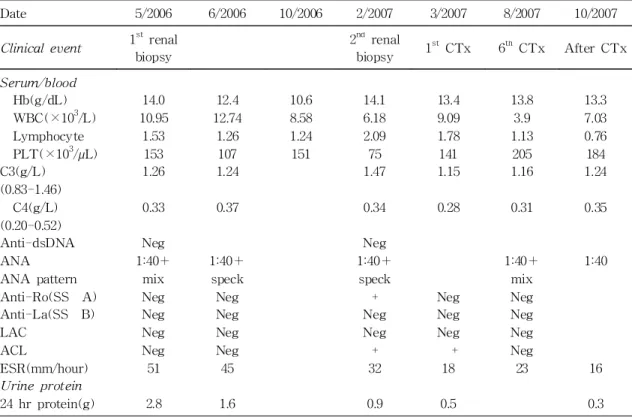
and her family had no remarkable medical hi- story or symptoms. Her blood pressure was 115/65 mmHg and her chest X-ray, electro- cardiogram, and abdominal ultrasonography were unremarkable. Initial laboratory findings were not remarkable except positive antinu- clear antibody(ANA), abnormal urinary find- ings and positive serum mycoplasma antibody (Ab)(1:160). The laboratory findings of the patient are shown in Table 1.
At first, the patient fulfilled only 2 of 11 criteria of the American Rheumatism Asso- ciation for SLE: renal disorder and antinuclear antibody. Renal biopsy showed endocapillary and mesangial proliferation under light mic- roscopy(LM)(Fig. 1A). Global and segmental
scleroses were observed in 6 and 4 glomeruli, respectively(Fig. 1B). The interstitium was diffusely widened by severe lymphoplasma- cytic and neutrophilic infiltrate. Focal tubulitis and minimal atrophy were also observed. Im- munofluorescence(IF) microscopy showed segmental granular IgG(++) and C3(+/-), staining along the peripheral capillary wall and granular deposits of IgG(++), C3(++), C4(+/-), and fibrinogen(+/-) in the mesangi- um(Fig. 1C). Electron microscopy(EM) show- ed mesangial expansion and swollen endo- thelium with subepithelial electron dense de- posits, along with diffuse foot process effa- cement and intramembranous electron dense deposition(Fig. 1D). Because mycoplasma Ab
Table 1. Laboratory Findings
Date 5/2006 6/2006 10/2006 2/2007 3/2007 8/2007 10/2007
Clinical event 1st renal biopsy
2
ndrenal
biopsy 1
stCTx 6
thCTx After CTx Serum/blood
Hb(g/dL) WBC(×10
3/L) Lymphocyte PLT(×10
3/µL) C3(g/L)
(0.83-1.46) C4(g/L) (0.20-0.52) Anti-dsDNA ANA ANA pattern Anti-Ro(SS‐A) Anti-La(SS‐B) LAC
ACL
ESR(mm/hour) Urine protein 24 hr protein(g)
14.0 10.95
1.53 153 1.26
0.33
Neg 1:40+
mix Neg Neg Neg Neg 51
2.8
12.4 12.74
1.26 107 1.24
0.37
1:40+
speck Neg Neg Neg Neg 45
1.6
10.6 8.58 1.24 151
‐
‐
‐
‐
‐
‐
‐
‐
‐
‐
‐
14.1 6.18 2.09 75 1.47
0.34
Neg 1:40+
speck + Neg Neg + 32
0.9
13.4 9.09 1.78 141 1.15
0.28
‐
‐
‐ Neg Neg Neg + 18
0.5
13.8 3.9 1.13 205 1.16
0.31
‐ 1:40+
mix Neg Neg Neg Neg 23
‐
13.3 7.03 0.76 184 1.24
0.35
‐ 1:40
‐
‐
‐
‐
‐ 16
0.3
Abbreviations : CTx, chemotherapy; WBC, White blood cell count; PLT, Platelet count; Anti-dsDNA,
Anti-double stranded DNA; ANA, Antinuclear antibodies; LAC, Lupus anticoagulant; ACL, Anticar-
diolipin antibodies; ESR, Erythrocyte sedimentation rate; Neg, negative; mix, mixed; speck, speckled
was positive, she was given enalapril(7.5 mg/
day) and oral Roxithromycin(150 mg/day), but proteinuria and hematuria persisted.
After 6 months, her heavy proteinuria per- sisted at 1.8 g/day and creatinine clearance was 52.79 ml/min. There was no interval change in mycoplasma Ab(1:160). We started Deflazacort(72 mg/day) and stopped enalapril due to a decreased glomerular filtration rate.
She subsequently developed right cervical lymphadenitis with fever and neck node biop- sy revealed subacute necrotizing lymphadeni- tis.
After two more months, she developed a malar rash and her platelet count decreased to
86× 10
3per microliter. Repeated immunologic assays were positive in anti-cardiolipin Ab, anti SS-A/Ro Ab and VDRL. At this point, she fulfilled 5 of 11 criteria of the American Rheumatism Association for SLE: malar rash, renal disorder, antinuclear Ab, hematologic disorder (thrombocytopenia), and immunologic disorder. Second renal biopsy was performed 9 months after the first biopsy. Nine of 35 glomeruli(26%) were globally sclerotic and 17 (49%) showed synechia to Bowman's capsule.
Mesangial proliferation was persisted and the glomerular basement membrane was thicken- ed. The tubules show mild atrophy. Interstitial inflammation was diffuse and severe(Fig. 2A).
Fig. 1. (A) The first renal biopsy findings. Renal biopsy shows
endocapillary and mesangial proliferation, and the interstitium is
diffusely widened by severe lymphoplasmacytic and neutrophilic
infiltrate(×100). (B) Segmental sclerosis is observed(×200). (C)
Immunofluorescence microscopy shows segmental granular
deposition of IgG(++) along the peripheral capillary wall and
in the mesangium(×400). (D) Electron microscopy shows me-
sangial expansion, swollen endothelium with subepithelial elec-
tron dense deposits, diffuse effacement of foot process and in-
tramembranous electron dense deposition.
IF microscopy showed no glomeruli. EM revealed subepithelial electron dense deposits, increased mesangial matrix, basement mem- brane thickening, and focal foot process effacement(Fig. 2B). Findings of the second biopsy led to a diagnosis of class IV lupus ne- phritis(LN), and she was treated with pulse methylpredinisolone(500 mg/d intravenously for 3 consecutive days), followed by oral deflazacort and monthly intravenous cyclopho- sphamide pulse(1 g/m
2) for 6 months.
After cyclophosphamide pulse therapy, 24hr urinary protein levels decreased, platelet count increased, and anti-cardiolipin and anti SS-A/
Ro Abs were negative.
DISCUSSION
In this report, we describe a female patient with SLE who initially presented with protei- nuria and hematuria. Initial renal biopsy re- vealed suspected postinfectious glomerulone- phritis.
Renal involvement in SLE is known to pre- sent as a variety of morphological lesions.
However, certain features are usually found
only in LN, such as a fullhouse IF pattern (i.e., simultaneous detection of IgA, IgG, IgM, C1q, and C3 deposits), cytoplasmic tubuloreti- cular inclusions(TRI) on EM, and membranous nephropathy with mesangial deposits[6]. Gian- viti et al.[7] reported three patients who pre- sented with a glomerulopathy suggestive of LN without any other clinical findings of SLE.
Nakahara et al[8]. reported an 11-year-old girl who was found to have proteinuria by mass urinary screening and developed SLE 21 mon- ths later. They concluded that endothelial TRI on EM is a more significant early sign of SLE than "full-house" IF pattern, especially in pe- diatric cases. We reviewed the first renal bi- opsy findings again. There was no full-hou- se IF pattern, but we could not absolutely rule out LN based on pathologic findings.
In Korea, we perform annual mass urinary screenings so we can detect hematuria or pro- teinuria relatively early before patients develop clinical symptoms of lupus.
After 6 months, our patient presented with cervical necrotizing lymphadenitis. It has been reported that Kikuchi's disease(KD)-like lym- phadenitis occurs in SLE patients[9]. The co-
Fig. 2. (A) The second renal biopsy findings. The glomeruli are globally sclerotic and show synechia to Bowmans capsule.
(B) Electron microscopy shows subepithelial electron dense
deposits and increased mesangial matrix, basement membrane
thickening and focal effacement of foot process.
existence of lymphadenitis with SLE is mainly seen in necrotizing type KD. So the possibility of SLE should always be considered if a lym- ph node looks like the necrotizing type of KD.
The laboratory finding of this patient needs differentiation from some other connective tis- sue disease. Especially positive finding in anti SS-A/Ro Ab, antinuclear Ab can suggest Sjogren syndrome. The diagnosis of Sjogren syndrome is based on clinical features suppor- ted by biopsy of lip or glands but this patient had no ocular or oral symptom related to exo- crine disease.
And Arbuckle et al.[10] investigated the on- set and progression of autoantibody develop- ment before clinical diagnosis. They reported that some autoantibodies(antinuclear, anti-Ro, anti-La, and antiphospholipid Abs) usually precede the onset of SLE by many years.
Others(anti-Sm and antinuclear ribonucleo- protein Abs) typically appear only months be- fore diagnosis[10,11].
In conclusion, the presenting manifestations of SLE in children are diverse[12], so if ab- normal urinalysis and renal pathologic findings are persistent, LN should be kept in mind, and urinalysis and immunologic markers should be serially monitored to avoid a delay in diag- nosis. If abnormal immunologic findings are found on follow-up, patients should be routi- nely evaluated for the emergence of clinical features. Because delayed diagnosis of SLE might lead to an unfavorable outcome, early detection and treatment with steroidal and/or immunosuppressive agents may be important to minimize organ damage in children with LN.
한 글 요 약
학교 집단 요 검사 이상으로 추적검사 중 전신 홍반 루푸스로
진단된 1예
연세대학교 의과대학 소아과학교실, 병리학교실*