H2-M3의 세포 표면 발현이 NK 세포의 활성에 미치는 영향 분석
이상열·전태훈*,#
경원대학교 자연과학대학 생명과학과, *고려대학교 생명과학대학
(Received February 27, 2009; Revised March 17, 2009; Accepted May 11, 2009)
The Cell Surface Expression of H2-M3 Does Not Directly Effect on the Killing Activity of NK Cell
Sang Yeol Lee and Taehoon Chun*,#
Department of Life Science, College of Natural Sciences, Kyungwon University, 461-701, Korea
*College of Life Sciences and Biotechnology, Korea University, 136-701, Korea
Abstract
— H2-M3 (M3) is a unique antigen presenting molecule which provides N-formylated peptide to certain type of T cells. Previous observation indicated that NK cell activity is significantly diminished during listerial infection in H2-M3
-/-mice.
To explore the possibility that M3 expression directly effect on NK cell activity, we measured NK cell activity with or with- out stimulation of N-formylated peptide on antigen presenting cells. Results indicated that the expression of M3 is not directly influence on NK cell activity. Further study will be focused on the indirect effect of M3 on regulating NK cell activity.
Keywords □
antigen presenting molecule, H-2M3 (M3), Major histocompatibility complex molecule, NK cell, N-formy- lated peptide
주요조직적합성항원
(MHC; major histocompatibility complex)
은구조와기능에따라두가지로나뉜다
.
그중class Ia
는8
개 에서11
개로구성되어진peptide
항원을세포독성T
림프구에 전달해주며, class Ib
는기존의peptide
항원이아닌각각의독특한항원을인식하여각기특이적인
T
림프구에전달해준다.
1)H2-M3(M3)
는주요조직적합성항원class Ib
로분류되며생쥐(mouse)
에서그기능이발견되어진매우독특한항원인식물질이다
. M3
의경우항원인식부위가N-formylated peptide
에매우높 은특이성을가지고있으며,
이러한항원에대한반응성은어떠한항원인식물질도흉내내지못하는것이다
.
2)생체내에서N- formylated peptide
는마이토콘드리아나세균의단백질에서발견 되며,
따라서항원인식물질로서M3
의기능은세균의감염시세균특이적항원을
T
림프구에전달해주는것으로생각되어진다.
3)M3
의 발현은 주요항원인식세포(professional antigen pre- senting cell)
인수지상세포(dendritic cell),
대식세포(macrophage),
미성숙
(immature) B
림프구의표면에선택적으로발현되는것으로알려졌으며
,
따라서발현양상은MHC class II
물질과유사한것으로알려져있다
.
4)M3
가항원을전달해주는T
림프구는주로세포독성
T
림프구(cytotoxic T lymphocyte)
로알려져 있으며,
지금까지알려진M3
에결합하는항원은mitochondria
에서파생된항원과감염성세균에서파생된항원으로나뉜다
.
5)그중감염성세균에서파생된항원은리스테리아와결핵균에서 발견되었다
.
6,7)M3
에결합하는외부항원인리스테리아와결핵균에서파생된항원을인식하는
T
림프구는실제로리스테리아와결핵균감염 시이러한병원성세균을죽일수있고,
대표적인Th1 type
싸이토카인인
IFN-
γ를방출할수있다. M3
에의해항원을인식하고그에따라활성이일어나는세포독성
T
림프구의중요성에 대해서는리스테리아감염모델에서자세히연구되었다.
흥미로운것은리스테리아감염시
MHC class Ia
물질이결핍된생쥐에서는리스테리아에대한저항성이정상생쥐와비교하여전혀 저하되지않는다는것이다
.
이러한원인으로아마도리스테리아감염시
M3
에의해활성이일어나는세포독성T
림프구의역할이중요하기때문일것으로생각되어진다
.
8)또한리스테리아 감염시M3
에의해활성이일어나는세포독성T
림프구는MHC class Ia
물질에의해 활성이일어나는세포독성T
림프구보다먼저활성이일어나는것으로알려졌다
.
9)M3
의항원인식과이에따른세포독성T
림프구의활성이리#본논문에관한문의는저자에게로
(
전화) 02-3290-3069 (
팩스) 02-3290-3507
(E-mail) tchun@korea.ac.kr
스테리아감염시에중요한역할을한다는결정적인증거는
M3
결핍생쥐를이용한리스테리아감염모델에서입증되었다
.
10)리스테리아감염시
M3
결핍생쥐는리스테리아에상당한감수 성을나타내는것으로보여졌고,
이러한결과는M3
에의해활성이일어나는세포독성
T
림프구가결핍되어전체적인세포독 성T
림프구의활성에영향을끼치기때문이라고알려졌다.
10)더 욱이리스테리아감염시M3
결핍생쥐는NK
세포의활성도떨어지는것으로알려졌다
.
10)NK
세포의활성은활성증진신호와활성감소신호의두가지신호체계에의해조절된다
.
활성증진신호와활성감소신호가동시에
NK
세포에전달되면NK
세포는활성이일어나지않지만 활성증진신호만이NK
세포에전달되면NK
세포는활성이일 어나target
세포의세포사멸을유발한다.
11)지금까지발견된NK
세포의활성감소신호를전달하는물질중생쥐의
Qa-1
과인간의
HLA(human leukocyte antigen)-E
는MHC class Ib
로구분된다
.
12,13)그러나다른MHC class Ib
물질이NK
세포의활성감소신호를전달하는가에대해서는알려진바없다
.
앞에서언 급했듯이리스테리아감염시M3
결핍생쥐는NK
세포의활성도가떨어지는것으로알려져있으며
,
이러한원인을규명하기위해본연구에서는
M3
의발현과NK
세포에활성의연관성에 대해알아보았다.
실험방법
실험재료
정제된 항
M3
항체(mAb 130 clone)
4)와LemA(fMIGWII)
6)and TB4(fMFLIDV)
7)peptide
는Chung-Ru Wang
박사(North- western Unviersity, USA)
가제공하였다.
각각의항체isotype control
과secondary antibody
는BD Pharmingen(USA)
에서구 입하였다.
이외언급하지않은모든시약은Sigma(USA)
에서구입하였다
.
세포배양
생쥐
lymphoma
세포주인YAC-1
과생쥐macrophage
세포주 인P388D1
은ATCC(USA)
에서분양받아실험에사용하였다. P388D1
세포주는10% fetal bovine serum(FBS, Hyclone, USA)
을함유한DMEM(Invitrogen, USA)
배지에서37
oC, 5%
CO
2분압조건으로배양하였다. NK
세포확립Lymphokine activated killer cell(LAK)
세포는C57BL/6(B6)
비장세포를
10% fetal bovine serum(FBS, Hyclone, USA)
와500 Unit/m
l의재조합생쥐IL-2(BDPharmingen, USA)
를함유 한DMEM
배지에서5
일간세포배양하여얻었다.
14)배양된NK
세포는
T
세포를인식하는항체(anti-TCR
β)
와NK
세포표면인자를인식하는항체
(anti-NK1.1)
로염색하여유세포 분석기(flow cytometry)
에서분석하였다.
그후, NK
세포를B6
생쥐NK
세 포표면인자를인식하는항체가붙어있는microbead(Miltenyi Biotech, Germany)
를사용하여분리하고세포독성측정(cyto- toxicity assay)
에effector
로사용하였다.
또한poly(I : C) activated killer cell
을얻기 위해B6
생쥐에200
µg
의poly(I : C)(Sigma,
USA)
를복강내주사하고20
시간후에비장에서세포를분리하였다
.
그후DMEM
배지에서1
시간배양후, nonadherent cell
을
effector
로사용하였다.
15)이때, T
세포를인식하는항체(anti- TCR
β)
와NK
세포표면인자를인식하는항체(anti-NK1.1)
로염 색하여유세포분석기(flow cytometry)
에서분석하였다.
세포독성측정
세포독성측정은
lactase dehydrogenase(LDH)
방출을이용한non-radioactive cytotoxicity assay(Promega, USA)
를사용하여측정하였다
.
우선effector
와target cell
을count
하여ratio
를정 하였다.
그 후U bottom 96 well plate
에target cell(1×10
4cells)
과ratio
를정한effector(LAK
와poly(I : C) activated killer cell)
를넣고4
시간동안37
oC, 5% CO
2분압조건으로배양하였 다.
이때total volume
은200
µl로하였다.
그후,
상층액50
µl 를LDH substrate
와반응시켜서absorbance 490 nm
에서상대적인세포독성을측정하였다
.
유세포분석기
(flow cytometry)
분석YAC-1
세포주와P388D1
세포주를유세포분석기로분석하기 위해PBS
로3
번세척하였다.
그후,
세포를2% fetal bovine serum
을함유한PBS 50
µl에현탁한후, 4
oC
에서anti-M3
항체에
30
분간방치하였다.
그후세포와항체가들어있는반응액 을PBS
로3
번 세척하였고secondary antibody
인 항FITC- conjugated hamster-IgG
를동일한조건에서반응시켰다. Flow cytometry
분석은FACSCalibur(BD Pharmingen, USA)
를이용 하였고PBS 400
µl에세포를현탁하여M3
의세포표면발현을측정하였다
.
실험결과 및 고찰
NK
세포확립및기능고찰본연구는
MHC class Ib
물질중의하나인M3
의발현이NK
세포활성에직접적으로영향을미치는가에대한고찰이다
.
우 선NK
세포를확립하기위해LAK
와poly(I : C) activated killer cell
두종류의NK
세포를확립하였다. NK
세포를확립하는방법은지금까지여러가지방법이개발되었는데
,
본연구에서사 용되어진방법은고전적인방법인생쥐비장세포에다량의IL-2
(500 unit/m
l)
를처리하는방법14)과생쥐의복강내poly(I : C) (200
µg/mouse)
를주사하는방법을사용하였다.
15)따라서한가지방법에서파생된
NK
세포보다좀더다양한조건에서파생 된NK
세포를사용하여M3
가NK
세포활성에미치는영향을평가하였다
.
고전적인
NK
세포확립방법인생쥐비장세포에다량의IL-2 (500 unit/m
l)
를처리하는방법에의해만들어진LAK
는두가지표현형으로나뉜다
.
즉NK1.1
+TCR
+처럼T
세포수용체(T cell receptor)
를가진LAK
와일반적인NK
세포의표현형을가진NK1.1
+TCR
-의두가지표현형으로나뉜다.
16)실험결과Fig. 1
에서보이는것같이
NK1.1
+TCR
- 세포형과NK1.1
+TCR
+세포 형이거의1 : 1
로파생되는데, NK1.1
+TCR
+세포형의경우대 부분CD8
+CD4
-세포형을가지며,
이때CD8
+세포형은CD8
αβheterodimer
를발현한다.
16)Poly(I : C)
의경우toll like receptor- 3(TLR-3)
의리간드로서강력한Th1
반응을유발한다.
17)또한1990
년대부터Poly(I : C)
를생쥐에주사하여NK
세포의활성을높이고있다
.
15)Poly(I : C)
를생쥐에주사하여얻어진비장세포 의NK
세포와T
림프구의분포도는Fig. 1
에서보이는것같이, Poly(I : C)
를주사하기전생쥐의NK
세포와T
림프구의분포도와비교해별다른차이가없으며
, Poly(I : C)
에의해자극받 은nonadherent cell
은세포독성을나타내는것으로알려졌다.
17)이러한두가지
NK
세포들을확립한후각각의NK
세포의활성을고찰하기위해세포독성실험을
YAC-1
세포주와P388D1
세포주로나누어실행하였다
. YAC-1
세포주는생쥐lymphoma
세포주로
NK
세포들의세포독성실험에가장널리사용되는세포주이며
, P388D1
은생쥐대식세포주로서대식세포는주요항원인식세포로
M3
를발현할수있는세포이다. LAK
의경우세포독성실험에
NK
세포표면인자(NK1.1
+)
를인식하는항체가붙어 있는
microbead
를 사용하여 분리 하여,
두 가지 세포형(NK1.1
+TCR
- 세포형과NK1.1
+TCR
+세포형)
을모두사용하였다
. Poly(I : C)
에의해활성화한NK
세포는nonadherent cell
을effector
로사용하였다.
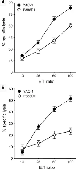
17)Fig. 2A, B
에서보는것같이두가지NK
세포들은YAC-1
세포주와P388D1
세포주모두에게세포 독성을나타내었으며, YAC-1
세포주를P388D1
세포주보다더효과적으로살해하였다
.
또한LAK
의경우percent specific lysis
가거의
80~90%
를보였는데반해, Poly(I : C)
에의해활성화한NK
세포의경우percent specific lysis
가60%
를보였다.
따라서LAK
가Poly(I : C)
에의해활성화한NK
세포보다세포독성에 대한활성이높았는데,
이러한이유는LAK
의경우NK
세포표 면인자만을발현하는세포를effector
로사용했기때문일것이다.
Fig. 1 −
Establishment of LAK and poly I : C activated killer cells.
Generated LAK and poly I : C activated killer cells were stained with antibody against TCR
βchain and NK1.1.
Numbers indicate percentages of cells in the corresponding quadrant. The results are representative of three separate experiments.
Fig. 2 −
The function of LAK and poly I : C activated killer cells.
Percent specific lysis of target cells (YAC-1 and P388D1)
were measured to access the function of LAK (A) and poly
I : C activated killer cells (B). Three independent experi-
ments were done and results are shown as mean±SE.
M3
발현이NK
세포활성에미치는영향고찰생체내면역반응에서
NK
세포는두가지중요한역할을한다
.
첫째로,
감염초기에병원성미생물에대한생체내저항성 을나타내며,
둘째로암세포를죽이는역할을한다.
따라서최근에는
NK
세포의활성조절을이용한면역요법들이활발히연 구되어지고있다.
최근에는몇몇MHC class Ib
물질,
특히생 쥐의Qa-1
과 인간의HLA(human leukocyte antigen)-E
가NK
세포의활성감소신호를전달하는물질들임이밝혀졌다
.
12,13)그 러나아직M3
와NK
세포의활성에대한연구는이루어진바 없다. Fig. 2
에서 보는 것 같이LAK
와poly(I : C) activated killer cell
은target
세포들인YAC-1
세포주와P388D1
세포주를 효과적으로살해할수있다.
따라서,
두가지세포주에서우선M3
의항원인 N-formylated peptide
를처리한후M3
의세포표면발현을유도한후
, M3
의발현유무가이들NK
세포들의세 포독성활성에미치는영향에대해서알아보았다.
실험에 사용되어진 N
-formylated peptide
는LemA
와TB4 peptide
로LemA peptide
는listeria
에서파생된항원으로M3
에 가장높은affinity
를가진항원이다.
6)Fig. 3
에서보는것과같이LemA
를처리하였을시에는M3
의세포표면발현을유발한다.
또한
TB4 peptide
는결핵균에서파생된항원으로LemA
보다는M3
에대한affinity
가높진않지만, TB4 peptide
역시M3
의세포표면발현을유발한다
(Fig. 3).
7)흥미로운점은LemA
나TB4 peptide
가YAC-1
세포주에서는M3
의세포표면발현을못시킨 다는것이다(Fig. 3).
지금까지알려진M3
의세포표면발현은주요항원인식세포들인수지상세포
,
대식세포,
미성숙B
림프구 들에서만관찰되었다.
따라서본연구에서는처음으로YAC-1
세포주에
high affinity
N-formylated peptide
를제공해도M3
의발현이거의유도가안된다는점을처음으로밝혔다
.
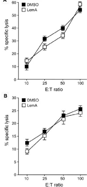
그반면Fig. 4 −
The induction of M3 on cell surface of target cell does not influence cytotoxic activity of NK cells. P388D1 cells were incubated with LemA peptide (10 mM) or negative control (0.1% DMSO) for overnight. Percent specific lysis of target cells (DMSO and LemA) were measured to access the cytotoxic activity of LAK (A) and poly I : C activated killer cells (B). Three independent experiments were done and results are shown as mean±SE.
Fig. 3 −
Induction of M3 on the cell surface by N -formylated
peptides. (A) YAC-1 and P388D1 cells were incubated with
LemA (fMIGWII) and TB4 (fMFLIDV) peptides at a
concentration of 10 mM overnight. Then, Flow cytometric
analysis was assessed the induction of M3 on the cell
surface. The results are representative of three separate
experiments. (B) YAC-1 and P388D1 cells were incubated
with LemA and TB4 peptides with varying concentrations
of M3-binding peptides. The range of concentrations and
the corresponding hatchmarks are shown. Bars represent
mean fluorescence intensity after staining with anti-M3
antibody as described. Three independent experiments
were done and results are shown as mean±SE.
대식세포주인
P338D1
에서는Fig. 3B
에서보는것같이농도의존적으로
M3
의세포표면발현이유도되었다(Fig. 3).
그후
, P388D1
에high affinity
N-formylated peptide
인LemA
를처리하고
, LAK
와poly(I : C) activated killer cell
의세포독성효능을측정하였다
.
이때음성대조구로는0.1% DMSO
를사용 하였다(Fig. 4).
실험결과그림4
에서보는것같이DMSO
를처리한세포와
LemA
를처리한세포주의세포독성효능은별다른차이가없음이나타났다
.
결국M3
의발현은NK
세포의활성에 직접적으로영향을끼치지는않는것으로사료된다.
10)이러한결과로미루어볼때
, M3
유전자적중생쥐의리스테리아감염시NK
세포의활성이감소하는것은T
림프구의활성이줄어들어보이는간접적인결과로생각된다
.
앞으로는M3
유전자적중 생쥐의리스테리아감염시, NK
세포의활성을조절할수있는Th1 type
주요cytokine
인IFN-
γ또는IL-12
의발현수준을살 펴볼예정이다.
결 론
본연구에서는N
-formylated peptide
를세포독성T
림프구에전달해주는
M3
의발현이NK
세포의활성에어떤영향을주는 가에대해고찰하였다.
그결과M3
의발현은NK
세포의활성에직접적으로영향을끼치지는않는것으로사료된다
.
이러한결과로미루어볼때
, M3
유전자적중생쥐의리스테리아감염시
NK
세포의활성이감소하는것은간접적인결과로생각된다
.
앞으로는M3
유전자적중생쥐의리스테리아감염시, NK
세포의활성을조절할수있는
Th1 type
주요cytokine
인IFN-
γ또는
IL-12
의발현수준을살펴볼예정이다.
감사의 말씀
이논문은
2006
년도한국학술진흥재단의지원에의해서연구되었음에감사를드립니다
(KRF-2006-311-E00282).
참고문헌
1) Rodgers, J. R. and Cook, R. G. : MHC class Ib molecules bridge innate and acquired immunity. Nat. Rev. Immunol.
5, 459 (2005).
2) Lindahl, K. F., Byers, D. E., Dabhi, V. M., Hovik, R., Jones, E. P., Smith, G. P., Wang, C. R., Xiao, H. and Yoshino, M. : H2- M3, a full-service class Ib histocompatibility antigen. Annu.
Rev. Immunol.
15, 851 (1997).
3) Busch, D. H., Kerksiek, K. and Pamer, E. G. : Processing of Listeria monocytogenes antigens and the in vivo T-cell response to bacterial infection. Immunol. Rev.
172, 163 (1999).
4) Chiu, N. M., Chun, T., Fay, M., Mandal, M. and Wang, C. R. : The majority of H2-M3 is retained intracellularly in a peptide- receptive state and traffics to the cell surface in the presence of N-formylated peptides. J. Exp. Med.
190, 423 (1999).
5) Colmone, A. and Wang, C. R. : H2-M3-restricted T cell response to infection. Microbes Infect.
8, 2277 (2006).
6) Lenz, L. L., Dere, B. and Bevan, M. J. : Identification of an H2- M3-restricted Listeria epitope: implications for antigen presentation by M3. Immunity
5, 63 (1996).
7) Chun, T., Serbina, N. V., Nolt, D., Wang, B., Chiu, N. M., Flynn, J. L. and Wang, C. R. : Induction of M3-restricted cytotoxic T lymphocyte responses by N-formylated peptides derived from Mycobacterium tuberculosis. J. Exp. Med.
193, 1213 (2001).
8) Seaman, M. S., Wang, C. R. and Forman, J. : MHC class Ib- restricted CTL provide protection against primary and secondary Listeria monocytogenes infection. J. Immunol.
165, 5192 (2000).
9) Kerksiek, K. M., Busch, D. H., Pilip, I. M., Allen, S. E. and Pamer, E. G. : H2-M3-restricted T cells in bacterial infection:
rapid primary but diminished memory responses. J. Exp. Med.
190
, 195 (1999).
10) Xu, H., Chun, T., Choi, H. J., Wang, B. and Wang, C. R. : Impaired response to Listeria in H2-M3-deficient mice reveals a nonredundant role of MHC class Ib-specific T cells in host defense. J. Exp. Med.
203, 449 (2006).
11) Biassoni, R. : Natural killer cell receptors. Adv. Exp. Med. Biol.
640