역류성 식도염 랫트에 미치는 金銀花 물 추출물의 치료 효과
이영준․박지하1․노성수1*
대구한의대학교 한의과대학 한방예방의학교실, 1 : 본초학교실
Effects on Rats with Reflux Esophagitis Treated with Lonicerae Flos Extract
Young Jun Lee, Ji Ha Park1, Seong Soo Roh1*
Department of Clinical Laboratory Science, 1 : Department of Herbology, College of Oriental Medicine, Daegu Haany University,
Because Lonicerae Flos has effects of antiinflammatory and antioxidant, we studied an effect of Lonicerae Flos on reflux esophagitis (RE) through those effects. Rats were treated with three different dosages of LF (500, 250 and 125 mg/kg) orally for 14 days before pylorus and forestomach ligation. Six hrs after pylorus and forestomach ligation, we dissected a stomach and examined a stomach volume, gastric acid output, pepsin release in the stomach, total hexose, sialic acid in stomach tissue and histamine contents of sera. The results were compared with an α-tocopherol (once orally, 1hr before operation, 30 mg/kg) treated group in which the effects on RE were already confirmed.
Lonicerae Flos extract (LE) reduced gastric volumes compared to RE control. This indicate that LE protect a stomach mucosa by depressing of gastric acid release and corresponse with a reducing histamine content of serum. And LE decreasd a volume of pepsin in stomach compraed to RE control, LE increased contents of total hexose and sialic acid based on esophageal and gastric mucus. This indicated that an increased mucus by LE protected inflammation of esophagus mucosa and gastric mucosa induced by gastric acid. So, LE suppressed a gasric acid by decreasing a pepsin release in stomach, suppressed an injury of esophagus inducted by gastric acid with increasing esophageal mucus and a minimum dose of LE to RE was 250 mg/kg. The results suggest that antioxidant effects of LF could attenuate the severity of reflux esophagitis and prevent the esophageal mucosal damage, and validate its therapeutic use in esophageal reflux disease.
Key words : Lonicerae Flos, reflux esophagitis, pepsin, hexose, sialic acid, histamine
* 교신저자 : 노성수, 경북 경산시 유곡동 대구한의대학교 2호관 306호
․E-mail : ddede@dhu.ac.kr, ․Tel : 053-819-1459
․접수 : 2010/09/28 ․수정 : 2010/11/03 ․채택 : 2010/12/01
서 론
역류성 식도염은 위 내용물 (주로 산과 펩신)이 식도로 역류 하여 속쓰림과 상복부 통증 등 다양한 임상 증상과 점막의 변화 를 가져오는 질환으로, 산에 노출되는 시간이 길수록 심한 병변 을 보이며 만성적인 경과를 밟는다. 전형적인 역류성 식도염은 식도의 과다한 산과 펩신의 노출이 주된 원인인 반면 비전형적인 역류성 식도염은 산과 펩신의 노출정도는 정상이나 식도의 감각 이 이에 비정상적으로 과민해져 발생하는 것으로 알려져 있다 1) . 역류성 식도염의 치료는 일반적으로 생활습관의 조절이 가 장 기본적인 치료법이다. 즉, 과식을 피하고, 중력을 이용하여 위
식도 역류를 억제하는 방법, 그리고 수면제, 칼슘 채널 길항제 등
의 복용을 금하는 등의 많은 방법이 있다 2) . 약물요법으로서는 보
편적으로 제산제 (antacids)나 위 장관 운동 조절제 (prokinetics)
를 가장 많이 사용해 왔다 2,3) . 이 후 histamine type 2 receptor
antagonist 및 proton pump inhibitor (PPI) 제제가 보다 더 효과
적으로 그 증상을 개선시키는 약물로 소개되면서, 많이 사용되고
있다 3,4) . 그 외 수술적인 방법이 있는데, 수술은 약물 치료에 반응
이 없는 경우이거나, 반응이 있어도 지나치게 자주 재발하는 경
우 및 합병증 (식도 협착, 하부식도 괄약근 이상 등)으로 인한 경
우에 사용되나, 여전히 부족한 점이 많다 4-6) . 역류성 식도염의 치
료에 있어 많이 사용되고 있는 histamine type 2 receptor
antagonist 및 PPI 제제는 모두 산 분비를 억제하는 약물로, 위
점막의 벽 세포에서의 산 분비량을 줄여 위산 역류시 발생되는
증상을 완화시키며, 손상된 식도 점막을 치유케 하고 합병증을
막아주는 역할을 한다. 그러나 최대 40-60%에 이르는 환자의 경 우는 충분한 기간의 산 분비 억제제의 투여에도 불구하고 증상 의 완전한 소실이 이루어지지 않으며 오히려 협착이 나 암 등의 합병증이 발생하기도 한다 2-6) .
金銀花는 인동과에 속한 다년생 木質藤本 인동 Lonicera japonica Thunb의 花蕾를 건조한 것으로 여름철 꽃이 피기 전에 채취하여 건조한다 7) .
性味는 寒하고 甘하며, 心, 肺, 胃經에 歸經하여 淸熱解毒, 凉散風熱, 凉血止痢의 效能으로 癰腫疔瘡, 喉痺, 丹毒, 熱毒血痢, 風熱感冒, 溫病發熱 等의 證狀에 常用되고 있다 7) .
약리작용으로는 항균, 항바이러스 작용으로 in vitro에서 황 색포도상구균, 용혈성연구균, 결핵구균, 백일해균 등 그람양성균 에 대해 억제작용이 있으며, 이질균, 장티푸스균, 대장균, 녹농균, 결핵균, 수막염균, 임균 등 그람음성균과 Leptospira에 대해서도 억제효과가 있다. 이러한 향균효과 성분으로는 caffeic acid, chlogenic acid, isochlogenic acid, Luteolin 등이다. 항내독소작 용이 있는데, 이는 세균의 내독소에 대한 활성을 일시적으로 억 제하는 것이 아니라 이에 대해 직접적으로 파괴하는 작용이 있 기 때문이다. 소염, 해열작용으로 金銀花는 염증의 삼출과 악화 를 억제하는데, 인동 총사포닌은 염증 초기의 모세혈관 투과성 증가나 염증성 삼출수종 등 여러 방법으로 유발한 염증반응에 대하여 억제작용이 있다. 면역 증강작용으로 金銀花 전탕액과 정 맥주사액은 백혈구의 탐식작용을 촉진하고 염증성 세포의 탐식 작용도 향상시키며 T림파구의 기능을 자극하여 림프구의 전화율 을 높인다 8) .
이외에 항산화작용 9) , 간보호작용 10) , 항암작용 11) , 항과민반응
12) 등이 연구 보고되었다. 이에 저자는 金銀花의 歸經이 胃經이 며, 식도는 胃經이 속하고 약리적으로 뛰어난 항염증작용과 항 산화작용이 있으므로, 金銀花가 역류성 식도염에 유효할 것으로 판단되었다.
이에 저자는 역류성 식도염 랫트 모델을 이용하여 치료효과 를 연구하였으며, 유의한 결과를 얻었기에 보고하는 바이다.
재료 및 방법
1. 재료 1) 동물
실험 동물은 SLC (Japan)에서 분양받은 6주령의 랫트를 2주 일 동안 실험실 환경에 적응시킨 후 실험에 사용하였다. 동물 사 육실의 조건은 conventional system으로 22±2℃, 1일 중 12시간 은 200~300 Lux로 조명하고, 12시간은 모든 빛을 차단하였다.
사료는 고형사료 (조단백질 22.1% 이상, 조지방 8.0% 이하, 조섬 유 5.0% 이하, 조회분 8.0% 이하, 칼슘 0.6% 이상, 인 0.4% 이상, 삼양사, 항생제 무첨가)와 물을 충분히 공급하였다.
2) 시료
본 실험에 사용한 金銀花 (Lonicerae Flos)은 옴니허브 (영 천) 제약회사에서 구입한 것을 대구한의대학교 한의과대학 본초 학교실에서 관능검사상 약전에 합격한 것만을 정선하여 사용하
였다.
2. 방법 1) 시료 추출
金銀花 (Lonicerae Flos;LF) 200 g에 증류수 1,000 ㎖를 가 하여 열탕 추출기에서 3시간 추출하여 얻은 액을 흡입 여과하고, 이를 감압 추출장치로 농축하였다. 위 과정을 3회 반복하였고, 3 회 농축한 것을 모아서 다시 동결 건조기를 이용하여 완전 건조 시켜 金銀花 추출물 (24 g, extract of Lonicerae Flos)을 제조하였 으며 수율은 12%였다. 이를 냉동 (-84℃) 보관하면서 적당한 농 도로 증류수에 희석하여 실험시에 사용하였다.
2) 역류성 식도염 랫트 모델
실험동물인 6주령의 랫트는 일본 SLC에서 공급 받아 실험 당일까지 고형사료 (항생제 무첨가, 삼양사료 Co., korea)와 물을 충분히 공급하고 온도 22 ± 2℃, 습도 40 ± 5%, 12시간-12시간 (light-dark cycle)의 환경에서 1주간 적응시킨 후 실험에 사용하 였다. 수술 전 24시간 절식한 후, Zoletile mixture (Vibrac, France)을 25 mg/kg 복강주사하여 마취하였다. 마취한 뒤, 2센 티 정도 개복을 하였고, 대만부위를 실크실로 묶었으며, 인접한 날문부위를 다른 실크실로 묶었다. 수술 후 봉합을 하고 회복 챔 버에 머무르게 한 후 케이지로 이동시켰다.
3) 시료 투여
랫트는 6마리씩을 한 군으로 하여 정상군 (Intact), 치료제를 처리하지 않은 대조군 (Re-C), α-tocopherol (30 ㎎/㎏) 처리군, LE 처리군 (500 ㎎/㎏, 250 ㎎/㎏, 125 ㎎/㎏)으로 나누어 약물 투여를 시작하였다. 정상군과 대조군은 생리식염수를 매일 1회 각각 매일 경구 투여하였고, 양성대조군은 α-tocopherol, LE 처 리군은 2주 동안 매일 오전 11시에 경구투여 하였다.
4) 위내용물 양 (Gastric volume)의 측정
Rao와 Vijayakumar의 방법 13) 에 따라, 유문 및 전위부 결찰 수술 6시간 후, 정상 대조군을 제외한 모든 실험동물에서 결찰 한 위부분에 형성된 내용물을 수집하여 5분간 2000 g에서 원심 분리한 다음, 하기의 공식과 같이 실험동물의 체중 kg으로 환산 하였다.
Gastric volume =
Collected amount of gastric contents × 1000 Body weight of animal (g)
5) 위산 분비도 (gastric acid output) 측정
Rao와 Vijayakumar의 방법 13) 에 따라, 원심분리한 위 내용물 에서 0.01 N NaOH와 phenolphthalein 을 발색제로 이용하여 위 산분비도를 μEq/6hrs로 측정하였다.
6) 펩신 분비도 측정
Sairam 등의 방법 14) 에 따라, 원심분리한 위 내용물에서 hemoglobin 을 이용하여 발색제로 이용하여 Pepsin 분비도를 μ mol of tyrosine/6hrs로 측정하였다.
7) Hexose 함량 측정
탄수화물에 Sulfuric acid 및 orcinole (5-metyl orcinole)를
반응시켜 유발시킨 발색 반응을 열량학적 (calorimetrically) 으로 측정하여, total hexose 함량을 측정하였다 15) . 즉, 0.2 ml의 식도 homogenate를 3.4 ml의 1.6% orcinol 용액과 4 ml의 60%
sulfuric acid를 혼합한 다음, 10분간 끓인 다음 냉각시키고, optical density를 흡광도 425 nm에서 측정하여, total hexose 함 량을 galactose-mannose 표준 곡선과 비교·측정하였다.
8) Sialic acid 함량의 측정
단백질에 결합되어 있는 sialic acid 함량은 thiobarbituric acid assay 법을 이용하여 Warren의 방법 16) 으로 측정하였다. 식 도 homogenate를 90% ethanol에 침전시킨 다음 침전물을 0.2 N sulphuric acid에 용해시켜, periodic acid로 37℃, 30분간 incubate하여 산화시키고, sodium arsenate, 6% thiobarbituric acid 및 cyclohexan을 첨가하였다. 이후 분홍색의 cyclohexane이 분리될 때까지 원심분리하고, 550 nm에서 흡광도를 측정하여, μ g/100 mg tissue 단위로 측정하였다.
9) 혈중 histamine 함량의 측정
최종 희생일에 안와 정맥총 (supraorbital plexus)에서 약 1 ml의 혈액을 heparin (180 unit/ml of blood; Sigma, MO, USA) tube에 채취하고, 원심분리하여, 혈장 (plasma)을 분리하였다. 분 리된 혈장에 0.2M perchloric acid를 첨가하여 4℃이하에서 10,000×g로 30분간 원심분리를 실시하였다. 이후 얻어진 상층액 에서 high performance liquid chromatography 17) 를 이용하여 IU/mg protein 단위로 histamine 양을 측정하였다.
10) 통계처리
모든 수치는 평균 ± 표준편차로 표시하였으며, 다중비교검 증을 이용하여 통계처리를 실시하였고, 분산동질성을 Levene test를 실시하여 검증 하였다. 등분산일 경우, one way ANOVA test를 실시한 다음 least-significant differences (LSD) test로 사 후 검증을 실시하여 실험군 간의 유의성을 측정하였다. 비등분산 일 경우에는 비모수 검증인 Kruskal-Wallis H test를 실시하여 유의성이 인정된 경우에는, Mann-Whitney U test 를 실시하여 실험군 간의 유의성을 검증하였다. 모든 통계처리는 SPSS for Windows (Release 14.0K, SPSS Inc., USA)를 이용하여 평가하였 으며, p-value가 0.05 이하인 경우 통계적 유의성을 인정하였다.
결 과
1. 위 내용물 양 측정
역류성 식도염 수술을 시행 한 후, 증류수만 투여한 대조군 은 20±1.51 ml/kg이었고, 토코페롤 투여군은 13.43±1.58 ml/kg, 金銀花 추출물 500 mg/kg 투여군은 8.66±1.58 ml/kg, 金銀花 추 출물 250 mg/kg 투여군은 13.41±2.62 ml/kg, 金銀花 추출물 125 mg/kg 투여군은 15.93±3.01 ml/kg으로 각각 감소되었다. 따라 서 항산화제인 토코페롤 투여군과 金銀花 추출물 투여군의 위 내용물 양은 증류수만 투여한 대조군에 비해 유의성있게 감소되 었다(Fig. 1).
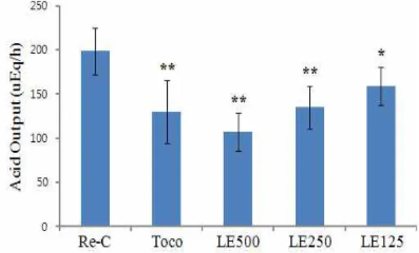
2. 위산 분비도 측정
역류성 식도염 수술을 시행 한 후, 증류수만 투여한 대조군 은 198.34±26.7 μEq/h이었고, 토코페롤 투여군은 129.67±35.88 μ Eq/h, 金銀花 추출물 500 mg/kg 투여군은 106.8±21.35 μEq/h, 金銀花 추출물 250 mg/kg 투여군은 134.6±24.76 μEq/h, 金銀花 추출물 125 mg/kg 투여군은 158.61±21.36 μEq/h로 각각 감소되 었다. 따라서 항산화제인 토코페롤 투여군과 金銀花 추출물 투여 군의 위산 분비도는 음성 대조군에 비해 유의성있게 감소되었다 (Fig. 2).
Fig. 1. Changes of the gastric volumnes of Re-C, α-tocopherol, LF 500, 250 and 125 mg/kg treated rats.
Re-C: Rats had a pylorus and forestomach ligation operation not treated with drug; Toco: Rats had a pylorus and forestomach ligation operation treated with α-tocopherol (30 mg/kg); and LF extract: Rats had a pylorus and forestomach ligation operation treated with extract of LF (respectively 125, 250 and 500 mg/kg). Values are expressed Mean ± SD of six rats; ** p<0.01 compared to RE control.Fig. 2. Changes of the acid output of Re-C, α-tocopherol, LF 500, 250 and 125 mg/kg treated rats.
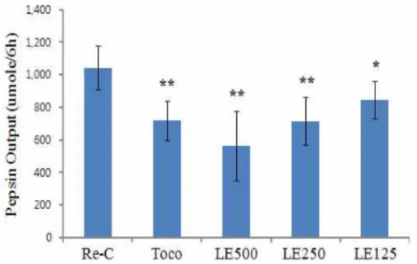
Values are expressed Mean ± SD of six rats; **p<0.01, *p<0.05 compared to RE control.3. 펩신 분비도 측정
역류성 식도염 수술을 시행 한 후, 증류수만 투여한 대조군 의 펩신 분비도는 1042.8±136.5 μmole/6h이었고, 토코페롤 투여 군은 718.1±123 μmole/6h, 金銀花 추출물 500 mg/kg 투여군은 561.8±211.1 μmole/6h, 金銀花 추출물 250 mg/kg 투여군은 713.9±146.1 μmole/6h, 金銀花 추출물 125 mg/kg 투여군은 844.6±116.1 μmole/6h로 각각 감소되었다. 따라서 항산화제인 토코페롤 투여군과 金銀花 추출물 투여군의 위산 분비도는 음성 대조군에 비해 유의성있게 감소되었다(Fig. 3).
4. 위 보호 효과
1) Hexose 변화량 측정
개복만 하고 역류성 식도염 수술을 하지 않은 정상 랫트의 위조직 내 hexose 함량은 2736.5±843.8 μg/100 mg이었고, 역류 성 식도염 수술을 시행 한 하고 증류수만 투여한 대조군의 위 조 직 내 hexose 함량은 1036.6±167 μg/100 mg이었고, 토코페롤 투 여군의 hexose 함량은 1387±248.8 μg/100 mg, 金銀花 추출물 500 mg/kg 투여군은 1570.2±364.7 μg/100 mg, 金銀花 추출물 250 mg/kg 투여군은 1396.7±157.5 μg/100 mg, 金銀花 추출물 125 mg/kg 투여군은 1303.7±123.4 μg/100 mg으로 각각 감소되 었다. 따라서 항산화제인 토코페롤 투여군과 金銀花 추출물 투여 군의 hexose 함량은 음성 대조군에 비해 유의성있게 감소되었다 (Fig. 4).
Fig. 3. Changes of the pepsin output of Re-C, α-tocopherol, LF 500, 250 and 125 mg/kg treated rats.
Values are expressed Mean ± SD of six rats; **p<0.01, *p<0.05 compared to RE control.Fig. 4. Changes of the total hexose content in gastric tissue on Re-C, α-tocopherol, LF 500, 250 and 125 mg/kg treated rats.
Intact:Normal rats had a laparotomy not pylorus and forestomach ligation operation;
Re-C: Rats had a pylorus and forestomach ligation operation not treated with drug; Toco: Rats had a pylorus and forestomach ligation operation treated with α-tocopherol (30 mg/kg); and LF extract: Rats had a pylorus and forestomach ligation operation treated with extract of LF (respectively 125, 250 and 500 mg/kg).
Values are expressed Mean ± SD of six rats; **p<0.01 and *p<0.05 compared to RE control.; #p<0.05 and ##p<0.01 compared to Intact control.
2) Sialic acid 변화량 측정
개복만 하고 역류성 식도염 수술을 하지 않은 정상 랫트의 위조직 내 sialic acid 함량은 165.5±32.0 μg/100 mg이었고, 역류 성 식도염 수술을 시행 한 하고 증류수만 투여한 대조군의 위 조 직 내 sialic acid 함량은 54.04±14.5 μg/100 mg이었고, 토코페롤 투여군의 sialic acid 함량은 80.7±17.1 μg/100 mg, 金銀花 추출 물 500 mg/kg 투여군은 101.6±17.1 μg/100 mg, 金銀花 추출물
250 mg/kg 투여군은 80.7±19.8 μg/100 mg, 金銀花 추출물 125 mg/kg 투여군은 76.8±13.8 μg/100 mg으로 각각 감소되었다. 따 라서 항산화제인 토코페롤 투여군과 金銀花 추출물 투여군의 sialic acid 함량은 음성 대조군에 비해 유의성있게 감소되었다 (Fig. 5).
Fig. 5. Changes of the sialic acid content in gastric tissue on Re-C, α -tocopherol, LF 500, 250 and 125 mg/kg treated rats.
Intact: Normal rats had a laparotomy not pylorus and forestomach ligation operation; Re-C: Rats had a pylorus and forestomach ligation operation not treated with drug; Toco:Rats had a pylorus and forestomach ligation operation treated with α-tocopherol (30 mg/kg); and LF extract: Rats had a pylorus and forestomach ligation operation treated with extract of LF (respectively 125, 250 and 500 mg/kg). Values are expressed Mean ± SD of six rats; **p<0.01 and *p<0.05 compared to RE control.; #p<0.05 and ##p<0.01 compared to Intact control.
Fig. 6. Changes of the histamine content in serum on Re-C, α -tocopherol, LF 500, 250 and 125 mg/kg treated rats.
Intact: Normal rats had a laparotomy not pylorus and forestomach ligation operation; Re-C: Rats had a pylorus and forestomach ligation operation not treated with drug; Toco:Rats had a pylorus and forestomach ligation operation treated with α-tocopherol (30 mg/kg); and LF extract: Rats had a pylorus and forestomach ligation operation treated with extract of LF (respectively 125, 250 and 500 mg/kg). Values are expressed Mean ± SD of six rats; **p<0.01 and *p<0.05 compared to RE control.; #p<0.05 and ##p<0.01 compared to Intact control.
3) 혈중 histamine 변화량 측정
개복만 하고 역류성 식도염 수술을 하지 않은 정상 랫트의
혈중 histamine 함량은 179.8±40.8 IU였고, 역류성 식도염 수술을
시행 한 하고 증류수만 투여한 대조군의 혈중 histamine 함량은
448.8±91.6 IU이었고, 토코페롤 투여군의 혈중 histamine 함량은
293.1±91.5 IU, 金銀花 추출물 500 mg/kg 투여군의 혈중
histamine 함량은 231.2±52.7 IU, 金銀花 추출물 250 mg/kg 투여
군의 혈중 histamine 함량은 275.4±110.5 IU, 金銀花 추출물 125
mg/kg 투여군의 혈중 histamine 함량은 311.1±62 IU으로 각각
감소되었다. 따라서 항산화제인 토코페롤 투여군과 金銀花 추출
물 투여군의 sialic acid 함량은 음성 대조군에 비해 유의성있게 감소되었다(Fig. 6).
결론 및 고찰
위산은 역류성 식도염의 가장 중요한 유발인자 중 하나로 18) , 역류성 식도염 및 이와 관련된 위점막 손상을 치료하기 위해서 는 위의 pH 조절이 필수적이다 19) . 특히, 사람과 실험동물에서, histamine H2-receptorantagonists와 PPI 제제 등 위 분비 억제제 에 의해 역류성 식도염의 감소 효과가 이미 잘 알려져 있다 20) .
Pepsin은 분문위의 주세포에서 분비되는 소화효소의 일환으 로 벽세포에서 염산 분비를 촉진하여, 위의 pH를 감소시킨다 21) . 따라서 Pepsin 분비도는 역류성 식도염의 유발에 매우 중요한 위치를 차지하며, Pepsin의 분비감소는 역류성 식도염 치료에 주 표적이 되어왔다 22,23) .
역류성 식도의 정도는 위산에 대한 노출과 함께 식도벽을 구성하고 있는 collagen fiber의 유동성 과 장력의 변화 역시 중 요한 위치를 차지하고 있으며 24) , 역류성 식도염의 진행 정도를 판단하는 데 중요한 인자로 사용되고 있다 25) .
식도 점막부위에 존재하는 샘들은 점액을 분비하여, 식도 점 막의 습도와 연하운동을 유지시켜 준다. Sialic acid를 포함한 당 단백질이 이들 점액의 주요성분이며, 당단백질은 일반적으로 total hexose로 측정할 수 있어 26) , 식도 점막의 손상 정도를 간접 적으로 나타내 준다. 또한 Histamine에 의한 위산 분비 촉진에 의한 식도 점막 세포의 파괴는 이미 잘 알려져 있다 27,28) .
저자는 역류성 식도염에 대한 금은화의 효능을 알아보기 위 해, 양성대조군으로 토코페롤을 투여하였으며, 이는 토코페롤이 가장 잠재력있는 항산화제이며 생물학적 막에서 지질부분에서의 자유기 체인연결의 증식을 억제할 수 있고, 산화 스트레스로 인 한 위궤양을 예방할 수 있으며 29) , Chandana and Madhavan α -tocopherol이 식도염에 효과가 있다고 발표하였다 30) .
金銀花 추출물이 위 내용물 분비량을 대조군에 비해 유의성 있게 감소시켰으며(Fig. 1), 이는 위산 분비 억제를 통해 위점막 의 손상을 방지할 수 있다고 생각된다. 이는 점막의 염증을 억제 하여 혈중 histamine에 의한 위산분비 촉진을 억제시킨 결과와 도 부합되고 있다(Fig. 2, Fig. 6). 金銀花추출물이 위 점막을 보호 하는데 펩신의 분비가 대조군에 비해 감소된 점에서 알 수 있으 며(Fig. 3), 이 효소의 분비를 金銀花가 억제했다고 생각된다. 또 한 hexose, sialic aicd의 분비량 증가 결과로 보아(Fig. 4, Fig. 5), 식도점막을 보호할 수 있는 점액의 분비를 증가시켜 위산에 의 한 염증을 억제시켰다고 생각된다. 따라서 金銀花는 위 점막의 펩신 효소 분비를 감소시켜 위산 분비를 억제하였고, 식도에서 점액 분비를 증가시켜 위산에 의한 손상을 억제시켰다고 사료되 며 유효한 투여 용량은 최소 250 mg/kg으로 판단된다.
참고문헌