Sandalwood Essential Oil의 iNOS 발현과 염증성사이토카인의 생성에 미치는 영향
박정숙#·정숙희*
남부대학교 대체의학과, *남부대학교 향장미용학과
(Received November 15, 2012; Revised February 26, 2013; Accepted February 27, 2013)
Effects of Sandalwood Essential Oil on the iNOS Expression and Proinflammatory Cytokine Production
Jeoung-Suk Park# and Sook-Heui Jung*
Department of Alternative Medicine Nambu University of Gwangju 506-606, Korea
*Department of Cosmotology, Nambu University of Gwangju 506-606, Korea
Abstract — The present study was designed to determine the effect of the Sandalwood Essential Oil (Santalum album) on pro-inflammatory factors such as NO, iNOS expression and IL-1β, IL-6, TNF-α in lipopolysaccharide (LPS) - stimulated RAW264.7 macrophages cells. The cell toxicity was determined by MTS assay. To evaluate of anti-inflammatory effect of Sandalwood Essential Oil, amount of NO was measured using the NO detection kit and the iNOS expression was measured by western blot analysis and reverse transcriptase polymerase chain reaction (RT-PCR). And proinflammatory cytokines were measured by ELISA kit. As a result, Sandalwood Essential Oil reduced NO, iNOS expression and IL-1β, IL-6, TNF- α production without cytotoxicity. Our results suggest that the Sandalwood Essential Oil may have an anti-inflammatory property through suppressing inflammatory mediator productions and appears to be useful as an anti-inflammatory oil.
Keywords □ sandalwood essential oil, iNOS, IL-1β, IL-6, TNF-α
Sandalwood Essential Oil(Santalum album)은 단향과 (Santalaceae)에 속하는 12~15미터크기의 상록교목으로 India, Malaysia, Australia, Indonesia 등에서 자생하거나 재배하며,1) 연 한 노란색에서 노란색을 띤 점성액체로 매우 부드럽고 달콤한 나 무향과 약간의 발삼향이 나타나며 향의 지속성 때문에 상당한 시 간동안 일정한 향을 유지한다.2)인도는 Sandalwood Essential Oil의 주요생산국이며, 오일 제조를 위해서는 30년 이상 된 나무 의 심재만을 사용한다.3) Sandalwood Essential Oil의 연구보고 를 보면 아토피 피부염의 주 증상인 가려움증과 피부염증을 경 감시킨다는 보고와,4)항산화 효과,5)항균 효과,6)방등은 ICR 마 우스에게 Sandalwood Essential Oil처리시 세포내 reactive oxygen species(ROS)와 지질과산화도(Malondialdehyde, MDA) 가 억제됨을 보고하였다.7)윤 등8)은 급성산화증을 일으킨 마우 스에서 항산화 및 항염증 효과를 보고하였으며, 신 등은
Sandalwood Essential Mixed Oil의 피부병변과 염증, 아토피피 부염의 효과가9)있음을 보고하고 있다.
염증은 다양한 경로를 통한 외부 감염, 생체 내 대산산물 등 내외적 자극에 대한 생체 내 방어기전의 발현으로서, 세포 내 다 양한 염증 조절인자들이 매개체로서 관여하며, 알레르기, 아토피, 관절염, 심장병, 뇌질환, 순환기 장애뿐만 아니라 암 등과 같은 다양한 질환의 원인을 제공하게 된다.9)염증은 신체방어기전이 고, 신체방어 기전 중 가장 중요하며,10)염증의 증상으로는 홍반, 부종, 열, 통증 그리고 기능 상실 등이 나타난다.11)또한 염증반 응은 세균감염과 같은 외부 자극이나 생체내 대사산물과 같은 내 부자극에 대한 생체조직의 방어기전으로12)세포내 다양한 염증조 절인자들인 TNF-α, IL-1β, IL-6, IL-8 등과 같은 proinflammatory cytokines, prostagrandin, lysosomal enzyme, free radicals 등 다양한 매개물질이 관여한다.13)특히 대식세포에서 cytokines, tumor necrosis factor(TNF-α), lipopolysaccharide(LPS)와 같은 자극에 의해 염증 반응의 전사 인자를 활성화시키며 그 결과 inducible nitric oxide synthase(iNOS), cyclooxygenase-2(COX- 2)를 발현시켜 nitric oxide(NO)와 Prostaglandin E2(PGE2)를 생
#본 논문에 관한 문의는 저자에게로
(전화) 062-970-0167 (팩스) 062-970-0162 (E-mail) [email protected]
종설
성하여 염증을 일으킨다.14,15)또한 NO가 필요이상으로 생성되 면 염증 반응의 항진, 과도한 혈관 확장에 의한 패혈성 쇼크 유 발, 상처 치유의 억제, 신경조직의 손상 등을 일으켜 생체에 유 해한 작용을 나타낸다.16)다양한 염증성 질환에서 주된 치료제 인 스테로이드와 비스테로이드성 항 염증제(NSAIDs)가 사용된 다. 그러나 이 제제들은 좋은 항염증 효과도 있지만 부작용 발생 우려로 장기간 사용이 어렵다. 최근 이러한 제제들을 대신 할 수 있는 소재를 개발하기 위하여 천연물에서 유효성분을 얻으려는 노력들이 시도 되고 있다.17,18)
또한 최근에는 천연물에서 추출한 오일의 적용이 활발해지고 있으며 특히 Chamomile Essential Oil, Lavender Essential Oil 의 성분들은 소염작용, 진통효과, 면역증진작용, 강장 작용 등 다 양한 효과를 발휘하는 것으로 보고되고 있다.19,20)본 연구에서는 기능성소재로 사용되고 있는 Sandalwood Essential Oil의 항염 증성과 기초적인 생리활성에 관한 연구는 보고된 바가 없어 Sandalwood Essential Oil을 이용하여 RAW264.7 세포에서 NO 의 생성, iNOS의 단백질과 유전자 발현정도와 염증성 cytokine 인 IL-1β, IL-6, TNF-α 분비량 등을 조사하여 Sandalwood Essential Oil의 항염증성을 검토하고자 하였다.
재료 및 방법
시료
실험에 사용한 Sandalwood Essential Oil(Sandalum album)은 유기농 인증기관으로부터 인정받은(ECOCERT-F-32600) 100%
순도의 천연 에션셜 오일인 NEW DIRECTIONS LABORATORY LTD사(Australia) 제품을 사용하였으며 에탄올과 essential oil을 4 : 1로 먼저 희석한 후 세포배양용 배지에 희석하여 사용하였다.
시약
세포 배양액인 Dulbecco's Modified Eagle Medium(DMEM) 과 fetal bovine serum(FBS), streptomycin-penicillin 등의 세포 배양용 시약들은 Gibco BRL사(Grand Island, USA), Lipopoly- saccharide(LPS)는 Sigma-Aldrich사(St. Louis, MO, USA), Cell Titer 96® AQueous One Solution은 Promega사(Madison, USA) 에서 구입하여 사용하였다. IL-1β, IL-6, Tumor necrosis factor-α (TNF-α), ELISA kit는 eBioscience사(San Diego, CA, USA)에 서 구입하였고 NO(Nitric Oxide) detection kit는 iNtRON Biotechnology사(Suwon, Korea)에서 구입하여 사용하였다. Total RNA를 분리시 Easy Blue®시약(iNtRON Biotechnology, Korea)을 이용하였고 분리된 RNA를 QuantiTect® Reverse Transcription Kit(Qiagen, USA)를 구입하여 cDNA 합성에 사용하였다. 이외에 실험에 사용된 모든 시약은 Sigma Chemical사(St. Louis, MO, USA)에서 구입하여 모두 분석용 시약 특급을 사용하였다.
세포배양
실험에 사용한 마우스의 대식세포주인 RAW264.7 세포는 한 국세포주은행(KCLB)에서 분양 받았으며, 세포배양을 위해 10%
FBS와 1% penicillin-streptomycin을 포함하는 DMEM(Dulbecco's Modified Eagle Medium) 배지를 사용하였다. 세포는 CO2배양 기(37oC, 5% CO2)에서 배양하였다.
MTS assay
Sandalwood Essential Oil의 세포에 대한 독성은 Desai 등의 방법18)에 준하여 5-(3-caroboxymeth-oxyphenyl)-2H-tetrazolium inner salt(MTS) assay 방법으로 측정하였다. 이는 Mitochondrial dehydrogenases에 의하여 MTS가 formazan으로 전환되는 것을 측정하는 것으로 96 well plate에 1.0×10⁵cells/well의 RAW264.7 세포를 분주하고 18시간 동안 배양한 후 Sandalwood Essential Oil를 125 µg/ml, 250 µg/ml, 500 µg/ml 처리하고 24시 간 동안 배양하였다. 20 µl의 MTS solution을 첨가한 후 CO2배 양기(37oC, 5% CO2)에서 4시간 반응시킨 후, 450 nm에서 흡광 도의 변화를 측정하여 대조군에 대한 세포 생존율을 백분율로 표 시하였다.
Nitric oxide 측정
NO의 농도는 배양액 내의 nitrite농도를 Wang 등의 방법4019) 에 따라 Griess Reagent System을 이용하여 측정하였다.
RAW264.7 세포를 96 well plate에 1.0×10⁵cells/well이 되도록 분주하고 18시간 동안 배양한 후 Sandalwood Essential Oil를 125µg/ml, 250 µg/ml, 500 µg/ml 전처리하고 1시간 후에 LPS 10µg/ml 처리한 후 24시간 동안 배양하였다. 배양액과 동량의 Griess Reagent를 가하고 10분간 상온에서 반응시킨 후 540 nm 에서 흡광도를 측정하였다. sodium nitrite의 농도별 표준곡선을 이용하여 배양액의 NO 농도를 결정하였다.
Western blot analysis
전기영동을 위한 단백질 시료의 추출은 처리 시간 별로 세포 를 Ice-cold tris buffered saline(TBS; 20 mM Tris-HCI, pH 8.0, 137 mM NaCl)으로 3회 세척한 후, lysis buffer(TBS, 1%
NP-40, 1 mM sodium orthovanadata, 10µg/ml aprotinin, 10 µg/
ml leupeptin 및 1 mM PMSF)를 넣어 4oC에서 30분간 반응시 키고 12,000 rpm에서 10분간 원심 분리하여 상층액을 모았다. 동 일한 양의 단백질을 sodium dodecyl sulfate-polyacrylamide gel electrophoresis(SDS-PAGE)로 분리시킨 후, 단백질을 nitrocellulose membrane에 transfer하였다. 이 membrane을 항체의 비특이적 결함을 차단하기 위하여 blocking buffer(5% non-fat milk와 0.1% Tween 20을 함유한 TBS용액)에서 1시간 동안 반응시킨 후, 각 검증 단백질에 대한 항체(anti-iNOS)를 가하여 1~2시간
동안 반응시켰다. 이어서 0.1% Tween 20을 함유한 TBST 용액 으로 40분간 세척한 다음, secondary antibody로 반응시켰다. 이 어서 ECL system으로 반응시킨 후 X-ray film 상에서 단백질을 확인하였다. 각 시료의 단백질 정량은 Bradford protein assay kit를 사용하여 595 nm에서 흡광도를 측정하여 실시하였다.
Reverse transcription polymerase chain reaction(RT- PCR)
RAW264.7 세포에서 발현되는 iNOS의 유전자 발현에 대한 Sandalwood Essential Oil의 효과를 조사하기 위해 RT-PCR을 수행하였다. 먼저 RAW264.7 세포를 60 mm dish에 4.0×10⁶cells/
Well이 되도록 분주하고 18시간 동안 배양하였다. Sandalwood Essential Oil를 125 µg/ml, 250 µg/ml, 500 µg/ml 전처리하고 LPS 10µg/ml 처리 후 6시간 동안 배양하였다. 세포를 수거하여 4oC에서 2,000 rpm으로 5분간 원심분리한 후 Easy Blue®시약 (iNtRON Biotechnology, Korea)을 이용하여 total RNA를 분리 하였다. 분리된 RNA를 QuantiTect® Reverse Transcription Kit(Qiagen, USA)를 이용하여 cDNA를 합성하였다. 합성된 cDNA에 대한 PCR을 수행하기 위해 cDNA 1 µg에 primers (sense, anti-sense) 1µl 및 10×buffer(10 mM Tris-HCl, pH 8.3, 50 mM KCl, 0.1% Triton X-100), 250µM dNTP, 1U Tag polymerase를 혼합한 후 denaturation을 위해 94oC에서 45초, annealing을 위해 55~60oC에서 45초 및 extension을 위해 72oC 에서 60초 조건으로 30 cycles을 수행하였다. PCR 반응의 표준 대조구로 GAPDH를 사용하였다(Table I). 이후 증폭된 DNA산 물을 1.5% agarose gel를 사용하여 100 volt에서 30분간 전기 영 동하여 UV에서 관찰하였다.
세포배양액 내의 Cytokine 측정
RAW264.7 세포를 96 well plate에 1.0×10⁵cells/well이 되도 록 분주하고 18시간 동안 배양한 후 Sandalwood Essential Oil 를 125 µg/ml, 250 µg/ml, 500 µg/ml 전처리하고 1시간 후에 LPS 10µg/ml 처리한 후 24시간 동안 배양한 후 세포배양액을 얻은 다음 배양액에 함유된 IL-1β, IL-6, TNF-α을 ELISA kit을 이용하여 측정하였다.
통계적 분석
실험결과는 평균±표준편차(Mean±SD)로 계산하였고, 각군간
의 유의성 검증은 students' t-test를 사용하였다. p<0.05 일 경 우에 유의성이 있는 것으로 판정하였다.
결과 및 고찰
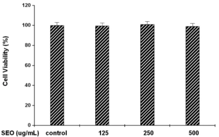
Sandalwood Essential Oil의 RAW264.7 세포에 대한 독성 Sandalwood Essential Oil의 세포독성을 보기 위하여 RAW264.7 대식세포에 Sandalwood Essential Oil을 125 µg/ml, 250µg/ml, 500 µg/ml 농도로 처리한 후 MTS assay를 실시하여 cell viability를 측정하였다. 실험 결과 각각 99.6±2.98%, 100.9±3.02%, 99.0±2.97%로 세포독성을 나타내지 않았다(Fig.
1). 이는 Sandalwood Essential Oil이 보여주는 항염효과가 세포 생존률의 감소가 아닌 시료의 약리활성으로 사료된다.
Sandalwood Essential Oil의 NO 생성에 미치는 영향 NO는 NO 합성효소에 의해 혈관이완, 혈액응고, 신경전달, 신 장 기능, 염증 및 항종양 기능 등의 다양한 생물학적 성질을 가 지는 것으로 알려져 있다.28)활성산소의 일종으로 최근 염증유 발에 중요한 역할을 하는 것으로 알려진 NO는 높은 반응성을 가진 생체 생성분자로서,29) NOS(Nitric oxide synthase)에 의해 L-arginine으로부터 생성되는데, 특히 iNOS(inducible NOS)가 염 증반응에 관여하며, TNF-α, lipopolysaccharide(LPS)와 같은 염 증성 사이토카인의 자극이 있을 때 발현된다.30)염증 유발물질 로 사용되는 LPS를 이용하여 RAW264.7 세포의 NO 생성에 대 한 Sandalwood Essential Oil의 효과를 측정한 결과(Fig. 2), RAW264.7 세포만 배양한 대조군에서 NO의 농도는 2.88±0.42 µM로 매우 낮게 측정되었으며, LPS를 처리한 군에서 NO의 농도는 53.23±0.51 µM로 현저히 증가되었다. Sandalwood
Table I− The Primers for PCR Primer Sequences
iNOS Sense 5'-GAC CAG ATA AGG CAA GCA C-3' Antisense 5'-CTT GTC TTT GAC CCA GTA GC-3' GAPDH Sense 5'-CTC GTG GAG TCT ACT GGT GT-3' Antisense 5'-GTC ATC ATA CTT GGC AGG TT-3'
Fig. 1− Effects of Sandalwood Essential Oil on the cell viability of RAW264.7 cells. Results of three independent experiments were averaged mean value of three independent experiments (SD=bars), and are shown as percentage cell viability compared with the viability of untreated control cell. SEO:
Sandalwood Essential Oil.
Essential Oil을 125 µg/ml, 250 µg/ml, 500 µg/ml 처리한 실험군 은 각각 37.49±2.98 µM, 34.62±2.89 µM, 21.42±3.58 µM 측 정되어 농도 의존적으로 NO 생성이 억제되는 것을 관찰할 수 있었으며 500 µg/ml에서 유의한 억제를 보였다.
Fig. 3− Inhibitory of LPS-induced iNOS protein expression by Sandalwood essential oil. The expression levels of iNOS were determined by western blotting as described in Materials and Methods. β-actin levels were used as intemal marker for loading variation. Results of the experiments were the mean values of three independent experiments and asterisks indicate the significant differences (*: p<0.05 compared to LPS; **: p<0.01 compared to LPS).
Fig. 2− Inhibitory effects of Sandalwood Essential Oil on NO Production in LPS-stimulated RAW264.7 Cells. RAW264.7 cells were treated with or without LPS (10µg/ml) and then with Sandalwood Essential Oil and incubated for 24 h. The nitrite concentrations in medium were determined by NO Detection Kit. Results of three independent experiments were averaged mean value of three independent experiments (SD=bars), and asterisks indicate significantly different from treatment with LPS alone (*: p<0.05 compared to LPS).
Fig. 4− Inhibitory effects of Sandalwood Essential Oil on iNOS Expression in LPS-stimulated RAW264.7 Cells. Results of the experiments were the mean values of three independent experiments and asterisks indicate the significant differences (*: p<0.05 compared to LPS: **: p<0.01 compared to LPS).
Quantification of iNOS mRNA expression was measured by densitometric analysis. the values were expressed as a percentage of maximal band intensity in culture treated with LPS alone. Data are the mean±SD of iNOS/GAPDH.
Sandalwood Essential Oil의 iNOS 발현에 미치는 영향 iNOS 단백질 발현조사를 위하여 Western blot analysis를 이용 하여 세포내에서의 단백질발현을 확인하였다. RAW264.7 세포에 LPS를 처리시 iNOS 단백질 발현이 증가 되었으나 Sandalwood Essential Oil를 처리한 군에서는 iNOS 단백질 발현이 농도 의 존적으로 감소하였으며, 250 µg/ml, 500 µg/ml에서 유의한 억제 를 보였다(Fig. 3). Sandalwood Essential Oil에 의한 iNOS 단백 질 발현 저하는 NO 생성 저해와 유사한 경향을 보임으로 NO 생성 억제는 iNOS 단백질 발현 저하 기전을 경유한 것으로 사 료된다. iNOS는 세포내에 존재하지 않으나 자극에 의해 유도가 되면 NO를 생성하며 생성된 NO는 혈관확장, 세포독성, 조직손 상과 같은 작용을 하며 염증을 심화시키는 것으로 알려져 있다.31) LPS에 의해 활성화된 RAW264.7 세포로부터 생성되는 NO의 합 성효소인 iNOS의 유전자 발현에 대한 Sandalwood Essential Oil 의 효과를 조사하기 위하여 RT-PCR을 수행하였다. 그 결과(Fig.
4), RAW264.7 세포만 배양한 대조군에서는 iNOS의 유전자발현 이 나타나지 않았으나 LPS 10 µg/ml를 처리한 군에서는 iNOS의 유전자 발현이 대조군에 비하여 현저히 증가하였다. Sandalwood Essential Oil를 처리 한 실험군은 농도 의존적으로 iNOS의 발 현이 125 µg/ml, 250 µg/ml, 500 µg/ml 모두에서 유의한 억제를 보였다. 이런 결과는 Sandalwood Essential Oil이 LPS에 의해 유도되는 염증효소인 iNOS 유전자의 발현을 효과적으로 억제시 켜 NO의 생성을 억제 시킨 것으로 사료된다.
Sandalwood Essential Oil의 IL-1β, IL-6, TNF-α 분비에 미치는 영향
염증에는 많은 매개물질이 관여하는데 활성화된 림프구 및 대 식세포 등의 여러 세포에서 분비되는 cytokine을 보면, 염증반응 에 관계하는 cytokine으로는 TNF-α와 IL-1β, IL-6, IL-8 등이 있
다.31)염증성 사이토카인은 염증을 나타내는 중요한 지표로 Sandalwood Essential Oil이 염증성 사이토카인인 TNF-α, IL-1β, IL-6의 생성에 미치는 영향을 살펴보기 위해 RAW 264.7 세포에 LPS(10µg/ml) 단독 처리 또는 LPS와 Sandalwood Essential Oil을 125 µg/ml, 250 µg/ml, 500 µg/ml 농도 의존적으로 처리한 후, 배지에 생성된 TNF-α, IL-1β, IL-6의 농도를 ELISA방법으 로 측정하였다. TNF-α는 LPS 반응의 주요 매개체로 선천면역 반응에 있어서 중요한 역할을 하며 Macrophage와 mast cell에 서 분비되는 TNF-α는 tumer cell에서 세포독성을 나타내며 만 성염증과 관련되어 있다.32) LPS에 의해 유도되는 TNF-α 생성 에 미치는 영향을 조사한 결과(Fig. 5A), 농도 의존적으로 억제 되는 것을 관찰할 수 있었으며 특히 500 µg/ml에서는 유의한 수 준이었다. LPS에 의해 유도되는 IL-1β, IL-6의 생성에 미치는 영 향을 조사한 결과(Fig. 5B and C), 농도 의존적으로 감소하는 경 향은 보였으나 유의성은 없었다.
결 론
기능성소재로 활용할 수 있는 Sandalwood Essential Oil에 대 한 항 염증성과 그 면역기전에 대한 기초적인 생리 활성을 측정 하였다. 실험결과 실험에 사용된 Sandalwood Essential Oil은 세 포독성을 나타내지 않았다. 세포 독성이 없음을 확인 한 후 LPS 로 처리한 RAW 264.7 세포에서 Sandalwood Essential Oil의 NO 생성억제를 평가한 결과 RAW264.7 세포만 배양한 대조군 에서 NO의 농도는 매우 낮게 측정되었으며, LPS를 처리한 군 에서 NO의 농도는 현저히 증가되었다. 그러나 Sandalwood Essential Oil을 처리한 실험군은 NO 생성이 유의성 있게 억제 되었으며, RAW 264.7 세포에 LPS로 처리 시 iNOS의 단백질과 유전자가 발현되어 NO를 생성하게 되므로 iNOS 단백질과 유전 자 발현에 미치는 영향을 평가 하였다. LPS 처리시 iNOS 단백 질과 유전자 발현이 유의하게 증가 되었으나, Sandalwood Essential Oil을 전 처리한 실험군에서 iNOS 단백질과 유전자의 발현이 감소하였다. Sandalwood Essential Oil에 의한 iNOS 단 백질과 유전자의 발현 억제는 NO 생성억제를 유도하였음을 의 미한다. Proinflammatory cytokines인 TNF-α, IL-1β, IL-6 분비 억제를 살펴본 결과 Sandalwood Essential Oil를 처리한 군에서 TNF-α 생성에 미치는 영향을 조사한 결과 농도 의존적으로 억 제되는 것을 관찰할 수 있었으며 특히 500 µg/ml에서는 유의한 수준이었다. 그러나 IL-1β, IL-6의 생성에 미치는 영향을 조사한 결과농도 의존적으로 감소하는 경향은 보였으나 유의성은 없었다.
이상과 같은 결과로 Sandalwood Essential Oil은 RAW 264.7 세포에서 LPS에 의해 유도되는 NO의 생성억제, iNOS 단백질 과 유전자의 발현을 억제시켰으며, proinflammatory cytokines인 TNF-α의 분비량도 억제시켰다. 이러한 실험 결과들은 기능성소 Fig. 5− Inhibitory effects of Sandalwood Essential Oil on the
production of TNF-α (A), IL-1β (B) and IL-6 (C) in LPS- stimulated RAW264.7 cells. Results of the experiments were the mean values of three independent experiments (SD=bars) and asterisks indicate the significant differences (*: p<0.05 compared to LPS).
재로 사용되고 있는 Sandalwood Essential Oil의 항 염증성과 그 면역기전에 대한 기초적인 생리 활성을 객관적으로 입증한 것으 로 사료된다.
참고문헌
1) Chana, J. : Sandalwood Production. The International Journal of Aromatherapy 6, 11 (1994).
2) 권소영, 김성은, 김은정, 김준홍, 유강목. : 살바토레의 아로마테 라피 완벽가이드. 현문사 (2008).
3) Kerr, J. : Essential Oil Profile - Australian Sandalwood Oil.
Aromatherapy Today 15, 8 (2000).
4) 양희태, 최화정 : 아토피 피부염 환자에서 기능성 한방 추출액의 효과. 한국식품영양학회지 380 (2005).
5) Scartezzini, P. and Speroni, E. : Review on some plants of indian traditional medicine with antioxidant activity. J.
Ethnopharmacol. 71, 23 (2000).
6) Jagetia, G. C. and Baliga, M. S. : The evaluation of nitric oxide scavenging activity of certain indian medicinal plants in vitro:
a preliminary study. J. Med. Food 7, 343 (2004).
7) 방민정, 윤미영, 송향희, 노영희, 정광조 : 샌달우드 오일의 항산 화 억제 단백질로서의 효과. 한국미용학회지 15, 1202 (2009).
8) 윤미영, 방민정, 진정화, 정광조 : 샌달우드 오일의 항산화 및 항 염증에 관한 효과. 대한피부미용학회지 7, 263 (2009).
9) 신길란, 김양원 : Chamomile German 오일도포가 아토피성 피 부염을 가진 NC/Nga 생쥐모델의 혈청 IgE와 IgG1양 변동에 미치는 영향. 한국생활과학회지 18, 501 (2009).
10) Rocca, B. and FitzGerald, G. A. : Cyclooxygenases and prostaglandins: shaping up the immune response. Int.
Immunopharmacol. 2, 603 (2002).
11) Craing, C. : Introduction to CNS Pharmacology. In Craing C.
Stitzel R(eds.), Modern Pharmacology, 4th ed. Boston: Little, Brown & Co. 329-336 (1994).
12) Kang-Rotondo, C. H., Major, S., Chiang, T. M., Myers, L. K.
and Kang, E. S. : Upregulation of nitric oxide synthase in cultured human keratinocytes after ultraviolet B and bradykinin.
Photodermatol Photoimmunol Photomed. 12, 57 (1996).
13) Seo, S. J., Choi, H. G., Chung, H. J. and Hong, C. K. : Time course of expression of mRNA of inducible nitric oxide synthase and generation of nitric oxide by ultraviolet B in keratinocyte cell lines. Br. J. Dermatol. 147, 655 (2002).
14) Shew, R. L., Papka, R. E., McNeill, D. L. and Yee, J. A. : NADPH-diaphorase-positive nerves and the role of nitric oxide in CGRP relaxation of uterine contraction. Peptides. 14, 637 (1993).
15) Kwqamata, H., Ochiai, H., Mantani, N. and terasawa, K. : Enhanced expression of inducible nitric oxide synthase by Juzen-taiho-to in LPS activated RAW 264.7 cells, a murine macrophage cell line. Am. J. Chin. Med. 28, 217 (2000).
16) Weller, R. : Nitric oxide - a newly discovered chemical transmitter in human skin. Br. J. Dermatol. 137, 665 (1997).
17) Shimada, S. and Katz, S. I. : The skin as an immunologic organ.
Arch. Pathol. Lab. Med. 112, 231 (1998).
18) 박정숙, 김미혜 : 미강에탄올추출물의 RAW264.7 세포에서 항염 증효과. 약학회지 55, 456 (2011).
19) 이순희, 민경진, 이경옥, 신정섭, 김영철 : 아토피 피부염에 German Chamomile 오일 도포가 혈청 IgE 양 변동에 미치는 영향. 한국미용학회지 13, 337 (2008).
20) 안영희, 최정숙 : Chamomile, Bergamot의 방향성 아로마 성분 분석연구. 한국미용학회지 9, 47 (2003).
21) Desai, A., Vyas, T. and Amiji, M. : Cyroroxicity and apoptosis enhancement in brain tumor cells upon coadministration of paclitaxel and ceramide in nanoemulsion formulations. J.
Pharm. Sci. 97, 2745 (2008).
22) Wang, S., Chen, Y., He, D., He, L., Yang, Y., Chen, J. and Wang X. : Inhibition of vascular smooth muscl cell proliferation by serum from rats treated orally with Gastrodia and Uncaria decoction, a traditional Chinese formulation. J. Ethnopharmacol.
114, 458 (2007).
23) Wang, M. T., Honn, K. V. and Nie, D. : Cycloxygenases, prostanoids and tumor progression. Cancer Metastasis Rev. 26, 525 (2007).
24) Weisz, A., Ciatiello, L. and Esumi, H. : Regulation of the mouse inducible-type nitric oxide synthase gene promoter by interferon-gamma, bacterial lipopolysaccharide and NG- monomethyl-L-arginine. Biochem. J. 316, 209 (1996).
25) Moncada, S., Palmer, R. M. and Higgs, E. A. : Nitric oxide physiology. Pathophysiology, and pharmacology. Pharmacol.
Rev. 43, 109 (1991).
26) Chae, S. W., Kim, J. S., Kang, K. A., Bu, H. D., Lee, Y. and Hyun, J. W. : Antioxidant activity of Jionoside D from Clerodendendron trichotomum. Biol. Pharm. Bull. 27, 1504 (2004).
27) 대한병리학회. 병리학(3판). 서울 : 고문사 (1997).
28) Lee, A. K., Sung, S. H., Kim, Y. C. and Kim, S. G. : Inhibition of lipopolysaccharide-inducible nitric oxide synthase, TNF-α and COX-2 expresstion by sauchinone effects on I-κBα phosphorylation, C/EBP and AP-1 activation. British Journal of Pharmacology 139, 11(2003).