Tuberc Respir Dis 2009;66:437-443
CopyrightⒸ2009. The Korean Academy of Tuberculosis and Respiratory Diseases. All rights reserved.
Usefulness of the Pleural Fluid Adenosine Deaminase with Lymph- ocyte/Neutrophil Ratio in the Diagnosis of Tuberculous Pleurisy for a Region of Intermediate Prevalence of Tuberculosis
1
Department of Internal Medicine and Sejong Medical Research Institute, Sejong General Hospital, Bucheon,
2Department of Internal Medicine, Hallym University College of Medicine, Chuncheon, Korea
Chang Hwan Kim, M.D.
1, Eun Kyung Mo, M.D.
2, Sung Hoon Park, M.D.
2, Yong Il Hwang, M.D.
2, Seung Hun Jang, M.D.
2, Yong Bum Park, M.D.
2, Cheol Hong Kim, M.D.
2, Dong-Gyu Kim, M.D.
2, Myung Goo Lee, M.D.
2, In Gyu Hyun, M.D.
2, Ki-Suck Jung, M.D.
2중등도 결핵 유병률 지역에서 결핵성흉막염 진단에 있어 흉수 아데노신 탈아미 노효소와 림프구/호중구 비의 유용성
김창환1, 모은경2, 박성훈2, 황용일2, 장승훈2, 박용범2, 김철홍2, 김동규2, 이명구2, 현인규2, 정기석2
1
세종병원 내과 및 세종의학연구소,
2한림대학교 의과대학 내과학교실
연구배경: 결핵성 흉막염의 진단에 있어 여러 보조 지표들이 이용되고 있으나 흉수 아데노신 탈아미노효소만큼 시행이 용이하고 저렴하면서 높은 특이도를 보이는 검사법은 없는 실정이다. 하지만 위양성이 문제가 될 수 있으 며, 질병의 유병률에 따라 진단율이 좌우될 수 있다. 본 연구는 중등도 결핵 유병률을 보이는 지역에서 결핵성 흉막염의 진단에 있어 흉수 아데노신 탈아미노효소와 림프구/호중구 비의 유용성을 확인해보고자 하였다.
방 법: 흉수천자에서 삼출성 흉수가 확인된 총 388명의 환자를 대상으로 하였다. 이들을 흉수 아데노신 탈아미노 효소 활성도 및 림프구/호중구 비에 따라 구분한 후 최종 진단 결과와 비교 분석하는 후향적 연구를 시행하였다.
결 과: 388명 중 108명의 환자가 결핵성 흉막염으로 진단되었다. 그 중 102명에서 흉수 아데노신 탈아미노효소 활성도가 40 IU/L 이상이었다. 결핵성 흉막염의 진단 기준을 흉수 아데노신 탈아미노효소 40 IU/L 이상으로 하였 을 때 민감도는 94.4%, 특이도는 87.5%였으며, 사후확률은 74.5%였다. 하지만 흉수 림프구/호중구 비 0.75 이상을 결핵성 흉막염의 진단 기준으로 함께 고려하였을 때 특이도와 사후확률은 각각 97.5%, 93%로 현저히 증가하였으 며, 양성우도비도 39.5로 높게 나타났다. 흉수 아데노신 탈아미노효소와 림프구/호중구 비가 높았던 다른 원인으 로는 림프종, 전이암종이 있었으며, 이들의 경우 흉부단순촬영이나 CT에서 종괴형 병변을 관찰할 수 있었다.
결 론: 중등도 결핵 유병률을 보이는 지역에서 삼출성 흉수의 원인을 평가하는데 있어 흉수 아데노신 탈아미노효 소와 림프구/호중구 비를 우선 측정하고, 그 결과가 높고 임상적, 방사선학적으로 종양이 의심되지 않는다면 결핵 성 흉막염으로 진단할 수 있겠다.
Key Words: Adenosine deaminase, Diagnosis, Tuberculous pleurisy
Address for correspondence: Eun Kyung Mo, M.D.
Division of Pulmonary and Critical Care Medicine, Department of Internal Medicine, Kangdong Sacred Heart Hospital, 445, Gil-dong, Gangdong-gu, Seoul 134-701, Korea
Phone: 82-2-2224-2561, Fax: 82-2-478-6925 E-mail: [email protected]
Received: Jun. 17, 2009 Accepted: Jun. 22, 2009
Introduction
According to the WHO report in 2007, South Korea is the region of intermediate prevalence of tuberculosis (TB). Tuberculous pleurisy (TBpl) is the most common cause of lymphocyte-dominant, exudative pleural effu- sion in Korea, but the diagnosis can be difficult because of the low sensitivities and specificities of the traditional
bacteriologic diagnostic methods. One third of the pa- tients with TBpl have a negative tuberculin test, only about 5% are detected by Ziehl-Neelsen stain, and only 25∼37% are identified by the culture of
Mycobacterium tuberculosis
in pleural fluid samples, which furthermore takes 2∼6 weeks1. Meanwhile, diagnostic yields of needle pleural biopsy are about 80%. Needle biopsy done with both histological and microbiological studies can improve the results, and thoracoscopic biopsy dem- onstrates better diagnostic yields of over 90%2. Howev- er, pleural biopsy might cause serious complications such as hemothorax, and it can't be easily performed in uncoorperative patients. Hence, there are an abun- dance of studies regarding the ancillary utilities for diag- nosing TBpl.Measuring the level of adenosine deaminase (ADA) activity in pleural fluid is the best-known marker of TBpl. The pathogenesis in development of pleural effu- sion in patients with TBpl is considered as a delayed hypersensitivity to the protein of acid-fast bacilli3, and it is suggested that macrophages and activated CD4+ T lympocytes take part in the process1. ADA is known to contribute to the differentiation and maturation of these T-lymphocytes and macrophages4. Since 1978, when Piras et al5 first reported the utility of its determination in pleural fluid as a means of diagnosing TBpl, many studies have investigated the usefulness of ADA for the rapid diagnosis of TBpl6,7. A recent meta-analysis of original articles pertaining to the diagnostic value of ADA found that overall sensitivity and specificity of ADA was as high as 92.2%8. It was also suggested that pleural biopsy might not be necessary in the regions where the prevalence of TB is high and if lymphocytes predominate in the pleural fluid8,9. Additionally, a report displayed that using pleural fluid ADA combined with the lymphocyte/neutrophil (L/N) ratio increased the specificity up to 95% in diagnosing TBpl10. Thereafter, many researchers tried to make up for the shortage of ADA by integrating other clinical data such as age, the level of lactate dehydrogenase (LDH), and the pleural fluid lymphocyte/neutrophil ratio in pleural fluid with ADA11-14.
Attempts have been made to use other ancillary markers in diagnosing TBpl, but actually ADA is the most useful owing to its convenience, higher sensitivity, and lower cost. However, it has some problems related with false-positivity and variable diagnostic values ac- cording to the prevalence of TB. Therefore, we ob- served the differences in sensitivity, specificity, negative predictive value, likelihood ratio, and post-test proba- bility among the different levels of ADA. We also reevaluated the possibility of improving diagnostic effi- ciency when we applied the L/N ratio combined with pleural fluid ADA in diagnosing TBpl in a region of in- termediate prevalence of TB.
Materials and Methods
Among the patients who were admitted into Kang- dong Sacred Heart Hospital between January 1, 2004 and December 31, 2005, we selected 487 patients on whom diagnostic thoracentesis was performed and the level of pleural fluid ADA activity was measured. We excluded 14 patients because the differential count of pleural fluid leukocyte had not been examined. 85 pa- tients were additionally excluded because their pleural effusions were transudates according to Light’s criteria.
After all, we collected data from 388 patients with exu- dative pleural effusion and reviewed their medical his- tories, clinical features, and radiologic findings. The re- sults from our diagnostic method using pleural fluid ADA activity combined with the L/N ratio were com- pared with their final diagnoses. The review of patients’
charts was approved by the institutional review board of the hospital.
Pleural effusions were diagnosed as TB if they corre- sponded with these 4 subclasses: (1) identification of TB in pleural fluid or biopsy specimen by stain, culture, or PCR (polymerase chain reaction); (2) presence of granulomas in pleural biopsy tissue; (3) positive sputum stain, culture, or PCR for
Mycobacterium tuberculosis
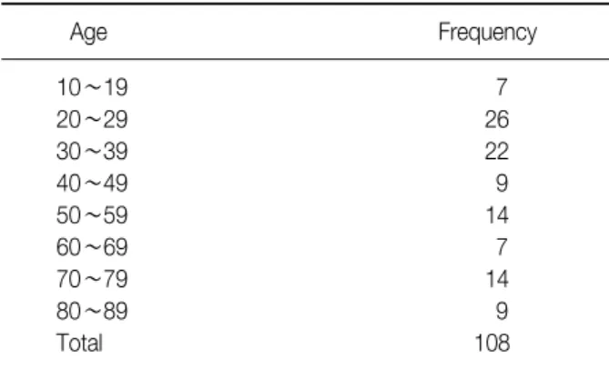
in the presence of clinical or radiologic evidence for TB in the absence of any other obvious cause associated with pleural effusions; (4) clinical or radiologic suspi-Table 1. Age distribution of the cases diagnosed as tuber- culous pleurisy
Age Frequency
10∼19 7
20∼29 26
30∼39 22
40∼49 9
50∼59 14
60∼69 7
70∼79 14
80∼89 9
Total 108
Table 2. Age and pleural fluid ADA activity in patients with tuberculous pleurisy
Number of ADA ADA
Age
patients (mean, IU/L) (SD*, IU/L)
<65 79 87.2 35.8
≥65 29 87.2 28.2
Total 108 87.2 33.8
*SD: standard deviation.
Table 3. The comparison of ADA activity alone (A) vs. ADA in combination with the L/N ratio ≥0.75 (B) as a means for diagnosing tuberculous pleurisy
(A)
ADA SN% SP% +LR NPV% PTP%
≥40 94.4 87.5 7.56 97.6 74.5
≥41 94.4 88.2 8.01 97.6 75.6
≥42 94.4 88.2 8.01 97.6 75.6
≥43 94.4 88.9 8.53 97.6 76.7
≥44 93.5 89.6 9.03 97.3 77.7
≥45 93.5 90.0 9.35 97.3 78.3
≥46 93.5 90.4 9.70 97.3 78.9
≥47 92.6 90.7 9.97 96.9 79.4
≥48 92.6 91.1 10.37 97.0 80.0
≥49 91.7 91.1 10.27 96.6 79.8
≥50 91.7 91.1 10.27 96.6 79.8
(B)
ADA SN% SP% +LR NPV% PTP%
≥40 89.8 97.5 35.93 96.1 93.3
≥41 89.8 97.9 41.91 96.1 94.2
≥42 89.8 97.9 41.91 96.1 94.2
≥43 89.8 98.2 50.30 96.2 95.1
≥44 88.9 98.2 49.78 95.8 95.0
≥45 88.9 98.2 49.78 95.8 95.0
≥46 88.9 98.2 49.78 95.8 95.0
≥47 88.0 98.6 61.57 95.5 96.0
≥48 88.0 98.6 61.57 95.5 96.0
≥49 87.0 98.6 60.93 95.2 95.9
≥50 87.0 98.6 60.93 95.2 95.9
ADA: adenosine deaminase (IU/L); SN: sensitivity; SP: specific- ity; +LR: positive likelihood ratio; NPV: negative predictive val- ue; PTP: post-test probability.
cion for TB in the absence of any other obvious cause associated with pleural effusions and related to a pos- itive response to antituberculous therapy.
The level of pleural fluid ADA activity was measured by an autoanalyzer (model 7170; Hitachi, Tokyo, Japan). The unit of ADA was given in IU/L. All stat- istical data were presented as average±standard devia- tion. Data were analyzed with SPSS Windows version 10.0.7 (SPSS Inc., Chicago, IL, USA). We used Indepen- dent Samples T-test to compare differences. Correla- tions were quantified by means of the Pearson Correla- tion Coefficient. Differences were considered signi- ficant at p<0.05 in all statistical tests.
Results
108 out of a total of 388 patients were finally diag- nosed as having TBpl. Ages of the cases were dis- tributed from 15 to 89. The mean age was 45.6±21.7, and 73% of the patients were less than 65. A relatively
higher frequency of TBpl occurred in patients in their 20∼30’s and 50∼70’s (Table 1), and the number of male patients was slightly larger (61%).
Concerning the method of diagnosis,
M. tuberculosis
was identified in pleural fluid by culture or PCR in 4 cases, granulomas were found in pleural biopsy tissue in 56 cases, sputum culture or PCR were positive in 26 cases, and the other 22 cases were clinically suspected and improved by antituberculous therapy.The mean level of pleural fluid ADA activities in 108 patients of TBpl was 87.2±33.8 IU/L. There was no dif- ference in the mean level of ADA between patients over
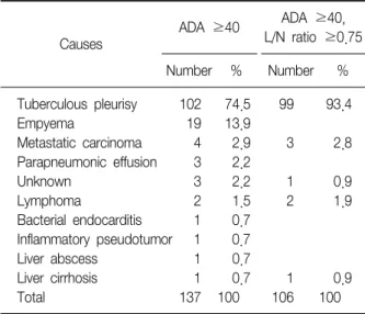
Table 4. The causes of high pleural fluid ADA activity
Causes
ADA ≥40 ADA ≥40, L/N ratio ≥0.75
Number % Number %
Tuberculous pleurisy 102 74.5 99 93.4
Empyema 19 13.9
Metastatic carcinoma 4 2.9 3 2.8
Parapneumonic effusion 3 2.2
Unknown 3 2.2 1 0.9
Lymphoma 2 1.5 2 1.9
Bacterial endocarditis 1 0.7 Inflammatory pseudotumor 1 0.7
Liver abscess 1 0.7
Liver cirrhosis 1 0.7 1 0.9
Total 137 100 106 100
ADA: adenosine deaminase (IU/L); L/N: lymphocyte/neutrophil.
Figure 1. The receiver operator characteristic (ROC) curve for pleural fluid ADA activity. The area under the curve is 0.943.
65 and less than 65 (Table 2).
When we regarded pleural fluid ADA activity ≥40 IU/L as a diagnostic criterion, the sensitivity was 94.4%, specificity 87.5%, negative predictive value 97.6%, and post-test probability 74.5%. Using the criterion of ADA activity ≥45 IU/L, the sensitivity fell slightly to 93.5%, and negative predictive value was much the same, but specificity and post-test probability increased to 90%
and 78.3%, respectively. Considering ADA ≥48 IU/L, the positive likelihood ratio was over 10 (Table 3). The area under the ROC curve for pleural fluid ADA activity was 0.943. It would be considered as ‘very good’ for separating TBpl from the other causes of exudative pleural effusion (Figure 1).
However, when we considered ADA activity ≥40 IU/L combined with L/N ratio ≥0.75 as diagnostic cri- teria, the specificity and post-test probability rose con- siderably to 97.5% and 93%, respectively. Also, the pos- itive likelihood ratio was 35.9, which was far more than 10. Using the criteria of ADA ≥45 IU/L and L/N ratio
≥0.75, the specificity and post-test probability increas- ed a little more, but the sensitivity tended to decline.
In brief, considering pleural fluid ADA activity com- bined with the L/N ratio demonstrated a higher positive likelihood ratio and better specificity and post-test prob- ability in spite of the lower level of pleural fluid ADA
(Table 3).
The other causes of exudative pleural effusions with elevated ADA level (≥40 IU/L) except TB were empye- ma, parapneumonic effusion, metastatic carcinoma, lymphoma, bacterial endocarditis, inflammatory pseudo- tumor, liver abscess, and liver cirrhosis. However, when combined with the L/N ratio, all of the patients with infectious causes such as empyema, parapneumonic ef- fusion, and liver abscess were completely excluded. In other words, using the criteria of ADA ≥40 IU/L and L/N ratio ≥0.75, 93.4% patients had real TBpl, and oth- er patients had malignancy including lymphoma (Table 4). Among them, of 2 patients diagnosed as having lym- phoma, mass lesions were detected on the chest radio- graphs and CT scans. In all the 4 patients with meta- static carcinoma, we also could find mass-like lesions on their chest or abdominal CT scans which were finally identified as primary lung cancer, gastric cancer with lung metastasis, and rectal cancer with hepatic metasta- sis.
Meanwhile, there were 6 patients who were finally diagnosed as TBpl but had pleural fluid ADA activities below 40 IU/L. They accounted for 5.6% of the 108 pa- tients with TBpl. Three of them were diagnosed by pleural biopsies, one by culture of sputum, another one by TB-PCR of sputum, and the other was diagnosed
clinically on the basis of a positive response to anti- tuberculous therapy.
Discussion
The definite diagnosis of TBpl requires either identi- fication of
Mycobacterium tuberculosis
in cultures of pleural fluid or pleural biopsy tissue, or observation of granulomas in the latter. However, the efficiency of cul- ture is quite low, and the procedure of pleural biopsy is somewhat invasive. Owing to these shortcomings, di- verse ancillary markers were evaluated to be used in diagnosing TBpl such as ADA, lysozyme, interferon-γ, and TB-PCR. However, the cost of TB-PCR was high and its sensitivity was very low. The sensitivity of lyso- zyme also was not high. The sensitivity and specificity of interferon-γ were as high as ADA activity, yet it is much more expensive to quantify than ADA1,2,15,16. Several cytokines and receptors were investigated as po- tential markers of TBpl, but until now, the determi- nation of ADA activity in pleural fluid has been the most useful method in regard to the aspects of sensitivity, specificity, and cost.For the differentiation of the lymphoid cells, partic- ularly T lymphocytes, and the maturation of monocytes- macrophages because of the metabolic requirement, it is necessary to rely on the presence of ADA4,17. For that reason, ADA has been looked on as a marker of cell-mediated immunity, which is the key mechanism of tuberculous pleural effusion. Meanwhile, age-associated immune decline is characterized by decreases in both B and T lymphocyte function, and the former may be largely a result of the latter. Therefore, pleural fluid ADA activity would be lower in old rather than in young patients with TBpl18. However, the level of pleu- ral fluid ADA among HIV-positive patients complicated with TBpl did not differ from the level among HIV-neg- ative patients19. In our study, there was no significant relationship between age and pleural fluid ADA activity.
Several reports describe the sensitivity and specificity of ADA level in the patients with pleural effusion. The sensitivity is 90∼100% and the specificity is 89∼100%.
Unfortunately, knowledge of the sensitivity and specific- ity of the test offers little clinical utility when evaluating individual patients. The utility was determined by the prevalence of TBpl20,21. In the area with high prevalence of TB, the proportion of false-positive results is lower, but positive predictive value decreases. We would like to clarify which parameters could be used as alternative to invasive pleural biopsy in countries with intermediate prevalence of tuberculous pleural effusion.
Clinical parameters such as age, sex, history of con- tact with patients with TB, and chest radiographic find- ings of tuberculous lesions held no predictive value al- though the data were not shown.
Likelihood ratio is the likelihood that a given test re- sult would be expected in a patient with the target dis- order compared to the likelihood that the same result would be expected in a patient without the target disorder. Likelihood ratio can combine the information about the prevalence of the disease and characteristics of the patient pool.
When we use the criterion of ADA activity ≥48 IU/L, the positive likelihood ratio was 10.37. Likelihood ratios of >10 or <0.1 generate large and often conclusive shifts from pretest to posttest probability (indicating high accuracy)22. Because we considered ADA ≥40 IU/L combined with the L/N ratio ≥0.75 as diagnostic criteria, specificity and post-test probability were exce- edingly high, and the positive likelihood ratio was even higher at 35.9. This value suggests that patients with TBpl have an approximately 36-fold more chance of be- ing ADA assay-positive compared with patients without TBpl.
There are other causes of exudative lymphocytic pleural effusions excluding TB such as malignancy, con- nective tissue disorders, sarcoidosis, chylothorax, and pulmonary thromboembolism23. Besides TBpl, ADA lev- els are also raised in empyema, rheumatoid pleurisy, and malignancy24,25. In this study, the other causes of exudative pleural effusions with elevated ADA were em- pyema, parapneumonic effusion, metastatic carcinoma, lymphoma, liver abscess, bacterial endocarditis, inflam- matory pseudotumor, and liver cirrhosis, and when
combined with the L/N ratio, the causes related to bac- terial infection were completely excluded. Like this, the combined approach with pleural fluid ADA and the L/N ratio proved to be useful in differentiating between TBpl and para-infective effusions10, but it was of no use for discerning TBpl from malignancy including lymphoma.
However, in the cases of malignancy, we could detect mass-like lesions on simple chest radiographs or CT scans.
As we put the results of our study and the literature cited together, it became clear that using pleural fluid ADA activity in combination with the L/N ratio is a mini- mally invasive, inexpensive, relatively accurate, and the most efficient method for diagnosing TBpl. Therefore, to evaluate the causes of exudative pleural effusions in a region of intermediate prevalence of TB, we recom- mend measuring pleural fluid ADA and the L/N ratio first. If the result is high enough, and malignancy is not suspected clinically or radiographically, it can be diag- nosed as TBpl.
There is a limitation because it is a retrospective ob- servational study. A prospective trial is needed on a larger group of patients. Especially, a larger proportion of malignant effusion will enhance the generalizability of our findings.
Summary
Background: The aim of this study was to consider the significance of pleural fluid adenosine deaminase (ADA) activity combined with lymphocyte/neutrophil (L/N) ratio in the diagnosis of tuberculous pleurisy (TBpl) in a region of intermediate prevalence of tuber- culosis (TB).
Methods: We collected data from 388 patients with exudative pleural effusions. The final diagnoses were compared to the results from our diagnostic method us- ing pleural fluid ADA and L/N ratio.
Results: 108 patients had a final diagnosis of TBpl;
102 cases had high levels of ADA (≥40 IU/L). When we considered ADA ≥40 IU/L as a diagnostic criterion, the sensitivity was 94.4%, specificity 87.5%, and post-
test probability 74.5%. However, when we considered ADA ≥40 IU/L combined with the L/N ratio ≥0.75 as a diagnostic criterion, the specificity and post-test prob- ability were rose to 97.5% and 93%, respectively. The other causes of high ADA and L/N ratios were lympho- ma and metastatic carcinoma, but mass-like lesions were found on the chest radiographs or CT scans.
Conclusion: To evaluate the causes of exudative pleu- ral effusions in a region of intermediate prevalence of tuberculosis, we recommend measuring the pleural fluid ADA and L/N ratio first. If the result is high and malig- nancies are not suspected, it may be diagnostic of TBpl.
References
1. Vald
é
s L, Pose A, San José
E, Martínez Vá
zquez JM.Tuberculous pleural effusions. Eur J Intern Med 2003;
14:77-88.
2. Ferrer J. Pleural tuberculosis. Eur Respir J 1997;10:942-7.
3. Berger HW, Mejia E. Tuberculous pleurisy. Chest 1973;
63:88-92.
4. P
é
rez-Rodriguez E, Jimé
nez Castro D. The use of ad- enosine deaminase and adenosine deaminase isoenzy- mes in the diagnosis of tuberculous pleuritis. Curr Opin Pulm Med 2000;6:259-66.5. Piras MA, Gakis C, Budroni M, Andreoni G. Adenosine deaminase activity in pleural effusions: an aid to differ- ential diagnosis. Br Med J 1978;2:1751-2.
6. Mo EK, Oh YM, Jung MP, Lee KY, Yoo CG, Kim YW.
A prospective study on the diagnostic value of adenos- ine deaminase activity in tuberculous pleural effusion.
Korean J Med 1995;48:625-32.
7. Vald
és
L, Alvarez D, San José
E, Penela P, Valle JM, Garcí
a-Pazos JM, et al. Tuberculous pleurisy: a study of 254 patients. Arch Intern Med 1998;158:2017-21.8. Goto M, Noguchi Y, Koyama H, Hira K, Shimbo T, Fukui T. Diagnostic value of adenosine deaminase in tuberculous pleural effusion: a meta-analysis. Ann Clin Biochem 2003;40:374-81.
9. Vald
é
s L, Alvarez D, San José
E, Juanatey JR, Pose A, Valle JM, et al. Value of adenosine deaminase in the diagnosis of tuberculous pleural effusions in young pa- tients in a region of high prevalence of tuberculosis.Thorax 1995;50:600-3.
10. Burgess LJ, Maritz FJ, Le Roux I, Taljaard JJ. Combined use of pleural adenosine deaminase with lympho- cyte/neutrophil ratio. Increased specificity for the diag-
nosis of tuberculous pleuritis. Chest 1996;109:414-9.
11. Diacon AH, Van de Wal BW, Wyser C, Smedema JP, Bezuidenhout J, Bolliger CT, et al. Diagnostic tools in tuberculous pleurisy: a direct comparative study. Eur Respir J 2003;22:589-91.
12. Ghanei M, Aslani J, Bahrami H, Adhami H. Simple method for rapid diagnosis of tuberculosis pleuritis: a statistical approach. Asian Cardiovasc Thorac Ann 2004;12:23-9.
13. Porcel JM, Vives M. Differentiating tuberculous from malignant pleural effusions: a scoring model. Med Sci Monit 2003;9:CR175-80.
14. Jeon EJ, Kwak HW, Song JH, Lee YW, Jeong JW, Choi JC, et al. Diagnostic value of ADA multiplied by lym- phocyte to neutrophil ratio in tuberculous pleurisy.
Tuberc Respir Dis 2007;63:17-23.
15. Roth BJ. Searching for tuberculosis in the pleural space.
Chest 1999;116:3-5.
16. Ferrer Sancho J. Pleural tuberculosis: incidence, patho- genesis, diagnosis, and treatment. Curr Opin Pulm Med 1996;2:327-34.
17. Pace E, Gjomarkaj M, Melis M, Profita M, Spatafora M, Vignola AM, et al. Interleukin-8 induces lymphocyte chemotaxis into the pleural space: role of pleural macrophages. Am J Respir Crit Care Med 1999;159:
1592-9.
18. Kim CJ, Yeon KM, Kim ST, Wang JH, Yoo KH.
Relationship between age and pleural fluid adenosine deaminase activity in patients with tuberculous pleural effusion. Tuberc Respir Dis 2002;52:608-15.
19. Riantawan P, Chaowalit P, Wongsangiem M, Rojanara- weewong P. Diagnostic value of pleural fluid ad- enosine deaminase in tuberculous pleuritis with refer- ence to HIV coinfection and a Bayesian analysis. Chest 1999;116:97-103.
20. Laniado-Laborin R. Adenosine deaminase in the diag- nosis of tuberculous pleural effusion: is it really an ide- al test? A word of caution. Chest 2005;127:417-8.
21. Kataria YP, Khurshid I. Adenosine deaminase in the di- agnosis of tuberculous pleural effusion. Chest 2001;120:
334-6.
22. Jaeschke R, Guyatt G, Lijmer J. Diagnostic tests. In:
Guyatt G, Rennie D, editors. Users' guides to the medi- cal literature: a manual for evidence-based clinical prac- tice. 1st ed. Chicago: AMA Press; 2002. p. 121-40.
23. Lee YC, Rogers JT, Rodriguez RM, Miller KD, Light RW.
Adenosine deaminase levels in nontuberculous lympho- cytic pleural effusions. Chest 2001;120:356-61.
24. Light RW. Diagnostic principles in pleural disease. Eur Respir J 1997;10:476-81.
25. Maskell NA, Butland RJ. BTS guidelines for the inves- tigation of a unilateral pleural effusion in adults. Thor- ax 2003;58 Suppl 2:ii8-17.