저작자표시-비영리-변경금지 2.0 대한민국 이용자는 아래의 조건을 따르는 경우에 한하여 자유롭게
l 이 저작물을 복제, 배포, 전송, 전시, 공연 및 방송할 수 있습니다. 다음과 같은 조건을 따라야 합니다:
l 귀하는, 이 저작물의 재이용이나 배포의 경우, 이 저작물에 적용된 이용허락조건 을 명확하게 나타내어야 합니다.
l 저작권자로부터 별도의 허가를 받으면 이러한 조건들은 적용되지 않습니다.
저작권법에 따른 이용자의 권리는 위의 내용에 의하여 영향을 받지 않습니다. 이것은 이용허락규약(Legal Code)을 이해하기 쉽게 요약한 것입니다.
Disclaimer
저작자표시. 귀하는 원저작자를 표시하여야 합니다.
비영리. 귀하는 이 저작물을 영리 목적으로 이용할 수 없습니다.
변경금지. 귀하는 이 저작물을 개작, 변형 또는 가공할 수 없습니다.
길 소 현
2 0 1 9 년 월2
석 사 학 위 논 문
Clinicopathological characteristics
계 명 대 학 교 대 학 원
의 학 과
길 소 현
지 도 교 수 이 재 호
2 0 1 9 년 2 월
and prognosis of TZAP mutation
in hepatocellular carcinomas
길 소 현
Clinicopathological characteristics
지 도 교 수 이 재 호
이 논 문 을 석 사 학 위 논 문 으 로 제 출 함
2 0 1 9 년 2 월
계 명 대 학 교 대 학 원
의 학 과
and prognosis of TZAP mutation
in hepatocellular carcinomas
길소현의 석사학위 논문을 인준함
주 심 최 인 장
부 심 이 재 호
부 심 이 현 수
계 명 대 학 교 대 학 원
2 0 1 9 년 2 월
Table of Contents
1. Introduction ··· 1
2. Materials and Methods ··· 3
3. Results ··· 8
4. Discussion ··· 16
5. Summary ··· 18
References ··· 19
Abstract ··· 23
국문초록 ··· 25
List of Tables
Table 1. Primer Sequences Used in This Study ··· 7
Table 2. Clinicopathological Characteristics of TZAP Mutation and TZAP mRNA Expressions and Telomere Length in HCCs ··· 10
Table 3. Correlation Between TZAP Expression and The Clinical
Parameters in Patients with HCC ··· 11
List of Figures
Figure 1. TZAP mutation (1272G/A) in HCCs ··· 12
Figure 2. Survival analysis of TZAP expression in HCCs ··· 13
Figure 3. Survival analysis of TZAP mutation in HCCs ··· 14
Figure 4. TCGA data of TZAP expression (ZBTB48) in HCCs ··· 15
1. Introduction
Telomeres are composed of 6 bp TTAGGG repeat sequences and they are nucleoprotein complexes capping each end of eukaryotic chromosomes [1,2]. In somatic cells, telomere is 5 to 15 kilobases and it becomes shorter about 30 to 300 base pairs during each cell division. Its shortening is counteracted by the reverse transcriptase telomerase in cancer and stem cells [3-5]. A telomere trimming mechanism is induced in the presence of overly long telomeres, which are cut as little length by rapid telomere trimming [6-8]. Though the detail mechanism of this process has not been clarified, recent studies have discovered a novel protein that is necessary for this process [9]. They identified the zinc finger protein ZBTB48, as a telomere-associated factor, and renamed it telomeric zinc finger-associated protein (TZAP). The study showed that overexpression of TZAP caused progressive telomere shortening. TZAP localizes to chromosome 1p36, a region that is cytologically changed in many cancers [10-12]. Genetic change in TZAP suggested be correlated with cancers, but TZAP has not been studied in any specific type of cancer.
Hepatocellular carcinoma (HCC) comprises the majority of primary hepatic tumors as a leading cause of cancer-related deaths worldwide [13,14]. Many researches focused on the clinical and prognostic values of telomeres in HCC [15,16]. They suggested that the regulation of telomere has an association with survival results in patients with HCC.
Here, I analyzed TZAP mutation and expression in patients with HCC
for the first time by determining their respective clinicopathological and
prognostic characteristics. Furthermore, based on previous studies [17], I
also confirmed whether genetic changes of TZAP induce the
deregulation of telomere length (TL).
2. Materials and Methods
2.1. Patients and tissue samples:
A total of 123 cases were identified from pathology reports of patients HCC surgery at the Kyungpook National University Hospital (KNUH) from January 2006 to October 2011. The clinical characteristics of each patient were analyzed by reviewing their medical records and slides.
Patients who received preoperative therapy, such as transarterial chemoembolization, radiofrequency ablation and systemic chemotherapy with sorafenib (Nexavar, Pittsburgh, PA, USA) were excluded from this study. In addition, patients with other malignancies or incomplete survival data were also excluded. This study was approved by the institutional review board (KNUH-2014-04-056-001).
Tumor tissues and matched non-malignant liver samples were fixed with formalin and paraffin. And then, paraffin blocks harboring the representative tumor tissues were selected by specialized pathologists for HCC diagnosis. Representative tissues were marked on the slide block and cored manually with a 3.0 mm diameter cylindrical device.
Total DNA samples of the tumor tissue and adjacent non-tumoral tissue were extracted using an extraction kit according to the instructions of manufacturer (Absolute™ DNA extraction Kit, BioSewoom, Korea). Total RNA was also extracted from same samples by TRIzol® Reagent (Molecular Research Center, Cincinnati, OH, USA).
The quantity and quality of extracted DNA and RNA were measured
by using NanoDrop 1000 (Thermo Fisher Scientific, Pittsburgh, PA,
USA).
2.2. TZAP mutation analysis:
TZAP exons were amplified by isolated DNA performing the polymerase chain reaction (PCR). It was carried out by using AmpliTaq Gold® DNA Polymerase (Applied Biosystems, Thermo Fisher Scientific, Waltham, MA, USA). The sequences of primer for all exons are presented in Table 1. Thermocycling was conducted under the following conditions: 40 cycles of 94 °C for 40 sec, 55-57 °C for 40 sec, and 72
°C for 50 sec. And then, the product was separated on a 2.0% agarose gel as electrophoresis, and then it was stained with ethidium bromide to confirm PCR result. Direct DNA sequencing of the TZAP was subsequently performed using an ABI 3730 DNA sequencer (Bionics, Seoul, South Korea).
2.3. Telomere length analysis:
Each telomere length (TL) was examined by quantitative real-time
PCR. To analyze quantitative TL relative to nuclear DNA (S), primers
were selected for telomere length using specific amplification (T) and
ß-globin for nuclear DNA (S). The primer sequences are presented in
Table 1. Real-time PCR was performed by LightCycler 480 II system
(Roche Diagnostics, Mannheim, Germany). TL was relatively presented
by calculating T/S values using the formula: T/S = 2 - ∆Ct, where ∆
Ct = average Ct telomere - average Ct ß-globin. Each measurement
was repeated three times and five serially diluted control samples were
included in each experiment.
2.3. TZAP expression analysis:
cDNA from each sample was synthesized from 1 µg of total RNA by using M-MLV Reverse Transcriptase (Promega, Madison, WI, USA) according to the protocol of manufacturer. To clarify the quality of mRNA extraction and reverse transcription, the products were subjected to qPCR amplification with the β‑actin primers (Bionics Inc., Seoul, Korea) using a LightCycler® 480 II system (Roche Diagnostics, Basel, Switzerland) with SYBR® Green PCR Master Mix (Toyobo, Osaka, Japan). The TZAP qPCR assay reactions were performed using primers as described in Table 1. TZAP expression levels were calculated as above method. Each experiment was repeated three times.
2.4. The Cancer Genome Atlas (TCGA) data analysis:
To investigate the clinical significance of TZAP, we used the TCGA database from OncoLnc and cBioPortal. TZAP mRNA expression data were downloaded from TCGA data portal on November, 2018 [18]. Its clinicopathological and prognostic values were analyzed.
2.5. Statistical analyses:
Chi-square test, the Mann–Whitney U test, Fisher’s exact test, and
simple correlation analysis were performed to analyze the associations
between the variables. Survival result by using the univariate
Kaplan-Meier estimators, were compared using the log-rank test.
Overall survival (OS) was set as the period between first diagnosis and
mortality. Disease-free survival (DFS) was defined as the period
between first diagnosis, and disease recurrence or the progression of
distant metastasis. The correlations between clinicopathologic parameters
were assessed with Pearson’s correlation coefficient analysis. A P value
of < 0.05 denoted significance for all statistical analysis performed in
this study.
Name Primer sequences
TZAP exon 1 F: CCAGACCTCAACAGCACAGA R: CACAGCCCACGAACCTAGTG TZAP exon 2 F: ATCCCATTGGCCGTTCTCT
R: CCGGCACAGTGAGAGGAT TZAP exon 3 F: TAGAGGCCAACTTCCCGTTT
R: CCTGGGCACAGTACCTCATT TZAP exon 4 F: CCTGCTGATTCATTTGGTGA
R: GGAATGGCAGACAGGAAAAG TZAP exon 5-1 F: GGAGGTGAGGAAGTTGACCA R: CCCTTCTAAGGGGAACAAGTG TZAP exon 5-2 F: GCTTGTCCCTGCACCTTAAC
R: GGAGAGGGCAACACATAACC TZAP exon 5-3 F: AGTCTGTCTGGGCCTGAGAA R: CCCTCCCTGTCACTTACTGC TZAP exon 6 F: CCCTTCCCTGCTCTCACC
R: AAGAGAGAACGGGCGACAC TZAP exon 7 F: GTCACTTCCCTTGGTGATGG
R: GAGGGGACCAGTGGTTTACA TZAP exon 8 F: CTGGGTGGCACTGGAGAG
R: CACGGGAACAGACTGTCAGG TZAP F: AAGGCCCTTAGAGGCTGAAG R: GACTCCCTCCTGGTCAGCAC β-Actin F: ACCCACACTGTGCCCATCTAC
R: TCGGEGAGGATCTTCATGAGG Telomere
length
F: CGGTTTGTTTGGGTTTGGGTTTGGGTTTGGG TTTGGGTT
R: GGCTTGCCTTACCCTTACCCTTACCCTTACCC TTACCCT
β-globin F: TGTGCTGGCCCATCACTTTG R: ACCAGCCA-CCACTTTCTGATAGG
Table 1. Primer Sequences Used in This Study
3. Results
3.1. TZAP mutation and TZAP expression in HCC:
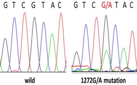
The sequences of TZAP regions were successfully analyzed in the 123 HCCs tissue samples. I found TZAP mutations in 10.6% (13/123) of these samples, all of which were c.1272G>A (L424L) (Figure 1). TZAP expression was analyzed in only 43 HCCs due to sample shortage, and as its expression in the HCC and paired non-cancerous tissues was similar (P = 0.53). The average TZAP expression in the HCC tissue was 1.64 ± 0.58, which contrasted with its expression in normal tissues.
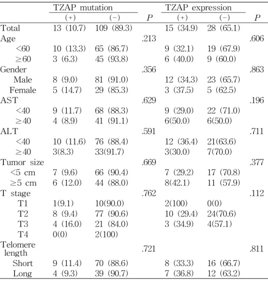
To further clarify the correlation between TZAP expression and the clinicopathological characteristics of HCC, patients were divided into two groups according to the median value of the Tumor/Non-tumor ratio (1.64). Clinicopathological significances associated with the TZAP mutation and expression are summarized in Table 2. There was no association between TZAP mutation and TZAP expression (P = 0.53).
Other clinicopathological parameters showed no association with the TZAP mutation and expressions.
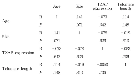
I also performed a quantitative correlation analysis with the clinical and genetic parameters including age, AST, ALT, tumor size, telomere length, and TZAP expression (Table 3). An association between age and tumor size was shown, however, it did not get a statistical significance.
And then, no quantitative correlation was observed with the other
parameters.
3.2. Prognostic value of TZAP mutation and TZAP expressions in HCC:
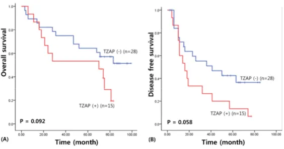
I assessed the survival results to confirm the prognostic value of the TZAP mutation and TZAP expression in patients with HCC. For survival analysis, the median follow-up term of HCC patients was 71.86 months (range: 3―105 months). The survival results of the univariate analysis was carried out by Kaplan–Meier curve, presented a poorer overall survival in HCC patients with TZAP expression (49.99 vs. 69.48 months, χ
2= 2.83, P = 0.092), albeit this result was not statistical significant (Figure 2). TZAP expression was also associated with poorer disease-free survival (26.60 versus 45.75 months, χ
2= 3.59, P = 0.058).
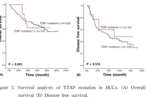
However, TZAP mutations did not have any prognostic value for the
patients with HCCs (overall survival: 65.92 vs. 73.42 months, χ
2= 0.001,
P = 0.995; Disease-free survival: 51.24 versus 47.86 months, χ
2= 0.348,
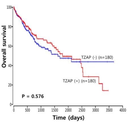
P = 0.555) (Figure 3). The Cancer Genome Atlas (TCGA) data analysis
showed no prognostic value of TZAP expression in HCC (P = 0.576)
(Figure 4).
TZAP mutation TZAP expression
(+) (-) P (+) (-) P
Total 13 (10.7) 109 (89.3) 15 (34.9) 28 (65.1)
Age .213 .606
<60 10 (13.3) 65 (86.7) 9 (32.1) 19 (67.9)
≥60 3 (6.3) 45 (93.8) 6 (40.0) 9 (60.0)
Gender .356 .863
Male 8 (9.0) 81 (91.0) 12 (34.3) 23 (65.7) Female 5 (14.7) 29 (85.3) 3 (37.5) 5 (62.5)
AST .629 .196
<40 9 (11.7) 68 (88.3) 9 (29.0) 22 (71.0)
≥40 4 (8.9) 41 (91.1) 6(50.0) 6(50.0)
ALT .591 .711
<40 10 (11.6) 76 (88.4) 12 (36.4) 21(63.6)
≥40 3(8.3) 33(91.7) 3(30.0) 7(70.0)
Tumor size .669 .377
<5 cm 7 (9.6) 66 (90.4) 7 (29.2) 17 (70.8)
≥5 cm 6 (12.0) 44 (88.0) 8(42.1) 11 (57.9)
T stage .762 .112
T1 1(9.1) 10(90.0) 2(100) 0(0)
T2 8 (9.4) 77 (90.6) 10 (29.4) 24(70.6) T3 4 (16.0) 21 (84.0) 3 (34.9) 4(57.1)
T4 0(0) 2(100)
Telomere
length .721 .811
Short 9 (11.4) 70 (88.6) 8 (33.3) 16 (66.7) Long 4 (9.3) 39 (90.7) 7 (36.8) 12 (63.2)
Table 2. Clinicopathological Characteristics of TZAP Mutation and TZAP
mRNA Expressions and Telomere Length in HCCs
Age Size TZAP
expression Telomere length
Age R 1 .141 -.073 .114
P .071 .642 .148
Size R .141 1 -.078 -.019
P .071 .626 .813
TZAP expression R -.073 -.078 1 -.053
P .642 .626 .736
Telomere length R .114 -.019 -.0053 1
P .148 .813 .736
Table 3. Correlation Between TZAP Expression and The Clinical
Parameters in Patients with HCC
Figure 1. TZAP mutation (1272G/A) in HCCs.
Figure 2. Survival analysis of TZAP expression in HCCs. (A) Overall
survival (B) Disease free survival.
Figure 3. Survival analysis of TZAP mutation in HCCs. (A) Overall
survival (B) Disease free survival.
Figure 4. TCGA data of TZAP expression (ZBTB48) in HCCs.
4. Discussion
In present study, I demonstrated gene mutation and expression of TZAP in cancer specifically HCC for the first time. The study about TZAP mutations have not been performed and little mutations were only reported in the International Cancer Genome Consortium data [19]. I showed that the c.1272G>A mutation in TZAP was frequently found in patients with HCC, which has been reported previously by studies;
however, this mutation was not found in 200 tissue samples of lung cancer and non-cancerous tissue, respectively. Therefore, TZAP mutation suggested to be HCC specific, though the hypothesis should be further confirmed.
TZAP expression was slightly increased in the HCC tissue samples.
To understand TZAP biology and the associated molecular mechanisms of HCC, telomere length were also analyzed and then, their correlations investigated based on the suggestions of previous research [17]. A previous study demonstrated that TZAP acts as a transcriptional activator for the mitochondrial fission regulator, MTFP1, providing evidence that a strong connection exits between telomere and mitochondrial homeostasis [17]. However, the study showed that there is no difference between HeLa TZAP knockout and TZAP wild type cells in terms of mitochondrial DNA level. More studies about the molecular mechanisms of telomere and TZAP with other cellular processes are warranted.
This study has presented a poorer survival result in HCC patients
with TZAP expression. However, TZAP mutation did not have any
clinical and prognostic values. The TCGA data also showed how overexpression of TZAP gene had a prognostic significance in some cancers, as colorectal, cervical and pancreatic cancers [18]. Interestingly, TZAP expression has an association with poorer prognosis in colorectal cancers, but a better prognosis in pancreatic and cervical cancers.
TCGA data demonstrated no prognostic value of TZAP expression in HCC [18-22]. I suggested that TZAP may have a different role and produce a different effect on telomere regulation and cancer cell survival according to cancer type.
In conclusion, I studied the clinicopathological and prognostic
characteristics of a TZAP mutation and TZAP expression in patients
with HCC. The result suggested that TZAP mutation and expression
may have an important role in hepatocellular carcinogenesis. The results
of my study warrant further study with larger samples to clarify the
underling mechanisms and signal pathway of TZAP gene and to
suggest its potential utility for clinicians.
5. Summary
A telomere is a repetitive sequences located at all chromosome end.
Average of telomere length in normal humans is from 10 kilobases at birth to less than 5 kilobases in older. Many studies suggest that telomere is an possible target for cancer treatment.
Zinc finger and BTB domain containing 48 (ZBTB48), renamed as
telomeric zinc-finger associated protein (TZAP) binds directly to the
double-stranded repeat sequence of telomeres. It has a competition with
the telomeric repeat binding factors, such as TRF1 and TRF2. TZAP
expression induced telomere trimming via telomere shortening for normal
telomere length. Therefore, I examined genetic study about TZAP
mutation and expression in hepatocellular carcinoma (HCC). The TZAP
mutation (c.1272G>A) was frequently shown in HCCs. TZAP expression
may be associated with a shorter overall survival, but, telomere length
did not show any predictable potential for HCCs. This result suggests
that mutation and expression of TZAP have a important role in
hepatocellular pathogenesis. It warrant future study to clarify signal
pathway and functional mechanisms of TZAP and to confirm its
potential role as a therapeutic target for HCC.
References
1. Harley CB, Futcher AB, Greider CW: Telomeres shorten during ageing of human fibroblasts. Nature. 1990; 345(6274): 458-60.
2. Blackburn EH, Greider CW, Szostak JW: Telomeres and telomerase:
The path from maize, Tetrahymena and yeast to human cancer and aging. Nat Med. 2006; 12(10): 1133-8.
3. Xin H, Liu D, Songyang Z: The telosome/shelterin complex and its functions. Genome Biol. 2008; 9(9): 232.
4. Shay JW, Wright WE: Role of telomeres and telomerase in cancer.
Semin Cancer Biol. 2011; 21(6): 349-53.
5. Jafri MA, Ansari SA, Alqahtani MH, Shay JW: Roles of telomeres and telomerase in cancer, and advances in telomerase-targeted therapies. Genome Med. 2016; 8(1): 69.
6. Pickett HA, Cesare AJ, Johnston RL, Neumann AA, Reddel RR:
Control of telomere length by a trimming mechanism that involves generation of t-circles. EMBO J. 2009; 28(7): 799-809.
7. Pickett HA, Reddel RR: The role of telomere trimming in normal telomere length dynamics. Cell Cycle. 2012; 11(7): 1309-15.
8. Sfeir A, de Lange T: Removal of shelterin reveals the telomere
end-protection problem. Science. 2012; 336(6081): 593-7.
9. Li JS, Miralles Fusté J, Simavorian T, Bartocci C, Tsai J, Karlseder J, Lazzerini Denchi E: TZAP: A telomere-associated protein involved in telomere length control. Science. 2017; 355(6325): 638-41.
10. Maris JM, Jensen SJ, Sulman EP, Beltinger CP, Gates K, Allen C, Biegel JA, Brodeur GM, White PS: Cloning, chromosomal localization, physical mapping, and genomic characterization of HKR3. Genomics.
1996; 35(2): 289–98.
11. White PS, Maris JM, Sulman EP, Jensen SJ, Kyemba SM, Beltinger CP, Allen C, Kramer DL, Biegel JA, Brodeur GM:
Molecular analysis of the region of distal 1p commonly deleted in neuroblastoma. Eur J Cancer. 1997; 33(12): 1957-61.
12. Maris JM, Jensen J, Sulman EP, Beltinger CP, Allen C, Biegel JA, Brodeur GM, White PS: Human Krüppel-related 3 (HKR3): a candidate for the 1p36 neuroblastoma tumour suppressor gene? Eur J Cancer. 1997; 33(12): 1991-6.
13. Siegel R, Ma J, Zou Z, Jemal A: Cancer statistics. CA Cancer J Clin. 2014; 64(1): 9-29.
14. Vauthey JN, Lauwers GY, Esnaola NF, Do KA, Belghiti J, Mirza
N, Curley SA, Ellis LM, Regimbeau JM, Rashid A, Cleary KR,
Nagorney DM: Simplified staging for hepatocellular carcinoma. J Clin
Oncol. 2002; 20(6): 1527-36.
15. Nault JC, Mallet M, Pilati C, Calderaro J, Bioulac-Sage P, Laurent C, Laurent A, Cherqui D, Balabaud C, Zucman-Rossi J: High frequency of telomerase reverse-transcriptase promoter somatic mutations in hepatocellular carcinoma and preneoplastic lesions. Nat Commun. 2013; 4(1): 2218.
16. Tornesello ML, Buonaguro L, Izzo F, Buonaguro FM: Molecular alterations in hepatocellular carcinoma associated with hepatitis B and hepatitis C infections. Oncotarget. 2016; 7(18): 25087-102.
17. Jahn A, Rane G, Paszkowski-Rogacz M, Sayols S, Bluhm A, Han CT, Draškovič I, Londoño-Vallejo JA, Kumar AP, Buchholz F, Butter F, Kappei D: ZBTB48 is both a vertebrate telomere-binding protein and a transcriptional activator. EMBO Rep. 2017; 18(6): 929-46.
18. The Cancer Genome Atlas. http://cancergenome.nih.gov/abouttcga
19. The COSMIC, the Catalogue Of Somatic Mutations In Cancer.
http://cancer.sanger.ac.uk/cosmic/mutation/overview?id=3705990
20. Hsu CP, Lee LW, Shai SE, Chen CY: Clinical significance of
telomerase and its associate genes expression in the maintenance of
telomere length in squamous cell carcinoma of the esophagus. World
J Gastroenterol. 2005; 11(44): 6941-7.
21. Oh BK, Kim H, Park YN, Yoo JE, Choi J, Kim KS, Lee JJ, Park C: High telomerase activity and long telomeres in advanced hepatocellular carcinomas with poor prognosis. Lab Invest. 2008;
88(2): 144-52.
22. Artandi SE, DePinho RA: Telomeres and telomerase in cancer.
Carcinogenesis. 2010; 31(1): 9-18.
Clinicopathological characteristics of TZAP mutation and TZAP expression in hepatocellular carcinomas
Kil, So Hyun Department of Anatomy
Graduate School Keimyung University
(Supervised by Professor Lee, Jaeho)
The zinc finger protein ZBTB48 is a telomere-associated factor and
renamed it as telomeric zinc finger-associated protein (TZAP). It binds
preferentially to long telomeres competing with telomeric repeat factor 1
and 2 (TRF1 and TRF2). However, its expression in cancers has not
been performed. In the present study, I analyzed TZAP mutation and
expression in 123 hepatocellular carcinomas (HCC) and their association
with telomere length was also investigated. TZAP mutations
(c.1272G>A, L424L) was found in 10.6% (13/123) and TZAP expression
level was not different between HCC and paired non-cancerous tissues.
There was no association between TZAP mutation and TZAP
expression (P = 0.53). TZAP mutation did not have any clinical and
prognostic values in HCC. And, TZAP expression tended to induce
poorer survival result (overall survival, χ
2= 2.83, P = 0.092;
disease-free survival, χ
2= 3.59, P = 0.058). However, The Cancer
Genome Atlas (TCGA) survival analysis showed no prognostic value of
TZAP expression in HCC (P = 0.576). This result suggested that TZAP
expression appears to be a possible prognosis marker in HCC.
간암에서 TZAP 돌연변이의 임상병리학적, 예후적인 특징
길 소 현
계 명 대 학 교 대 학 원 의학과 해부학 전공 (지도교수 이 재 호)