Veterinary Science
DOI: 10.4142/jvs.2010.11.1.9
*Corresponding authors
Tel: +82-62-530-2831; Fax: +82-62-530-2809
E-mail: [email protected]
†The first two authors contributed equally to this work.
Multidetector row computed tomography evaluation of the micropig kidney as a potential renal donor
Woong Yoon
2,†, Min Young Lee
1,†, Jung Min Ryu
1, Yong Ju Moon
2, Sang Hun Lee
1, Jae Hong Park
1, Seung Pil Yun
1, Min Woo Jang
1, Sung Su Park
3, Ho Jae Han
1,*
1
College of Veterinary Medicine, Biotherapy Human Resources Center (BK21), Chonnam National University, Gwangju 500-757, Korea
2
Department of Radiology, Chonnam National University Hospital, Chonnam National University Medical School, Gwangju 501-757, Korea
3
College of Veterinary Medicine, Seoul National University, Seoul 151-742, Korea
Multidetector row computed tomography (MDCT) provides anatomical information about the kidney and other internal organs. Presently, the suitability of 64-channel MDCT to assess the kidney of healthy micropigs was evaluated.
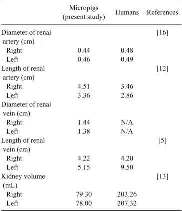
Morphological evaluations of the kidney and the major renal vessels of six healthy micropigs were carried out using MDCT, recording kidney volume and the diameter and length of renal arteries and veins. The mean diameters and lengths of the renal artery were 0.44 ± 0.05 and 4.51 ± 0.55 cm on the right side and 0.46 ± 0.06 and 3.36 ± 0.27 cm on the left side, respectively. The mean diameters and lengths of the renal vein were 1.44 ± 0.52 and 4.22 ± 1.29 cm on the right side and 1.38 ± 0.17 and 5.15 ± 0.87 cm on the left side, respectively. The mean volume of the right kidney was 79.3
± 14.5 mL and of the left kidney was 78.0 ± 13.9 mL. The data presented in this study suggest that the MDCT offers a noninvasive, rapid, and accurate method for the evaluation of the renal anatomy in living kidney donors. It also provides sufficient information about extra-renal anatomy important for donor surgery and determination of organ suitability.
Keywords: kidney, micropig, multidetector row computed tomography, renal vessels
Introduction
Transplantation is used to treat fulminate organ failure, but severe shortages in the availability of suitable human donors have limited the application of organ transplants [4]. This donor shortage has stimulated interest in the
possible use of animal organs for transplantation into humans.
Animal-to-human transplantation (xenotransplantation) would offer an unlimited supply of organs and tissue for transplantation. Pigs are the most likely source animals for xenotransplantation due to their anatomical and physiological similarities with humans [1]. Additionally, the pig can be raised to obtain large numbers of specific-pathogen-free animals. The reproductive properties of pigs such as early sexual maturity, short gestation time, and generation of large litters can allow a large pool of animal donors for xenotransplantation [18]. The ability to genetically modify the pig also allows modification of the targets of the human immune response and amelioration of some aspects of the rejection process without directly affecting the recipient’s immune system.
In order to transplant swine organs into humans, physiological or anatomical comparison and analyses are essential in the investigation of whether an individual donor organ is suitable to a prospective patient. However, an appropriate method for estimating micropig organs has not been established.
This study examined the feasibility of evaluating the kidney and its related major vessels using multidetector row computed tomography (MDCT) in micropigs. In recent years, major technological improvements have been achieved in CT.
The most significant development has been the introduction of MDCT, which has brought about substantial improvements in spatial and particularly temporal resolution [3,6]. The present study examined the feasibility of MDCT on evaluation of kidney and renal vascular system of micropig as a potential renal donor.
Materials and Methods Animals
All experimental protocols were approved by the Ethics