In contrast, DADS pretreatment significantly attenuated the AAP-induced nephrotoxic effects, including serum BUN level and expression of KIM-1 and NGAL proteins
8
0
0
전체 글
(2) Effects of diallyl disulfide on acetaminophen-induced nephrotoxicity. superfamily that is initially found in activated neutrophils, in accordance with its role as an innate antibacterial factor [10]. However, other cells, like kidney tubular cells, may also produce NGAL in response to various insults [11,12]. It has been demonstrated that KIM-1 and NGAL may be the most promising new biomarkers, and are specifically induced at the target site of toxicity in both animal and human studies involving acute injury of the proximal tubule epithelium [8,11,13-15]. Diallyl disulfide (DADS), which is an organosulfur component of garlic, is a potent compound that prevents toxicant-induced oxidative injury [16,17]. Recently, we have reported anti-oxidative, anti-inflammatory, and anti-apoptotic effects of DADS in various experimental systems [18,19]. There are also some reports on the ameliorating effect of DADS against nephrotoxicantinduced renal injury [16,17]. However, the protective capacity of DADS against the nephrotoxicity of AAP has not been explored. The aim of the present study was to evaluate the protective effects of DADS on AAPinduced nephrotoxicity and the possible application of KIM-1 and NGAL as newly identified biomarkers of AAP-induced acute kidney injury.. Materials and Methods Animals handling and environmental conditions. Male Sprague-Dawley rats (aged 9 weeks) were obtained from a specific pathogen-free colony at Samtako Co. (Osan, Korea) and were subjected to 1 week of quarantine and acclimatization before the experiments. Two rats per stainless wire mesh cage were housed in a room maintained under the following conditions: temperature of 23±3oC, relative humidity of 50±10%, artificial lighting from 08:00 to 20:00, and 13 to 18 air changes per hour. Rats were provided with tap water that had been sterilized by ultraviolet irradiation, and commercial rodent chow (Samyang Feed, Wonju, Korea) ad libitum. The Institutional Animal Care and Use Committee of Chonnam National University approved the protocols for animal study, and the animals were cared for in accordance with the Guidelines for Animal Experiments of Chonnam National University. Test chemicals and treatment. AAP was purchased from Sigma Aldrich Co. (St. Louis, MO, USA). DADS was purchased from Tokyo Kasei Chemical Co. (Tokyo, Japan). All other chemicals. 201. were of the highest grade that is commercially available. DADS was dissolved in corn oil. AAP was dissolved in a saline solution kept in warm boiling water bath, and was used after cooling to 37oC. These chemicals were prepared freshly before treatment. The daily application volumes of AAP (20 mL/kg body weight) and DADS (5 mL/kg body weight) were calculated in advance, based on the most recently recorded body weight of the individual animal. DADS (50 mg/kg/day) was gavaged to rats once daily for a period of 5 days. One hour after the final DADS treatment, the rats were injected with a single intraperitoneal dose of AAP (1,000 mg/kg). Experimental groups and dose selection. Twenty-four healthy male rats were randomly divided into four experimental groups (n=6 per group): (1) vehicle control, (2) AAP, (3) AAP&DADS, and (4) DADS. The selected AAP dose was based on a previous study that demonstrated significant acute renal injury in rats [3]. The effective dose of DADS was also based on a previous study [19]. Body weight changes and clinical signs. All animals were observed daily for any clinical signs of toxicity and mortality throughout the test period. Abnormal signs were recorded individually for type, observation day and time, and duration. The body weight of each rat was measured on test days 0 and 1. Necropsy, organ weight, and serum biochemistry. After 24 h of acute kidney injury induction, all male rats were euthanized by carbon dioxide for blood serum collection and exsanguination from the aorta. Serum samples were collected by centrifugation at 3,000 rpm for 10 min and stored in the −80°C freezer before they were analyzed. Serum creatinine and BUN were evaluated using a serum biochemical autoanalyzer (Dri-chem 4000i, Fujifilm Co., Japan). The absolute and relative (organ-to-body weight ratio) weights of the kidneys of all rats were also measured. Histopathological examination. The left kidney was fixed in 10% neutral buffered formalin solution for 1 week. The tissues were routinely processed, embedded in paraffin, sectioned at 4 µm thickness, deparaffinized, and rehydrated using standard techniques. The sections were stained with a hematoxylineosin (H&E) stain for microscopic examination. All Lab Anim Res | December, 2016 | Vol. 32, No. 4.
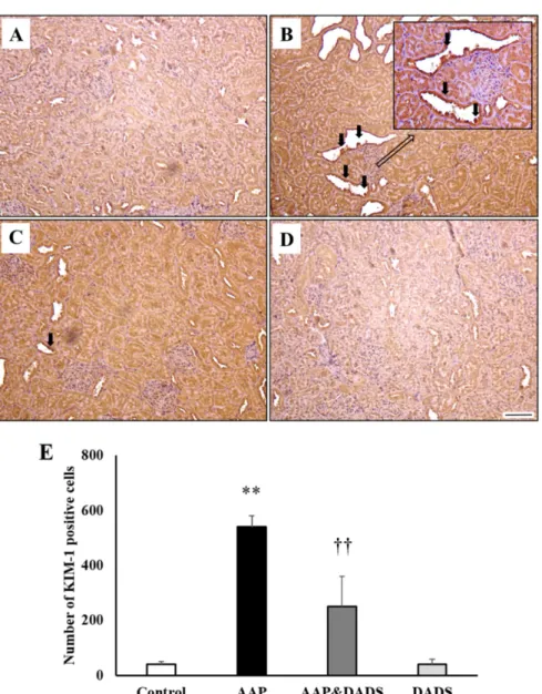
(3) 202. Jin-Young Shin et al.. sections were examined with a light microscope by a pathologist, who was blinded to the sample treatments. Immunohistochemistry (IHC). The paraffin-embedded sections were deparaffinized and rehydrated. After incubation with a protein block (Rabbit Specific HRP/DAB Detection IHC Kit; Abcam, Cambridge, MA, USA), the sections were incubated overnight with anti-KIM-1antibodies (1:200; LifeSpan Biosciences, Seattle, WA, USA) and anti-NGAL antibodies (1:500; Abcam) at 4oC. The expression of KIM-1 and NGAL was visualized using Rabbit Specific HRP/DAB Detection IHC Kit (Abcam), according to the manufacturer’s protocol. The sections were counterstained with Harris’s hematoxylin before mounting. The number of KIM-1 and NGAL positive cells was counted in 10 different fields in each section under 100× magnification. Western blot analysis. The frozen right kidney tissues were lysed in a RIPA lysis buffer (Cell Signaling Technology, Lexington, KY, USA), and centrifuged at 12,000×g at 4oC for 10 min to isolate the cellular proteins in the supernatant. The kidney tissues supernatants were separated by sodium dodecyl sulfate-polyacrylamide gel electrophoresis, transferred to a polyvinylidene difluoride membrane (Millipore, Bedford, MA, USA), and blocked in blocking buffer (150 mM NaCl in 10 mM Tris, pH 7.5 containing 5% non-fat dry milk) for 1 h at room temperature. The membranes were incubated with primary antibodies against KIM-1 (1:1,000; LifeSpan Biosciences) and NGAL (1:1,000; Abcam) for 18 h at 4oC, washed three times (20 mM Tris-HCl, pH 7.5, 137 mM NaCl, and. 0.1% Tween 20), incubated with horseradish peroxidaseconjugated secondary antibody (1:5,000, Thermo Scientific, Rockford, IL, USA) for 1 h at room temperature, washed three times, and then detected with an enhanced chemiluminescence method (Supersignal West Pico, Pierce, IL, USA). The protein concentration was determined using the BCA Protein Assay Kit (Pierce). Statistical analyses. The data are expressed as means±SD, and all statistical comparisons were made by means of one-way analysis of variance, followed by Tukey’s multiple comparison test. Statistical analyses were performed by comparisons of the treatment groups with the control group, using the GraphPad InStat v. 3.0 (GraphPad Software, La Jolla, CA, USA). Differences with a P-value of 0.05 or lower were considered to be statistically significant.. Results Effects of DADS on clinical sign, body weight, and kidney weight. No treatment-related mortality was observed in rats that were treated with AAP and/or DADS during the study period. However, the incidence and severity of clinical signs, such as decreased locomotor activity (n=4) and dull fur (n=3), increased in the AAP group, as compared with those in the control group. There were no significant differences in the body weights and kidney weights between the groups (data not shown). Effects of DADS on renal function. As shown in Figure 1A, the rats treated with 1,000 mg/. Figure 1. Serum blood urea nitrogen (A) and creatinine (B) in male rats treated with AAP and/or DADS. Values are presented as means±SD (n=6). *P<0.05 compared with the control group. †P<0.05 compared with the AAP group. Lab Anim Res | December, 2016 | Vol. 32, No. 4.
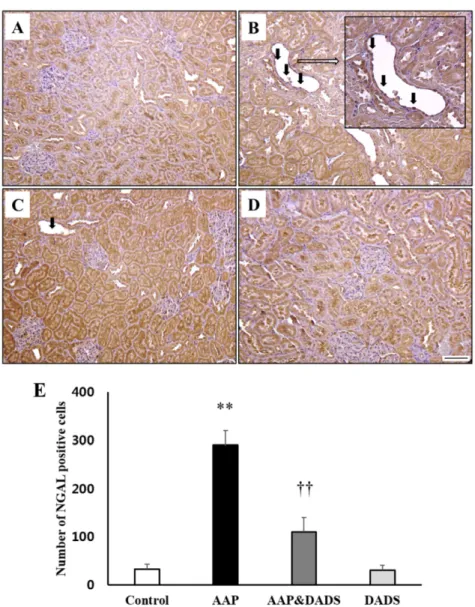
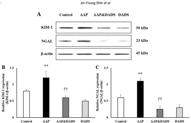
(4) Effects of diallyl disulfide on acetaminophen-induced nephrotoxicity. 203. Figure 2. Representative photographs of kidney sections treated AAP and/or DADS. Kidney from vehicle control (A) and DADS (D) treated rats showed a normal morphology. However, kidney from an AAP-treated rat (B) showed cast (black arrow), tubular dilation (white arrow), tubular necrosis (black arrow head), and tubular degeneration (white arrow head). Kidney from an AAP&DADStreated rat (C) showed nearly normal appearance. H&E stain. Bar=50 μm.. kg of AAP showed significantly increased serum BUN level, as compared with that in the control group. In contrast, the serum BUN level in the AAP&DADS group decreased significantly, as compared with that in the AAP group. There were no statistically significant differences in the serum creatinine levels between the groups (Figure 1B). Effects of DADS on renal histopathology. The results of the histopathological examination are presented in Figure 2. The control (Figure 2A) and DADS (Figure 2D) groups showed kidneys with normal morphology. However, kidney tissues from rats treated with AAP showed extensive injuries, characterized by cast, tubular dilation, tubular necrosis, and tubular degeneration (Figure 2B). Although these changes were also observed in the AAP&DADS group (Figure 2C), the incidence and severity of histopathologic lesions were significantly lower than those in the AAP group. Effects of DADS on KIM-1 and NGAL expression. and NGAL (Figure 4) in the kidneys, IHC for KIM-1 and NGAL was conducted. KIM-1 and NGAL positive cells were seldom observed in the control and DADS groups (Figures 3A, 3D, 4A, and 4D). However, rats treated with AAP manifested many KIM-1 and NGAL positive cells, which were mainly distributed in proximal tubular epithelial cells (Figures 3B and 4B). This increase was attenuated in rats treated with DADS (Figures 3C and 4C). Accordingly, the number of KIM1 and NGAL positive cells in the AAP group was significantly higher than that in the control group (Figures 3E and 4E). The number of KIM-1 and NGAL positive cells in the AAP&DADS group decreased significantly compared with that in the AAP group. The protein expression levels of KIM-1 and NGAL are shown in Figure 5. The expression levels of KIM-1 and NGAL in the AAP group increased significantly, as compared with those in the control group. In contrast, the expression levels of KIM-1 and NGAL in the AAP&DADS group decreased significantly, as compared with those in the AAP group.. To assess a possible contribution of KIM-1 (Figure 3). Lab Anim Res | December, 2016 | Vol. 32, No. 4.
(5) 204. Jin-Young Shin et al.. Figure 3. Representative photographs of immunohistochemical analysis of KIM-1 in kidney sections of (A) controls and rats treated with (B) AAP, (C) AAP&DADS, and (D) DADS. Arrows indicate KIM-1 positive cells (brownish-stained cells). Bar=50 μm. (E) The number of KIM-1 positive cells was counted in ten different fields in each section under 100× magnification. Results are presented as means±SD (n=6). **P<0.01 compared with the control group. ††P<0.01 compared with the AAP group.. Discussion It has been demonstrated that DADS has renoprotective effects in models of renal injury induced by gentamicin and cisplatin [16,17]. The results of the present study showed that DADS has a protective effect against acute kidney injury induced by AAP treatment in male rats. Several investigators have reported that AAP treatment leads to nephrotoxicity, characterized by poor renal function and increased serum creatinine and BUN levels [3,20]. In the present study, the nephrotoxicity observed in the AAP group included elevated BUN levels. These alterations correlated well with histopathological findings: Lab Anim Res | December, 2016 | Vol. 32, No. 4. increased incidence of cast, tubular dilation, tubular necrosis, and tubular degeneration. These findings observed in the AAP group may represent impaired renal function. However, DADS pretreatment effectively prevented the AAP-induced elevation in serum BUN levels, indicating the renoprotective activity of DADS against acute renal injury of AAP. This phenomenon was also confirmed by histopathological examination, which showed a decrease in the incidence and severity of renal histopathological lesions. Animal models are commonly used for toxicity evaluation of new therapeutics and assessment of potential chemical hazards in industry and environment..
(6) Effects of diallyl disulfide on acetaminophen-induced nephrotoxicity. 205. Figure 4. Representative photographs of immunohistochemical analysis of NGAL in kidney sections of (A) controls and rats treated with (B) AAP, (C) AAP&DADS, and (D) DADS. Arrows indicate NGAL positive cells (brownish-stained cells). Bar=50 μm. (E) The number of KIM-1 positive cells was counted in ten different fields in each section under 100× magnification. Results are presented as means±SD (n=6). **P<0.01 compared with the control group. ††P<0.01 compared with the AAP group.. Therefore, KIM-1 and NGAL may serve as useful biomarkers for drug safety and chemical hazard-related renal injury tests. Previous studies have shown that KIM-1 may function as an extracellular sensor or a receptor for adhesion/signaling in a variety cell-cell or cell-pathogen interactions [21,22]. The protein structure of this molecule suggests that KIM-1 may be an adhesion and/or protective molecule for the cell surface [23]. Therefore, it is considered that KIM-1 might alter cellular adhesion and/or modulate interactions between the injured epithelial cell and the luminal contents, and enhance mobility and proliferation of surviving epithelial cells [24]. The NGAL protein is expressed by neutrophils and various epithelial cells, and is found at very low. concentrations in various human tissues: kidney, trachea, lungs, stomach, and colon [12,25]. It has also been found to play a role in kidney development and tubular regeneration and repair after injury [12]. Many factors leading to tubular epithelial cell injury result in an increased expression of KIM-1 and NGAL proteins. Thus, reduction of KIM-1 and NGAL expression is a major target for renoprotective therapy. Although it is clear that expression of KIM-1 and NGAL protein is up-regulated by renal injury of ischemiareperfusion, ochratoxin A, and gentamicin in animal models [26-28], the response to AAP-induced nephrotoxicity was not clear. Therefore, we investigated the role of DADS on the expression of KIM-1 and NGAL induced Lab Anim Res | December, 2016 | Vol. 32, No. 4.
(7) 206. Jin-Young Shin et al.. Figure 5. Western blot analysis of KIM-1 and NGAL expressions in rats treated with AAP and/or DADS. Detection of β-actin expression was used as a loading control. The bar graphs show quantitative relative levels of KIM-1 (B) and NGAL (C) protein expressions for vehicle, AAP, AAP&DADS, and DADS-treated rats. Values are presented as means±SD (n=6). **P<0.01 compared with the control group. ††P< 0.01 compared with the AAP group.. by AAP treatment. In the present study, AAP caused a significant increase in KIM-1 and NGAL protein expression without a significant increase in serum creatinine level. Ichimura et al. [8] also reported that nephrotoxicants treatment increased the expression of KIM-1 and NGAL, before causing a measurable increase in serum creatinine level. Although the specific functions of KIM1 and NGAL are still unknown, up-regulation of these proteins is associated with proliferation/regeneration and repair in response to toxicity or disease [9,12,14]. Changes in the expression of marker proteins were confirmed by immunohistochemistry. In accordance with other studies [8,13-15], KIM-1 and NGAL occurred in proximal tubular epithelial cells. However, DADS pretreatment effectively inhibited AAP-induced KIM-1 and NGAL protein expression, suggesting that DADS-mediated improvement in nephrotoxicity might be mediated, at least in part, by its ability to reduce the KIM-1 and NGAL expression in the kidney. These results are in accordance with the decreased histopathological changes in the kidney. In conclusion, DADS has protective effects against AAP-induced nephrotoxicity in male rats. KIM-1 and NGAL could be useful in preclinical and clinical studies Lab Anim Res | December, 2016 | Vol. 32, No. 4. vital to drug development and evaluation. They may also serve in the monitoring of disease states that manifest as injury to the proximal tubule and be useful in guiding interventional strategies.. Acknowledgments This study was financially supported by Chonnam National University, 2014. Conflict of interests The authors declare that there is no financial conflict of interests to publish these results.. References 1. Kanno S, Tomizawa A, Hiura T, Osanai Y, Kakuta M, Kitajima Y, Koiwai K, Ohtake T, Ujibe M, Ishikawa M. Melatonin protects on toxicity by acetaminophen but not on pharmacological effects in mice. Biol Pharm Bull 2006; 29(3): 472-476. 2. Hengy B, Hayi-Slayman D, Page M, Christin F, Baillon JJ, Ber CE, Allaouchiche B, Rimmele T. Acute renal failure after acetaminophen poisoning: report of three cases. Can J Anaesth 2009; 56(10): 770-774. 3. Ghosh J, Das J, Manna P, Sil PC. Acetaminophen induced renal injury via oxidative stress and TNF-alpha production: therapeutic potential of arjunolic acid. Toxicology 2010; 268(1-2): 8-18. 4. Singh VP, Singh N, Jaggi AS. A review on renal toxicity profile of common abusive drugs. Korean J Physiol Pharmacol 2013; 17(4):.
(8) Effects of diallyl disulfide on acetaminophen-induced nephrotoxicity. 347-357. 5. Schnellman RG. Toxic responses of the kidney. In: Casarett & Doull’s Toxicology: The Basic Science of Poisons (Klassen CD, ed), 6th ed., McGraw-Hill Medical Publishing Division, New York, 2001; pp 491-514. 6. Mediæ B, Rovcanin B, Vujovic KS, Obradovic D, Duric D, Prostran M. Evaluation of Novel Biomarkers of Acute Kidney Injury: The Possibilities and Limitations. Curr Med Chem 2016; 23(19): 1981-1997. 7. Schrezenmeier EV, Barasch J, Budde K, Westhoff T, Schmidt-Ott KM. Biomarkers in acute kidney injury - pathophysiological basis and clinical performance. Acta Physiol (Oxf) 2016; 30: 12764. 8. Ichimura T, Hung CC, Yang SA, Stevens JL, Bonventre JV. Kidney injury molecule-1: a tissue and urinary biomarker for nephrotoxicant-induced renal injury. Am J Physiol Renal Physiol 2004; 286(3): F552-563. 9. Huo W, Zhang K, Nie Z, Li Q, Jin F. Kidney injury molecule-1 (KIM-1): a novel kidney-specific injury molecule playing potential double-edged functions in kidney injury. Transplant Rev (Orlando) 2010; 24(3): 143-146. 10. Bolignano D, Donato V, Coppolino G, Campo S, Buemi A, Lacquaniti A, Buemi M. Neutrophil gelatinase-associated lipocalin (NGAL) as a marker of kidney damage. Am J Kidney Dis 2008; 52(3): 595-605. 11. Mishra J, Ma Q, Kelly C, Mitsnefes M, Mori K, Barasch J, Devarajan P. Kidney NGAL is a novel early marker of acute injury following transplantation. Pediatr Nephrol 2006; 21(6): 856-863. 12. Soni SS, Cruz D, Bobek I, Chionh CY, Nalesso F, Lentini P, de Cal M, Corradi V, Virzi G, Ronco C. NGAL: a biomarker of acute kidney injury and other systemic conditions. Int Urol Nephrol 2010; 42(1): 141-150. 13. Han WK, Bonventre JV. Biologic markers for the early detection of acute kidney injury. Curr Opin Crit Care 2004; 10(6): 476-482. 14. Mishra J, Ma Q, Prada A, Mitsnefes M, Zahedi K, Yang J, Barasch J, Devarajan P. Identification of neutrophil gelatinaseassociated lipocalin as a novel early urinary biomarker for ischemic renal injury. J Am Soc Nephrol 2003; 14(10): 25342543. 15. Ding H, He Y, Li K, Yang J, Li X, Lu R, Gao W. Urinary neutrophil gelatinase-associated lipocalin (NGAL) is an early biomarker for renal tubulointerstitial injury in IgA nephropathy. Clin Immunol 2007; 123(2): 227-234. 16. Pedraza-Chaverrí J, González-Orozco AE, Maldonado PD, Barrera D, Medina-Campos ON, Hernández-Pando R. Diallyl disulfide ameliorates gentamicin-induced oxidative stress and nephropathy in rats. Eur J Pharmacol 2003; 473(1): 71-78. 17. Chiarandini Fiore JP, Fanelli SL, de Ferreyra EC, Castro JA.. 207. Diallyl disulfide prevention of cis-Diamminedichloroplatinuminduced nephrotoxicity and leukopenia in rats: potential adjuvant effects. Nutr Cancer 2008; 60(6): 784-791. 18. Kim SH, Lee IC, Baek HS, Shin IS, Moon C, Bae CS, Kim SH, Kim JC, Kim HC. Mechanism for the protective effect of diallyl disulfide against cyclophosphamide acute urotoxicity in rats. Food Chem Toxicol 2014; 64: 110-118. 19. Lee IC, Kim SH, Baek HS, Moon C, Kang SS, Kim SH, Kim YB, Shin IS, Kim JC. The involvement of Nrf2 in the protective effects of diallyl disulfide on carbon tetrachloride-induced hepatic oxidative damage and inflammatory response in rats. Food Chem Toxicol 2014; 63: 174-185. 20. Das J, Ghosh J, Manna P, Sil PC. Taurine protects acetaminopheninduced oxidative damage in mice kidney through APAP urinary excretion and CYP2E1 inactivation. Toxicology 2010; 269(1): 2434. 21. Kumanogoh A, Marukawa S, Suzuki K, Takegahara N, Watanabe C, Ch'ng E, Ishida I, Fujimura H, Sakoda S, Yoshida K, Kikutani H. Class IV semaphorin Sema4A enhances T-cell activation and interacts with Tim-2. Nature 2002; 419(6907): 629-633. 22. Monney L, Sabatos CA, Gaglia JL, Ryu A, Waldner H, Chernova T, Manning S, Greenfield EA, Coyle AJ, Sobel RA, Freeman GJ, Kuchroo VK. Th1-specific cell surface protein Tim-3 regulates macrophage activation and severity of an autoimmune disease. Nature 2002; 415(6871): 536-541. 23. Ichimura T, Bonventre JV, Bailly V, Wei H, Hession CA, Cate RL, Sanicola M. Kidney injury molecule-1 (KIM-1), a putative epithelial cell adhesion molecule containing a novel immunoglobulin domain, is up-regulated in renal cells after injury. J Biol Chem 1998; 273(7): 4135-4142. 24. Van Klinken BJ, Dekker J, Büller HA, Einerhand AW. Mucin gene structure and expression: protection vs. adhesion. Am J Physiol 1995; 269: G613-627. 25. Xu S, Venge P. Lipocalins as biochemical markers of disease. Biochim Biophys Acta 2000; 1482: 298-307. 26. Supavekin S, Zhang W, Kucherlapati R, Kaskel FJ, Moore LC, Devarajan P. Differential gene expression following early renal ischemia/reperfusion. Kidney Int 2003; 63(5): 1714-1724. 27. Vaidya VS, Ramirez V, Ichimura T, Bobadilla NA, Bonventre JV. Urinary kidney injury molecule-1: a sensitive quantitative biomarker for early detection of kidney tubular injury. Am J Physiol Renal Physiol 2006; 290(2): F517-529. 28. Hoffmann D, Adler M, Vaidya VS, Rached E, Mulrane L, Gallagher WM, Callanan JJ, Gautier JC, Matheis K, Staedtler F, Dieterle F, Brandenburg A, Sposny A, Hewitt P, EllingerZiegelbauer H, Bonventre JV, Dekant W, Mally A. Performance of novel kidney biomarkers in preclinical toxicity studies. Toxicol Sci 2010; 116(1): 8-22.. Lab Anim Res | December, 2016 | Vol. 32, No. 4.
(9)
수치


+2
관련 문서
