366
완전 좌각 차단에서 QRS폭의 좌심실 수축 및 이완 기능에 대한 예측성
가톨릭대학교 의과대학 순환기내과학교실
이동현·윤호중·최윤석·김지훈·박철수·오용석·정욱성·김재형·최규보·홍순조
Predictability of the Left Ventricular Systolic and Diastolic Function according to the QRS Duration for Left Bundle Branch Block Patients
Dong-Hyun Lee, MD, Ho-Joong Youn, MD, Yun-Seok Choi, MD, Ji-Hoon Kim, MD, Chul-Soo Park, MD, Yong-Seog Oh, MD, Wook-Sung Chung, MD,
Jae-Hyung Kim, MD, Kyu-Bo Choi, MD and Soon-Jo Hong, MD
Division of Cardiology, Department of Internal Medicine, College of Medicine, The Catholic University of Korea, Seoul, Korea
ABSTRACT
Background and Objectives:Left bundle branch block (LBBB) is an ECG finding that suggests a poor prog- nosis for patients suffering with cardiovascular disease. The aim of this study was to elucidate the relation between the QRS duration and the left ventricular function in the patients with LBBB. Subjects and Methods:95 patients (M : F=35 : 60, mean age: 69±13 yrs) with LBBB observed on ECG were recruited for this study. The relations among the QRS duration, the demographic·clinical findings and the conventional echocardiographic findings, including the LV systolic and diastolic function, were analyzed for these subjects. Results:The QRS duration was closely related to the LV ejection fraction (r=-0.613, p<0.005), the LV end-systolic volume (r=0.418, p<0.005), the LV end-diastolic volume (r=0.224, p=0.029), the mitral E wave velocity (r=0.210, p=0.041), the mitral A wave velocity (r=-0.223, p=0.030) and the mitral E/A ratio (r=0.278, p=0.006). The LV ejection fraction (odds ratio: 0.839[95% confidence interval: 0.751-0.937]) was associated with the supramedian QRS duration (150 msec) according to multivariate logistic regression analysis. The other parameters such as age, gender, the presence of diabetes mellitus and hypertension were not related to the QRS duration. The QRS duration ≥158 msec showed a sensitivity of 73% and a specificity of 77% for predicting a LV EF <35%.
Conclusion:The QRS duration is an important predictor that reflects the disease progression in patients with LBBB. Therefore, the QRS duration should be closely monitored in symptomatic patients with LBBB. (Korean Circulation J 2006;36:366-373)
KEY WORDS:
Bundle-branch block;Electrocardiography;Ventricular function.
서 론
1900년대 초에 Eppingger와 Rothenberger가 완전 좌각 차단(complete left bundle branch block, 이하 cLBBB)의 심전도 소견을 발견한 이래로, cLBBB는 아직도 말기 심부
전 환자에서 흔히 관찰되는 심전도 소견이며 때로는 기질 적인 심장질환이 없는 환자에서도 발견된다.
Fahy 등1)은 건강검진 선별검사에서 110,000명을 대상으 로 각 차단의 심전도 소견을 보인 310명(0.28%) 중 완전 우 각 차단(complete right bundle branch block, 이하 cRBBB) 논문접수일:2006년 2월 17일
수정논문접수일:2006년 4월 3일 심사완료일:2006년 4월 17일
교신저자:윤호중, 150-713 서울 영등포구 여의도동 62 가톨릭대학교 의과대학 순환기내과학교실 전화:(02) 3779-1325·전송:(02) 3779-1374·E-mail:[email protected]
이 0.18%(198명), cLBBB에서 0.1%(112명)를 보였다고 보고 하였으며, Hardarson 등2)은 75~79세의 연령층을 대상으 로 조사하여 남·여 각각 0.4%와 0.28%의 유병율을 보고 하였다. 국내 연구에서는 정 등3)이 건강검진을 받은 14,450 명을 대상으로 cLBBB의 유병율에 대해 40대 이상 군에서 는 0.1%, 65대 이상 군에서는 0.3%로 보고하였다. 이와 같 이 cLBBB의 유병율은 cRBBB에 비해 상대적으로 낮지만 심 부전증의 독립적 예후인자로서 그 의미가 크다. 즉 cLBBB 는 심근병의 진행 정도, 좌심실 기능의 악화 정도 및 심부전 의 예후와 사망률과 연관이 있다는 연구들이 많이 있다.1)3)
한편 최근 cLBBB를 가진 심부전증 환자에서 좌심실 재동 기화나 양심실 재동기화가 운동내성, 임상상태, 건강과 연 관된 삶의 질, 신경 호르몬적 지표를 향상 시킬 수 있으며, 병원 입원율을 감소시킬 수 있다고 보고되면서 cLBBB의 임상적 중요성이 더욱 강조되고 있다.4)
저자들은 cLBBB에서 QRS폭과 좌심실 수축기능 및 이완 기능 지표들과의 연관성을 알아 봄으로써 향후 cLBBB를 가 진 심부전증 환자에서 좌심실 재동기화나 양 심실 재동기 화 등5) 치료 후 그 효과 판정의 자료로 이용하고자 본 연구 를 시행하였다.
대상 및 방법
대 상
2000년 1월부터 2005년 6월까지 가톨릭의과대학 성모병 원 순환기 내과에 입원환자를 포함하여 외래에서 추적관찰 중인 cLBBB의 심전도 소견을 보인 환자를 대상으로 하였 으며, 진단기준6)에 합당한 총 95명의 예가 포함되었다.
역학적·임상적 자료
역학적 측면으로 나이, 성별, 체중, 신장, body mass index (BMI), body surface area(BSA), 음주력, 흡연력을 측정하 였으며, 나이는 다시 세분하여 10등급으로 나누어 평균값 을 구하였고, 성별은 남, 여로 구분하였다. BMI(body mass index)는 체중과 신장을 이용하였으며(BMI=weight(kg)/
height2(m2)), BSA(body surface area)는 duBois공식7)을 이용하여(BSA=0.007184×H0.725×W0.425) 구했으며, 그리고 음주력과 흡연력은 설문조사를 통해 이루어졌다. 증상 및 징 후, NYHA(New York Heart Association) class,8) 관상동 맥 위험인자, 당뇨병, 고혈압, 고지혈증에 대한 임상적 자 료는 의무기록을 기초로 하여 후향적으로 이루어졌다. 심부 전의 진단은 임상증상과 흉부 X선 사진상의 폐 울혈, 폐 부 종, 심장비대 소견 등을 근거로 하여 Framingham heart study에서 제시한 기준을 적용하였다.9) 2003년 미국 당뇨 병 학회(ADA)의 진단기준에 의거하여, 서로 다른날 측정하 여 1) 당뇨병의 고전적 증상(다뇨, 다음, 설명 안되는 체중 감소)가 있으면서 평상시 혈장 글루코스 농도가 200 mg/
dL이상 이거나, 2) 공복시 혈장 글루코스 농도가 126 mg/
dL이상 이거나 3) 식 후 2시간 혈장 글루코스 농도가 200 mg/dL이상 일 때를 당뇨병으로 정의 하였다.10) 고혈압은 2003년 제7차 국제협력모임(JNCⅦ) 보고서 따라 수축기혈 압 및 이완기 혈압이 각각 140 mmHg과 90 mmHg을 초과 하거나 고혈압을 치료력이 있는 경우로 정의하였다.11)
고지질혈증은 2001년 국제 콜레스테롤 교육 프로그램(N- CEP)의 성인 치료 지침 3판(ATP Ⅲ)12)에 따라 취침 전 8시 간 이상 공복 후 혈장 총콜레스테롤, 고농도 지단백, 중성 지방 농도를 효소 검사법으로 측정하였으며 저농도 지단백 은 Friedewald 공식13) 의해 구하였다.
심전도
안정시 심전도는 25 mm/sec의 속도로 1 mV를 10 mm로 조정하여 기록하였으며, 각차단, 축변이, 병적 Q파, 좌심실 비대, 심방 세동 등을 고려하여 순환기분과에 소속되어 있 는 숙련된 전문의에 의해 판독되었다.
cLBBB의 진단기준은 QRS폭이 120 ms 이상이고 leads I, V5와 V6에서 단순 넓은 R파와 Q파가 없는 경우, 그리고 주 QRS군 반대편에서 ST파와 T파의 변형이 있는 것을 포 함 하였다.6)
좌심실 비대는Sokolow-Lyon voltage 진단기준에 합당 하면서 2차 재분극 이상이 있는 경우로, 좌축변이는 QRS축 이 -30도와 -90도 사이일 경우로 정의하였으며,14) 안정시 심전도는 변형된 torso-based lead 방법을 적용 하였다.
또한 이 표준 12유도 심전도는 우축변이, 위양성의 측벽 경색, 첨부경색과 위음성의 하벽경색, 후벽경색을 포함하 였다.15)
심초음파
모든 환자에서 이학적 검사 후에 안정시 표준 12유도 심 전도와 2면성 컬러 도플러 심초음파를 시행하였다. 좌심방 내경, 좌심실 이완기말 및 수축기말 내경 그리고 이완기말 심실 중격벽 두께, 심실후벽 두께, 변형된 Simpson 방법을 이용하여 심첨 4방 및 2첨방 단면도에서 좌심실 이완기말 및 수축기말 용적 측정하여 좌심실의 구혈율(ejection fraction) 을 평가하였다.16)17)
간헐파형의 도플러 속도는 Same Volume을 심첨 4방 단 면도의 승모판 첨부 중앙에서 얻어 50 mm/sec로 기록하여 심장 이완 기능의 지표로 조기 이완기 최대 혈류속도(peak E velocity)와 심방 수축기 최대 혈류속도(peak A velocity) 를 측정하여 E/A 비를 구하였으며, 승모판 감속시간(mitral valve deceleration time: MVDT) 및 등용이완시간(isovolu- metric relaxation time: IVRT)을 측정하였다.18)19)
관상동맥 혈관조영술
관상동맥 혈관조영술은 공복시에 Judkins 방법을 이용하
였다. 우전사위, 좌전사위, 전후위, 측위 및 두미향 경사 등 의 여러 투사면하에서 좌우 관상동맥 혈관조영술을 시행하 여 50% 이상의 협착소견이 있을 때 의미가 있는 것으로 보 았다.
통계학적 분석
통계학적 검증을 위해 SPSS 13.0(Statistical package for the social sciences, SPSS Inc, Chicago, IL, U.S.A.)을 이 용하여 평균±표준편차(범위)로 표시하였고 QRS폭과 좌심 실의 구혈율 사이의 연관성을 조사하기 위해 Pearson ’s Cor- relation Test와 Linear Regression Test를 시행하였으며, 심전도상 QRS폭과 관련있는 독립인자를 도출하기 위해 QRS 폭의 중앙값 이상을 종속변수로 하는 다변성 논리 회귀분 석(multivariate logistic regression analysis)을 이용하였 다. 통계적 유의성은 p값이 0.05 미만인 경우로 하였다.
결 과
환자의 역학적·임상적 특성
총 95명의 대상 환자 중 남자는 35명(36.8%), 평균 연령 은 69±13세(범위: 25~102세)이었다. 당뇨는 22명(23.2%), 고지질혈증은 14명(14.7%)에서 동반되었으며, 흡연력이 있 는 환자는 44명( 46.3%)이었다.
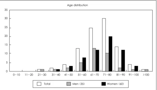
심부전증이 동반된 경우는 75명(78.9%)이었으며 그 중 특 발성 확장성 심근병증 31명(32.6%), 고혈압성 심부전 15명 (15.8%), 허혈성 심장 질환 14명(14.7%), 판막 질환에 의한 심부전 2명(2.1%), 이외 원인이 불분명한 예가 13명(13.7%), 이었으며, 특히 어떠한 기질성 심장 질환을 동반하지 않은 고 립성 cLBBB 환자는 20명(21.1%)였다. 나이별 분포는 Fig. 1 과 같았다(Fig. 1). 성별 및 신장 체중, BMI등과 QRS폭과 는 유의한 상관관계가 없었으며, NYHA class에 따른 호흡
Table 1. Patient characteristics and QRS duration
Correlation with QRS duration Mean±SD*
r p
Demographic
Age (years) 68.51±13.38 -0.222 <0.050
Weight (kg) 64.08±10.23 -0.085 <0.414
Height (cm) 163.19±8.27--- -0.004 <0.968
Body mass index 24.04±2.63-- -0.150 <0.148
Body surface area 1.69±0.16- -0.48 <0.642
Clinical data
Blood pressure Systolic (mmHg) 131.34±21.34-- -0.021 <0.837
Diastolic (mmHg) 77.17±11.56 -0.107 <0.303
Lipid profiles TC (mg/dL) 210.37±53.43- -0.23 <0.824
TG (mg/dL) 155.05±67.395 -0.028 <0.784
HDL (mg/dL) 52.43±21.40 -0.271 <0.108
LDL (mg/dL) 127.31±52.61- -0.162 <0.119
*: p<0.05 versus control. TC: total cholesterol, TG: triglyceride, HDL: high density lipoprotein cholesterol, LDL: low density lipoprotein cholesterol
Fig. 1. Age distribution.
0-10 11-20 21-30 31-40 41-50 51-60 61-70 71-80 81-90 91-100 >100 35
30 25 20 15 10 5 0
Total Men (35) Women (60) Age distribution
곤란의 정도와 QRS폭과도 연관성은 없었다. 또한 수축기 및 이완기 혈압, 고지질혈증과 QRS 폭과도 연관성은 없었다 (Table 1).
심전도 지표와 QRS폭과의 관계
QRS폭과 축(Axis)의 비교에서는 P 파(r=-0.129, p=0.214), T 파(r=0.040, p=0.699)의 축과는 상관관계를 보이지 않았 지만 R 파(r=0.226, p<0.005)의 축과는 유의한 상관관계 를 보였다. QRS폭과 PR간격(r=0.266, p<0.001), QT간격 (r=0.280, p<0.001), QTc간격(r=0.513, p<0.001)은 유의한 양의 상관관계를 보였다. QRS폭과 각 유도의 전압(mV)과의 관계는 흉부유도 V3(r=0.018, p=0.243), V4(r=0.290, p=
0.004), V5(r=0.226, p=0.028) 들은 유의한 상관관계를 보 였지만 그 외 다른 유도에서는 유의한 상관관계가 없었다 (Table 2).
심초음파 지표와 QRS폭과의 관계
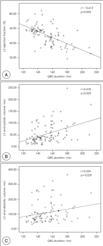
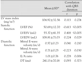
QRS폭과 좌심실 구혈율(LV ejection fraction)과는 통계 적으로 유의한 음의 상관관계를 보였다(r=-0.613, p<0.005).
좌심실 수축기말 용적 및 이완기말 용적(r=0.418, p<0.005) (r=0.224, p=0.029)(Fig. 2)과는 양의 상관관계를 보였으며 이 밖에 도플러 심초음파를 이용한 심실 이완 기능의 지표로 서 승모판에서 조기 이완기 최대 혈류속도(peak E velocity) (r=0.210, p=0.041)와 심방 수축기 최대 혈류속도(peak A velocity)(r=-0.223, p=0.030) 및 E/A비(r=0.278 p=0.006) (Fig. 3)와도 유의한 상관관계를 보였다(Table 3).
QRS duration과 연관된 다변량 분석 결과 95% CI에서 심
초음파 인자중 좌심실 구혈율(odds ratio, 0.839[95% con- fidence interval, 0.751~0.937])만이 독립적인 인자로 작 용하는 것으로 나타났다(Table 4).
Table 2. Electrocardiographic parameters and QRS duration Correlation with
QRS duration Mean±SD*
r p
Axis P (°) -4.55±37.01 -0.129- <0.214 R (°) 123.47±77.640 0.226 <0.027 T (°) 73.64±13.90 0.040 <0.699 Duration PR (ms) 176.33±32.880 0.266 <0.010 QRS (ms) 152.40±18.460
QT (ms) 431.43±58.780 0.280 <0.010 QTc (ms) 475.29±39.790 0.513 <0.010 Voltage Lead I (mV) 9.31±4.06 0.244 <0.172 Lead II (mV) 9.41±4.08 0.144 <0.165 Lead III (mV) 10.51±4.470 0.198 <0.054 V 1 (mV) 24.29±9.830 0.146 <0.157 V 2 (mV) 35.03±12.68 0.010 <0.921 V 3 (mV) 34.85±13.25 0.243 <0.018 V 4 (mV) 23.15±12.47 0.290 <0.004 V 5 (mV) 15.74±10.18 0.226 <0.028 V 6 (mV) 15.47±8.200 0.063 <0.543
*: p<0.05 versus control
80.00
60.00
40.00
20.00
LV ejection fraction (%)
120 140 160 180 200 220 QRS duration (ms)
r=-0.613 p<0.005
A
250.00
200.00
150.00
100.00
50.00
0.00
LV end-systolic volume (mL)
120 140 160 180 200 220 QRS duration (ms)
r=0.418 p<0.005
B
Fig. 2. Echocardiographic Systolic function and QRS duration. A:
relationship between LVEF and QRS duration. B: relationship bet- ween LVESV and QRS duration and C: relation between LVEDV and QRS Duration. LVEF: left ventricular ejection fraction (%), LVESV: left ventricular end systolic volume (mL), LVEDV: left ventricular end diastolic volume (mL), LV: left ventricular.
400.00
300.00
200.00
100.00
0.00
LV end-diastolic volume (mL)
120 140 160 180 200 220 QRS duration (ms)
r=0.224 p=0.029
C
관상동맥 질환 정도와 QRS폭과의 관계
관상동맥 조영술을 시행한 33명 중에서 정상, 한 혈관, 두 혈관, 세 혈관 질환은 각각 17명(51.5%), 8명(24.2%), 4명
(12.1%), 14명(12.1%)을 보였으며 관상동맥 질환의 중등도와 QRS 폭과는 상관 관계가 없었다.
고 찰
cLBBB 환자의 역학적·임상적 특성
본 연구에서는 여성이 더 많았으며(63.1%), 연령이 증가 함에 따라 cLBBB의 수가 증가하였다. 이것은 연령의 증가 에 따라서 cLBBB의 발생빈도가 증가한다는 몇몇 문헌1-3) 보고와 유사한 소견이며, 최근 Heese 등20)은 관상 동맥 질 환이 의심되어 운동부하 검사를 시행한 환자들을 대상으로 6년 동안 추적 관찰 하였는데 cRBBB가 있는 환자는 남자와 관상동맥질환에서 더 많았지만, cLBBB는 오히려 여자에서 더 많이 나타나는 것으로 보고 하였다.
심부전증에 있어서 사망률과 QRS폭과의 연관성에 대한 많은 연구21-23)가 있었지만 그 결과에 있어서는 아직 논란이 많다. 심부전증 환자를 5년간 추적 관찰한 연구22)에서 QRS
Table 3. Echocardiographic parameters and QRS duration Correlation
with QRS duration Mean±SD*
r p LV mass index
(mg/m2) 104.92±32.560 -0.113 <0.274 Systolic
function LVEF (%) 50.69±12.70 -0.613 <0.005 LVESV (mL) 55.37±41.35 -0.418 <0.005 LVEDV (mL) 108.81±71.030 -0.224 <0.029 Diastolic
function
Mitral E-wave
velosity (m/s) 0.57±0.23 -0.041 <0.210 Mitral A-wave
velosity (m/s) 0.71±0.25 -0.223 <0.030 E/A ratio 1.01±0.76 -0.278 <0.006 DT (ms) 241.13±70.180 -0.093 <0.373
*: p<0.05 versus control. LV mass index: left ventricular mass index, LVEF: left ventricular ejection fraction, LVESV: left ventri-cular end systolic volume, LVEDV: left ventricular end diastolic volume, DT: deceleration time
Table 4. Multivariate analysis for variables associated with QRS du- ration greater than median (150 msec)
Odds ratio (95% CI) p Systolic
function LVEF (%) 0.839 (0.751-0.937) 0.002 LVESV (mL) 0.968 (0.904-1.037) 0.356 LVEDV (mL) 1.021 (0.987-1.055) 0.227 Diastolic
function
Mitral E-wave
velosity (m/s) 34.598 (0.218-5494.920) 0.171 Mitral A-wave
velosity (m/s) 0.013 (0.000-2.076) 0.093 E/A ratio 0.193 (0.018-2.073) 0.174 DT (ms) 0.997 (0.989-1.005) 0.486 CI: confidence interval, LVEF: left ventricular ejection fraction, LVESV: left ventricular end systolic volume, LVEDV: left ventricular end diastolic volume
Fig. 3. Echocardiographic diastolic function and QRS duration. A:
relationship between mitral E-wave velocity and QRS duration. B:
relation between mitral A-wave velocity and QRS duration, and C:
relation between E/A ratio and QRS Duration.
3.0
2.0
1.0
0.0
E/A ratio
120 140 160 180 200 220 QRS duration (ms)
r=-0.278 p=0.006
C
1.20 1.00 0.80 0.60 0.40 0.20 0.00
Mitral E wave velocity (m/s)
120 140 160 180 200 220 QRS duration (ms)
r=0.210 p=0.041
A
1.40 1.20 1.00 0.80 0.60 0.40 0.20 0.00
Mitral A wave velocity (m/s)
120 140 160 180 200 220 QRS duration (ms)
B
r=0.223 p=0.030
폭이 증가 할수록 사망률이 증가하며, QRS폭의 증가가 사 망률의 증가와 독립적인 예후인자로 나타났지만 또 다른 연 구23)에서는 이것을 증명하지 못하였다. 이처럼 서로 상반된 결과를 나타내는 것은 심부전증의 원인질환에 따른 차이점 과 확장성 심근병증이 있는 환자에서 엔지오텐신 전환효소 억제제를 더 많이 사용했고 허혈성 심장 질환 환자에서는 항 혈소판제 나 β-차단제를 더 많이 사용했다는 차이점도 이 러한 상충된 결과에 관여했으리라 보여진다.24)
본 연구에서는 심부전증 환자 75명 중 확장성 심근병증 과 허혈성 심장 질환이 각각 41.3%와 18.7%으로 나타났으 며 이는 향후 추적관찰을 통해 약제에 의한 사망률이나 예 후인자 등에 대한 연구가 필요하다.
심초음파 지표
본 연구에서 QRS폭은 좌심실 구혈율, 좌심실 수축기말 용 적 및 이완기말 용적 이외에도 승모판에서 조기 이완기 최 대 혈류속도(peak E velocity)와 심방 수축기 최대 혈류속 도(peak A velocity) 및 E/A 비등의 좌심실 이완 기능 지 표들과도 상관관계가 있었다. 그러나 여러 심초음파 인자 에 대한 다변량 분석에서는 오직 좌심실 구혈률(odds ratio, 0.839[95% confidence interval, 0.751~0.937])만이 의미 있는 변수로 나타났다.
최근 QRS폭이 120 ms 미만에서는 좌심실 수축기능 저 하와 연관성이 적었으나 QRS폭이 170 ms 이상에서는 수축 기능 저하와 함께 사망률이 현저히 증가함을 관찰하여 QRS 폭이 150 ms 초과하는 환자에서 심실 재동기화가 치료효 과가 있다고 보고하고 있다.25) 좌심실 수축기능장애나 심실
내 전도시간 지연26)27)이 있는, 약물치료에 반응하지 않는 심부전에 대해 최근 심실 재동기화는 새로운 치료 방법으 로 제시되고 있다. 아직까지 장기적인 연구결과가 미흡하 기는 하지만 진행중인 일부 연구에서 심실 재동기화가 2년 정도 지난 후에도 그 효과가 유지되고 있어 이뇨제, 1) 안 지오텐신 전환효소 억제제의 지속적인 약물치료에도 불구 하고 NYHA class III 또는 IV 이며, 2) 좌심실의 구혈율 35% 미만이면서, 좌심실 이완기말 직경 60 mm 초과하는 좌심실 수축기능 장애, 3) 표면 심전도에서 QRS 폭이 120 ms를 초과하는 심실 내 전도시간 지연이 있는 심한 심부전 증 환자에서 심실 재동기화를 고려해보는 것이 바람 직 할 것으로 보인다.5)
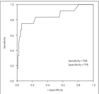
본 연구에서 좌심실의 구혈율을 35% 미만으로 예견하는 QRS폭은 158 msec 이상에서 73%의 민감도와 77%의 특이 도를 보였다는 것에 의미를 부여 할 수 있다(Fig. 4). 즉 이 러한 결과는 cLBBB 환자에서 QRS폭은 심부전의 진행 정도 와 밀접한 관련이 있음을 시사하고 있다.
제한점
본 연구의 제한 점으로 cLBBB가 나타난 모든 환자에서 관 상동맥 혈관조영술을 시행하지 않았다는 점이며 이를 위해 안정 시와 부하시의 cLBBB의 변화를 설명하기 위해 부하 초음파나 신티그라피와 같은 역동학적 검사를 추가적으로 시행해야 할 것이다.28) 두번 째로 본 연구는 한 시점에서 시 행된 cross-sectional study로서 전 예에서 환자의 임상 양 상, 심전도 검사 및 심장초음파 검사 등의 추적검사가 이루 어지지 않았으며 또한 proBNP(N-terminal-pro B-type natriuretic peptide) 등과의 연관성이 비교 되지 않았다.
Bruch 등29)은 심한 이완 기능 장애, 좌심실 충만압 증가, 높은 NT-proBNP와 연관이 있다 하였다. 하지만 본 연구 에서 심전도의 QRS폭(중앙값 150 msec)과 연관된 다변량 분석(Multivariate analysis, 95% CI: confidence interval) 에서 심초음파 지표중 좌심실 구혈율만이 독립적인 인자로 작용하는 것으로 나타났으며 이를 위해서는 추적 심초음파 를 통한 연구가 필요하다. O¨zdemir 등30)은 cLBBB에서 좌 심실 수축기능 및 이완기능을 동시에 반영하는 myocardial performance index(MPI)를 이용하여 MPI가 증가함에 따 라 cLBBB 환자에서 좌심실 기능이 악화됨을 보고하였는데 향후 MPI, tissue Doppler 등과 QRS폭과의 비교연구가 도 움이 될 수 있을 것으로 사료된다.
결론적으로 cLBBB를 갖는 환자에서 QRS폭은 좌심실기 능, 특히 좌심실 구혈율과 밀접한 연관이 있으며, 158 msec 이상의 QRS폭을 보이는 cLBBB 환자는 중증의 수축기능 장 애를 시사한다. 그러므로 수축기 및 이완기 심부전증의 진행 을 반영하는 중요한 예측 인자로서 추적 관찰 시 QRS폭의 평가가 꼭 포함되어야 할 것이다.
Fig. 4. ROC curve. The QRS duration ≥158 msec shows a sensiti- vity of 73% and a specificity of 77% for predicting of LV EF <35%.
ROC: receiver-operating characteristic.
Sensitivity=73%
Specificity=77%
1.0
0.8
0.6
0.4
0.2
0.0
0.0 0.2 0.4 0.6 0.8 1.0 1-Specifficity
Sensitivity
요 약
배경 및 목적:
완전 좌각 차단의 심전도소견은 심혈관계의 악화 예후인 자로 밝혀진바 이번 연구의 목적은 심전도에서 완전 좌각 차단을 보이는 환자에서 QRS폭과 좌심실의 기능과의 관계 를 규명하고자 하였다.
방 법:
완전 좌각 차단 환자 95명(남:여=35:60, 평균나이=69
±13세)을 대상으로 역학적·임상적 자료, 심전도 및 심초 음파를 이용한 수축기능과 이완기능의 지표를 분석하였다.
결 과:
QRS폭과 심실 수축기능의 지표인 좌심실의 구혈율(LV ejection fraction)과는 음의 상관관계(r=-0.613, p<0.005) 를, 좌심실 수축기말 용적(r=0.418, p=0.005) 및 이완기말 용적(r=0.224, p=0.029)과는 양의 상관관계를 보였으며 심 실 이완기능 지표로 승모판에서 조기 이완기 최대 혈류속도 (peak E velocity)(r=0.210, p=0.041)와 심방 수축기 최대 혈류속도 (peak A velocity)(r=-0.223, p=0.030) 및 E/A 비(r=-0.278, p=0.006)와도 유의한 상관관계를 보였다. QRS duration과 연관된 다변량 분석 결과 95% CI에서 심초음파 인자중 좌심실 구혈율(odds ratio, 0.839[95% confidence interval, 0.751-0.937])만이 독립적인 인자로 작용하는 것 으로 나타났다(Table 4).
좌심실의 구혈율(LV ejection fraction)을 35% 미만으로 예견하는 QRS폭은 158 msec 이상에서 73%의 민감도와 77%
의 특이도를 보였다.
결 론:
완전 좌각 차단에서 QRS폭은 좌심실의 수축기능과 밀접 한 관련이 있으며 QRS폭은 심부전증의 진행정도를 예측할 수 있는 중요한 인자이다.
중심 단어:완전 좌각 차단;QRS폭;좌심실 기능.
REFERENCES
1) Fahy GJ, Pinski SL, Miller DP, et al. Natural history of isolated bundle branch block. Am J Cardiol 1996;77:1185-90.
2) Hardarson T, Arnason A, Eliasson GJ, Palsson K, Eyjolfsson K, Sigfusson N. Left bundle branch block prevalence, incidence, follow-up and outcome. Eur Heart J 1987;8:1075-9.
3) Jeong GH, Kim JH, Park YH, et al. Incidence and risk factors for bundle branch block in adults older than 40 years. Korean J Intern Med 2004;19:171-8.
4) Cazeau S, Leclercq C, Lavergne T, et al. Effects of multisite biventricular pacing in patients with heart failure and intra- ventricular conduction delay. N Engl J Med 2001;344:873-80.
5) Gregoratos G, Abrams J, Epstein AE, et al. ACC/AHA/NASPE 2002 guideline update for implantation of cardiac pacemakers and antiarrhythmia devices: summary article: a report of the american college of cardiology/american heart association task force on practice guidelines (ACC/AHA/NASPE committee to update the 1998 pacemaker guidelines). Circulation 2002;106:
2145-61.
6) Chou T. Electrocardiography in Clinic Practice. 3rd ed. Phila- delphia: W.B. Saunders Company; 1991. p.87-98.
7) DuBois D, DuBois EF. A formula to estimate the approximate surface area if height and weight be known. Arch Intern Med 1916;17:863-71.
8) Goldman L, Hashimoto B, Cook EF, Loscalzo A. Comparative reproducibility and validity of systems for assessing cardiova- scular functional class: advantages of a new specific activity scale. Circulation 1981;64:1227-34.
9) Ho KK, Anderson KM, Kannel WB, Grossman W, Levy D. Sur- vival after the onset of congestive heart failure in Framingham heart study subjects. Circulation 1993;88:107-15.
10) Expert Committee on the Diagnosis and Classification of Dia- betes Mellitus. Report of the expert committee on the diagnosis and classification of diabetes mellitus. Diabetes Care 2003;26:
S5-20.
11) Chobanian AV, Bakris GL, Black HR, et al. The seventh report of the Joint National Committee on Prevention, Detection, Evalua- tion, and Treatment of High Blood Pressure. Hypertension 2003;
42:1206-52.
12) Expert Panel on Detection Evaluation, and Treatment of High Blood Cholesterol in Adults. Executive summary of the third report of the National Cholesterol Education Program (NCEP) expert panel on detection, evaluation, and treatment of high blood cholesterol in adults (Adult Treatment Panel III). JAMA 2001;285:2486-97.
13) Warnick GR, Knopp RH, Fitzppatrick V, Branson L. Estimating low-density lipoprotein cholesterol by the Friedewald equation is adequate for classifying patients on the basis of nationally re- commended cutpoints. Clin Chem 1990;36:15-9.
14) Sokolow M, Lyon TP. The ventricular complex in ventricular hypertrophy as obtained by unipolar precordial and limb leads.
Am Heart J 1949;37:161.
15) Sevilla D, Dohrmann ML, Somelofski CA, et al. Invalidation of the resting electrocardiogram obtained via exercise electrode sites as a standard 12-lead recording. Am J Cardiol 1989;63:35-9.
16) Tei C, Ling LH, Hodge DO, et al. New index of combined systolic and diastolic myocardial performance: a simple and repro-duci- ble measure of cardiac function: a study in normal and dilated cardiomyopathy. J Cardiol 1995;26:357-66.
17) Schiller NB, Shah PM, Crawford M, et al. Recommendations for quantitation of the left ventricle by two-dimensional echocardio- raphy. J Am Soc Echocardiogr 1989;2:358-67.
18) Gottdiener JS, Bednarz J, Devereux R, et al. American Society of Echocardiography recommendations for use of echocardiography in clinical trials. J Am Soc Echocardiogr 2004;17:1086-119.
19) Feigenbaum H. Hemodynamic Information Derived from Echo- ardiography. 5th ed. Philadelphia: Lea & Febiger; 1994. p.182- 215.
20) Hesse B, Diaz LA, Snader CE, Blackstone EH, Lauer MS. Com- lete bundle branch block as an independent predictor of all- cause mortality: report of 7,073 patients referred for nuclear exercise testing. Am J Med 2001;110:253-9.
21) Kashani A, Barold SS. Significance of QRS complex duration in patients with heart failure. J Am Coll Cardiol 2005;46:2183-92.
22) Huang X, Shen W, Gong L. Clinical significance of complete left bundle-branch block in dilated cardiomyopathy. Chin Med Sci J 1995;10:158-60.
23) Juilliere Y, Barbier G, Feldmann L, Grentzinger A, Danchin N, Cherrier F. Additional predictive value of both left and right ventricular ejection fractions on long-term survival in idiopathic dilated cardiomyopathy. Eur Heart J 1997;18:276-80.
24) al-Khadra AS, Salem DN, Rand WM, Udelson JE, Smith JJ,
Konstam MA. Antiplatelet agents and survival: a cohort analysis from the studies of left ventricular dysfunction (SOLVD) trial. J Am Coll Cardiol 1998;31:419-25.
25) Das MK, Cheriparambil K, Bedi A, et al. Prolonged QRS dura- ion (QRS ≥170 ms) and left axis deviation in the presence of left bundle branch block: a marker of poor left ventricular systolic function? Am Heart J 2001;142:756-9.
26) Lee MY, Rho TH, Yoo KD, et al. The relationship between QRS duration and LV systolic function in paced patient. Korean Circ J 2001;31:327-34.
27) Park CS, Youn HJ, Cho EJ, et al. Influence of left bundle branch block on left anterior aescending coronary artery flow: study
using transthoracic Doppler echocardiograph. Korean Circ J 2003;33:484-90.
28) Rlee KJ. Stress scintigraphy in patients with left bundle branch block. Korean Circ J 1993;23:702-6.
29) Bruch C, Stypmann J, Grude M, Gradaus R, Breithardt G, Wichter T. Left bundle branch block in chronic heart failure-impact in diastolic function, filling pressure, and B-type natriuretic peptide levels. J Am Soc Echocardiogr 2006;19:95-101.
30) Özdemir K, Altunkeser BB, Danis G, et al. Effect of the isolated left bundle branch block on systolic and diastolic functions of left ventricle. J Am Soc Echocardiogr 2001;14:1075-9.