• 검색 결과가 없습니다.

Effect of Lacidipine on Blood Pressure and Endothelial Function in Mild-to-Moderate Essential Hypertension Patients With Diabetes in Korea

N/A
N/A
Protected

Academic year: 2022

Share "Effect of Lacidipine on Blood Pressure and Endothelial Function in Mild-to-Moderate Essential Hypertension Patients With Diabetes in Korea"

Copied!
7
0
0

로드 중.... (전체 텍스트 보기)

전체 글

(1)

632

Open Access

Effect of Lacidipine on Blood Pressure and Endothelial Function in Mild-to-Moderate Essential Hypertension Patients

With Diabetes in Korea

Dae-Hee Kim, MD

1

*, Il-Young Oh, MD

1

, Hae-Young Lee, MD

1

, Yong-Jin Kim, MD

1

, Hyo-Soo Kim, MD

1

, Cheol-Ho Kim, MD

1

, Byung-Hee Oh, MD

1

, Kwon-Sam Kim, MD

2

, Doo-Il Kim, MD

3

, Young-Dae Kim, MD

4

, Kyu-Hyung Ryu, MD

5

,

Si-Hoon Park, MD

6

, Sang-Hong Baek, MD

7

, Dong-Gu Shin, MD

8

, Wan Joo Shim, MD

9

, Tae-Hoon Ahn, MD

10

, Seok-Kyu Oh, MD

11

, Seung-Hwan Lee, MD

12

, Sung-Yun Lee, MD

13

, Myung-Ho Jeong, MD

14

, Wook-Sung Chung, MD

7

, Jun-Young Jeong, MD

15

, So-Yeon Choi, MD

16

, Si-Wan Choi, MD

17

and Min-Su Hyon, MD

18

1

Department of Internal Medicine, College of Medicine, Seoul National University, Seoul,

2

Department of Internal Medicine, Kyung Hee University College of Medicine, Seoul,

3

Department of Internal Medicine, Inje University College of Medicine, Busan Paik Hospital, Busan,

4

Department of Internal Medicine, Dong-A University College of Medicine, Busan,

5

Department of Internal Medicine, Konkuk University College of Medicine, Seoul,

6

Department of Internal Medicine,

Ewha Womans University College of Medicine, Mokdong Hospital, Seoul,

7

Department of Internal Medicine, The Catholic University College of Medicine, Seoul St. Mary’s Hospital, Seoul,

8

Department of Internal Medicine, Yeungnam University College of Medicine, Daegu,

9

Department of Internal Medicine, Korea University College of Medicine, Anam Hospital, Seoul,

10

Department of Internal Medicine, Gachon University Gil Hospital, Incheon,

11

Department of Internal Medicine, Wonkwang University College of Medicine, Iksan,

12

Department of Internal Medicine, Yonsei University Wonju Christian Hospital, Wonju,

13

Department of Internal Medicine, Inje University College of Medicine, IIsan Paik Hospital, Goyang,

14

Department of Internal Medicine, Chonnam National University College of Medicine, Gwangju,

15

Department of Internal Medicine, Eulji University College of Medicine, Daejeon,

16

Department of Internal Medicine, Ajou University College of Medicine, Suwon,

17

Department of Internal Medicine, Chungnam National University College of Medicine, Daejeon,

18

Department of Internal Medicine, Soonchunhyang University College of Medicine, Seoul, Korea

ABSTRACT

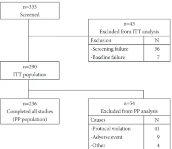
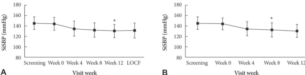
Background and Objectives: The aim of this study was to evaluate the efficacy of lacidipine in reducing blood pressure (BP) and to determine its effect on endothelial function in mild-to-moderate hypertensive patients with type 2 diabetes mellitus (DM). Subjects and Methods: This was a prospective, multicenter, open-label, single-arm study, enrolling 290 patients with mild-to-moderate hypertension and type 2 DM. Patients were initially treated with 2 mg lacidipine orally once daily for 4 weeks, which was then increased as necessary every 4 weeks to a maximal dose of 6 mg daily. The primary endpoint was the mean change in systolic blood pressure (SBP) from baseline after 12 weeks of treatment. Secondary endpoints included mean changes in diastolic blood pressure (DBP), flow-mediated vasodilatation (FMD), and serum concentrations of biochemical markers such as high-sensitivity C-reactive protein (hs-CRP), monocyte chemo-attractant protein-1 (MCP-1), matrix metal- loproteinase-9 (MMP-9), and plasminogen activator inhibitor-1 (PAI-1). Results: Lacidipine treatment significantly reduced SBP by -13.4±13.0 mmHg (p<0.001) and DBP by -6.2±9.3 mmHg (p<0.001). Lacidipine treatment did not improve endothe- lial-dependent vasodilatation, despite significantly improved nitroglycerin-induced, endothelial-independent vasodilatation.

MCP-1 levels significantly decreased from 283.66±110.08 pg/mL to 257.83±100.23 pg/mL (p<0.001); whereas there were no significant changes in the levels of hs-CRP, MMP-9, or PAI-1. Conclusion: Twelve weeks of treatment with lacidipine was ef- fective and well tolerated in mild-to-moderate hypertensive patients with type 2 DM. In spite of inducing a significant reduc- tion in MCP-1 levels, lacidipine did not improve endothelial function. (Korean Circ J 2010;40:632-638)

KEY WORDS: Lacidipine; Diabetes mellitus; Hypertension; Endothelium.

Received: March 3, 2010 / Accepted: May 27, 2010

Correspondence: Byung-Hee Oh, MD, Department of Internal Medicine, Seoul National University College of Medicine, 101 Daehak-ro, Jongno-gu, Seoul 110-744, Korea

Tel: 82-2-2072-3345, Fax: 82-2-2072-2577, E-mail: [email protected]

*Dr. Dae-Hee Kim is now affiliated with Department of Cardiology, Asan Medical Center, University of Ulsan College of Medicine, Seoul, Korea

cc

This is an Open Access article distributed under the terms of the Creative Commons Attribution Non-Commercial License (http://creativecommons.org/licens-

es/by-nc/3.0) which permits unrestricted non-commercial use, distribution, and reproduction in any medium, provided the original work is properly cited.

(2)

Introduction

The prevalence of hypertension in patients with diabetes mellitus (DM) is extremely high (20-60%).

1)

Moreover, comor- bid hypertension has been widely reported to increase the risk of cardiovascular events, which account for 86% of the deaths occurring among patients with DM; it is also known to in- crease the incidence of microvascular complications. How- ever, it has been reported that only 28% of diabetic patients have adequately controlled blood pressure (BP) under 130/80 mmHg.

2)

This control rate is even lower (21.6%) among Ko- rean patients.

3)

Although recent guidelines recommend an- giotensin receptor antagonists or angiotensin-converting enzy- me inhibitors as frontline antihypertensive agents in patients with DM, most patients eventually require more than 2 classes of antihypertensive agents to achieve target BP.

4)

Lacidipine, a long-acting dihydropyridine calcium channel blocker, has been reported to reverse endothelial dysfunction,

5)

and to cause regression of atherosclerosis.

6)

The European La- cidipine Study on Atherosclerosis demonstrated that 4 years of lacidipine treatment retarded the progression of athero- sclerosis more effectively than atenolol. Investigators suggest- ed that this effect was due to restoration of endothelial func- tion and reduction in oxidative stress.

7)

However, the effects of lacidipine in type 2 DM patients have been only reported from small-scale, nonblinded studies.

8)

Fur- thermore, there is a paucity of data regarding the effects of la- cidipine on endothelial dysfunction, which is observed in most patients with DM.

In this study, we evaluated the efficacy of lacidipine in reduc- ing BP, as well as its effect on endothelial function, in mild-to- moderate hypertensive patients with type 2 DM.

Subjects and Methods

Study population

A subject was only eligible for inclusion in this study if all the following criteria applied: 1) male or female 35 to 75 years of age at screening, 2) newly diagnosed essential hypertension or essential hypertension untreated in the 2 months prior to screening, and 3) type 2 DM (ADA criteria 2004). Any sub- jects taking antihypertensive medications had to undergo a 2-week washout period before enrollment. The definition of hypertension was a sitting systolic blood pressure (SiSBP) of

≥130 mmHg, measured with a BP cuff, because the SBP goal is less than 130 mmHg in patients with diabetes. Exclusion criteria were as follows: severe hypertension with SiSBP >180 mmHg; or any serious disorder that could limit the ability of the patient to participate in the study, including severe coro- nary artery disease, uncontrolled DM (hemoglobin A1C >11%), or secondary hypertension.

Study design

This was a multicenter, open-label, single-arm study per- formed at 20 sites in the Republic of Korea. The study proto- col was approved by the institutional review board at each site. Before entering the study, patients provided written, in- formed consent. A medical history was obtained during the screening phase, and a physical examination, 12-lead electro- cardiogram, complete blood count, serum biochemistry, and routine urinalysis were performed at each center. During the 12-week treatment period, patients were initially treated with lacidipine 2 mg orally once daily for 4 weeks, and then were ti- trated with increasing doses every 4 weeks to a maximal dose of 6 mg daily if the SiSBP did not decrease to <130 mmHg.

Blood pressure measurement

At each visit, the SiSBP, sitting diastolic blood pressure (Si- DBP), pulse rate, and body weight were recorded. To ensure consistency of trough values, these measurements were taken at the same time of day, before the next dose of lacidipine. At each center, a single investigator used the standard ausculta- tory technique for measuring BP in each patient’s arm, using a mercury sphygmomanometer produced by one company (W. A. Baum Co. Inc, Copiague, NY, USA) and an appropri- ately-sized BP cuff.

Measurement of flow-mediated dilatation of the brachial artery

The examination was conducted at each center by the same examiner throughout the study, using a standard technique.

9)

Briefly, the diameter of the artery was measured using high- resolution, 2-dimensional images obtained by ultrasound with a 7.5-MHz linear-array transducer. Subjects were asked not to smoke or drink coffee or tea for at least 2 hours before the scan was performed. The right brachial artery was scanned over a longitudinal section 3 to 5 cm above the right medial elbow for the first resting image. The diameter of the brachial artery was measured from the anterior to the posterior inter- face between the media and adventitia (“m line”) at a fixed dis- tance.

The mean brachial artery diameter was calculated from 4

cardiac cycles synchronized with R-wave peaks on the electro-

cardiography. A pneumatic tourniquet placed around the fo-

rearm distal to the target artery was then inflated to a pres-

sure of 250 mmHg, and inflation was maintained for 5 min-

utes. Increased flow was then induced by sudden cuff defla-

tion. A second scan was performed continuously for 60 se-

conds before, and for 120 seconds after cuff deflation. Then,

15 minutes later, another resting image was recorded. A ni-

troglycerin tablet (0.4 mg) was then administered sublingually,

and 3 minutes later the last scan was performed. All the im-

ages were stored in DICOM format for offline analysis, and

then sent to the core laboratory. Two expert sonographers an-

(3)

alyzed all the images. Interobserver coefficients of variation for measuring vessel diameters at baseline, after hyperemia, and after nitroglycerin were 3.3±0.25%, 2.9±0.28%, and 3.5±0.22%, respectively.

Blood tests

Blood samples were collected after at least 12 hours of fast- ing. Measurements of lipids, fasting glucose, and high-sensi- tivity C-reactive protein (hs-CRP) were performed in the local laboratories at each center. Other serum biochemical markers were analyzed in the central, core laboratory of GSK Korea using commercially available enzyme-linked immunosorbent assay kits from various vendors as follows: monocyte chemo- attractant protein-1 {(MCP-1); Bio-Rad Laboratories, Hercu- les, CA, USA}, matrix metalloproteinase-9 {(MMP-9); Amer- sham Biosciences, Uppsala, Sweden}, and plasminogen acti- vator inhibitor-1 {(PAI-1); IMUBIND, American Diagnostica, Stamford, CT, USA}.

Efficacy and safety variables

The primary efficacy variable in this study was the mean ch- ange in trough SiSBP from baseline after 12 weeks of treat- ment. Secondary efficacy variables included mean changes in SiDBP, flow-mediated dilatation (FMD), and biochemical mar- kers such as hs-CRP, MCP-1, MMP-9, and PAI-1 after 12 weeks of treatment.

Safety variables included the incidence of all adverse events (AEs), drug-related AEs, and the number of discontinuations.

AEs were evaluated at each visit by physical examination and direct questioning. The seriousness of AEs and their relation to the study drugs were determined by the chief investigator at each center. The results of all laboratory tests were assessed by the investigators for clinical significance and for their pos- sible relationship to the study drug.

Statistical analysis

Effectiveness was defined as >4.5 mmHg decrease in the mean SiSBP from baseline after 12 weeks. We calculated that 321 patients would be required to fulfill the following pa- rameters: standard deviation of 20 mmHg for SiSBP, overall 1-sided significance of 0.05, 90% power, and 20% exclusion rate. In terms of absolute benefit, data from the per-protocol (PP) population were used for the main analysis and those from the intention-to-treat (ITT) population were used as a supplement. The ITT population was used primarily to evalu- ate tolerability.

Continuous variables were analyzed using the t-test. Chan- ges in BPs and serum markers were analyzed using a paired t-test. All statistical analyses were performed with Statistical Package for the Social Sciences (SPSS) 12.0 (SPSS Inc, Chica- go, IL, USA), and p less than 0.05 were considered statistically significant.

Results

Baseline characteristics

A total of 333 patients were screened, and 290 patients (male : female=173 : 117) were enrolled in the study. Of them, 236 pa- tients (81.4%) completed the study. The flow diagram of study patients is summarized in Fig. 1. Eighty subjects (33.9%) were treated with 2 mg of lacidipine throughout the study, 108 (45.8%) were titrated up to 4 mg, and 48 (20.3%) were titrated up to 6 mg.

The mean age in the study group was 56.6±9.2 (35-79) years, and 41% of the patients were over 60 years of age. The most common concomitant medications at baseline and during the study were oral hypoglycemic agents (75%), lipid-lowering agents (29%), antiplatelet agents (40%), and antacids (13%).

The proportions of oral hypoglycemic agents were as follows:

sulfonylureas 58%, biguanides 37%, alpha-glucosidase inhibi- tors 13%, thiazolidinediones 14%, combination of more than two drugs 4%.

Changes in blood pressure from baseline

In the PP population (n=236), SiSBP was significantly de- creased by 13.4±13.0 mmHg (from 144.0±11.4 mmHg to 130.6±12.5 mmHg, p<0.0001) after 12 weeks of treatment.

In the ITT population (n=277), SiSBP also significantly de- creased by 13.0±13.3 mmHg (from 144.2±12.3 mmHg to 130.9±13.1 mmHg, p<0.0001) (Fig. 2).

SiDBP decreased by 6.2±9.3 mmHg (from 88.7±8.8 mmHg to 82.5±9.0 mmHg, p<0.001) in the PP population and by 6.1±9.1 mmHg (from 88.7±8.6 mmHg to 72.0±9.4 mmHg, p<0.001) in the ITT population.

The overall response rate (SiSBP <130 mmHg) after 12 weeks of treatment was 58.5%. In the subgroup with baseline SiSBP greater than 140 mmHg (PP population, n=132), the change in

n=333 Screened

n=43 Excluded from ITT analysis

Exclusion N

-Screening failure 36 -Baseline failure 07 n=290

ITT population

n=236 Completed all studies

(PP population)

n=54 Excluded from PP analysis

Causes N

-Protocol violation 41

-Adverse event 09

-Other 04

Fig. 1. Flow diagram of study patients. ITT: intention to treat, FMD:

flow-mediated dilatation, PP: per-protocol.

(4)

SiSBP was -18.1±13.3 mmHg (from 151.6±9.7 mmHg to 133.5±

13.6 mmHg, p<0.001); whereas in the subgroup with baseline SiSBP less than 140 mmHg (n=104), the change in SiSBP was -7.5±9.8 mmHg (from 134.4±3.0 mmHg to 126.9±10.0 mmHg, p=0.03).

Changes in endothelial-dependent and endothelial-independent vasodilatation

Changes in endothelial-dependent vasodilatation of the brachial artery were analyzed in the PP population. The mean baseline percentage of endothelial-dependent vasodilatation was not related to baseline body mass index, lipids, serum bio- markers, or SiSBP levels (Table 1). After 12 weeks of treatment with lacidipine, the percentage of endothelial-dependent va- sodilatation changed from 5.22±4.91% to 5.52±4.92%, but this did not represent a statistically significant change (p=0.313).

In contrast, endothelial-independent vasodilatation after ad- ministration of sublingual nitroglycerin significantly increased from 13.94±7.21% to 14.97±7.42% (p=0.02). The baseline per- centage of nitroglycerin-induced vasodilatation had a weak negative correlation with MCP-1 levels. All the subjects were divided into 2 subgroups. The FMD-responder group (n=

130, 55.1%) included those patients who had increased en- dothelial-dependent vasodilatation, and the FMD-nonre-

sponder group (n=106, 44.9%) included those patients who had no increase in endothelial-dependent vasodilatation. The FMD-responder group had significantly lower baseline tri- glyceride (TG) levels (150±97 mg/dL vs. 185±156 mg/dL, p=

0.02) and higher baseline high density lipoprotein-cholester- ol (HDL-C) levels (46±11 mg/dL vs. 49±12 mg/dL, p=0.01) (Table 2).

Changes in blood chemistry and biomarkers HbA1C and lipid profiles, including TGs, low density lipo- protein-cholesterol, and HDL-C levels, did not change after 12 weeks of lacidipine treatment. Among the biomarkers evalu- ated, the plasma levels of MCP-1 significantly decreased from 283.66±110.08 pg/mL to 257.83±100.23 pg/mL (p<0.001).

In contrast, hs-CRP did not show any significant change (from 0.51±1.07 mg/dL to 0.55±1.60 mg/dL, p=0.644). The results are presented in Table 2.

Tolerability

More than one AE event was reported in 40% of the pa- tients in the ITT population (a total of 116 events in 290 pa- tients). There were 105 events that were considered to be drug-related. The most common AEs thought to be drug-re- lated were headache (n=16, 5.52%), palpitations (n=6, 2.07%), Fig. 2. Effect of lacidipine in mean sitting cuff- systolic blood pressure (SiSBP) in patients with mild- to- moderate hypertension after 4, 8, and 12 weeks of treatment. The mean change from baseline was significant in intention-to-treat (A) and per-protocol (B) groups (*p<0.001).

LOCF: last observation carried forward.

180 160 140 120 100 80

180 160 140 120 100

Screening Week 0 Week 4 Week 8 Week 12 LOCF 80 Screening Week 0 Week 4 Week 8 Week 12

Visit week Visit week

Si SB P (m m H g) Si SB P (m m H g)

* *

A B

Table 1. Correlation coefficients between various baseline values and vasorelaxation

Baseline endothelial-dependent vasodilatation Baseline endothelial-independent vasodilatation

Correlation coefficient p Correlation coefficient p

BMI -0.037 0.573 -0.069 0.292

LDL-C -0.002 0.977 -0.147 0.52

TG -0.035 0.588 -0.080 0.222

FBS -0.023 0.729 -0.096 0.142

HbA1c -0.002 0.975 -0.117 0.073

MCP-1 -0.058 0.379 -0.146 0.025*

hs-CRP -0.172 0.070 -0.041 0.530

MMP-9 -0.088 0.181 -0.014 0.828

PAI-1 -0.025 0.698 -0.053 0.414

Systolic BP -0.026 0.687 -0.025 0.703

BMI: body mass index, LDL-C: low density lipoprotein-cholesterol, TG: triglyceride, FBS: fasting blood glucose, MCP-1: monocyte chemoat-

tractant protein-1, MMP-9: matrix metalloproteinase-9, PAI-1: plasminogen activator inhibitor-1

(5)

facial flushing (n=5, 1.72%), dizziness (n=4, 1.38%), and chest pain (n=4, 1.38%). There were 11 severe AEs reported in 9 pa- tients (3.10%). All of the severe events required hospitaliza- tion, and included 2 cases of chest pain, and 1 case each of la- ryngeal cancer, bladder cancer, back pain, hypoglycemia, he- morrhoids, cerebral hemorrhage, depression, benign prostatic hyperplasia, and impaired glucose tolerance. All severe AEs were thought to be unrelated to drug therapy.

Discussion

This study was performed to evaluate the efficacy and tol- erability of lacidipine in mild-to-moderate hypertensive pa- tients with type 2 DM. To the best of our knowledge, this is

the largest study evaluating the effect of calcium antagonists on endothelial function in hypertensive patients with DM.

Lacidipine treatment effectively reduced SBP and DBP.

When the dose was titrated up to 6 mg once daily, the over- all response rate after 12 weeks of treatment reached 58.5%.

Although this study did not have a comparative design, the extent of lacidipine-induced SiSBP reduction in our Korean population was comparable to the effects of lacidipine seen in other races,

10)11)

as well as to the effects of other antihyper- tensive agents, such as nifedipine,

12)

atenolol,

13)

thiazide,

14)

is- radipine,

15)

and amlodipine

16)

in Korean patients.

The most interesting findings in this study were related to the role of lacidipine in the improvement of endothelial dys- function in hypertensive patients with DM. Although a few Table 2. Baseline values and changes in the levels of glucose, lipids, serum markers, and SiSBP, stratified according to vasorelaxation

All

Endothelial-dependent vasodilatation Endothelial-independent vasodilatation Decreased (≤)

(n=130)

Increased (>) (n=106)

Decreased (≤) (n=141)

Increased (>) (n=95)

HbA1C (%) 0 week 7.12±1.25 6.9±1.1 07.3±1.3* 7.1±1.3 07.1±10.2

12 weeks 7.15±1.31 6.9±1.1 07.3±1.4* 7.1±1.4 7.2±1.3

p NS NS NS NS NS

TG (mg/dL) 0 week 165±111 185±125 150±97* 157±940 171±122

12 weeks 166±115 183±141 153±86* 169±133 164±103

p NS NS NS NS NS

LDL-C (mg/dL) 0 week 112±32 110±320 113±330 117±330 108±320

12 weeks 112±31 111±290 113±330 114±290 111±320

p NS NS NS NS NS

HDL-C (mg/dL) 0 week 48±12 46±11 049±12* 45±10 *49±12*

12 weeks 49±13 46±10 051±14* 47±11 50±13

p NS NS NS NS NS

hs-CRP (mg/dL) 0 week 0.51±1.10 0.39±0.59 0.60±1.33 0.60±1.48 0.45±0.67

12 weeks 0.55±0.10 0.40±0.83 0.68±2.01 0.66±2.11 0.48±1.14

p NS NS NS NS NS

MCP-1 (pg/mL) 0 week 283.7±7.200 292.1±115.2 276.8±105.7 275.6±95.10 289.1±119.2

12 weeks 257.8±6.500 261.7±92.60 254.7±106.3 259.0±95.10 257.0±103.9

p <0.001 0.005 0.024 0.06 0.002

MMP-9 (pg/mL) 0 week 626.1±372.7 644.5±383.1 611.0±364.8 614.2±320.0 634.2±405.5

12 weeks 611.7±324.4 592.3±348.7 627.6±303.4 565.7±296.4 645.2±339.6

p NS NS NS NS NS

PAI-1 (pg/mL) 0 week 29.6±27.9 27.6±26.5 31.2±29.1 28.4±27.5 30.1±28.3

12 weeks 31.7±26.4 33.1±27.7 30.7±25.3 31.0±26.6 32.3±26.4

p NS NS NS NS NS

SiSBP (mmHg) 0 week 144.0±11.40 143.9±11.20 144.2±11.60 144.2±11.70 143.8±11.00

12 weeks 130.6±12.50 129.3±13.60 131.7±11.60 129.9±12.90 131.1±12.30

p <0.001 <0.001 <0.001 <0.001 <0.001

ΔSiSBP (mmHg) -13.5±13.0- -14.5±14.1- -12.5±12.00 -14.0±12.90 -13.1±12.30

*p<0.05 between the increase group and the no increase group. TG: triglyceride, LDL-C: low density lipoprotein-cholesterol, hs-CRP: high-

sensitivity CRP, MCP-1: monocyte chemoattractant protein-1, MMP-9: matrix metalloproteinase-9, PAI-1: plasminogen activator inhibitor-1,

endothelial-independent vasodilatation group, SiSBP: sitting cuff-systolic blood pressure, ΔSiSBP: absolute amount of decrease in SiSBP

(6)

preclinical or small-scale clinical studies have suggested the beneficial effects of lacidipine on endothelial function in non- diabetic subjects,

5)

this is the first study evaluating the effects of lacidipine on endothelial function in diabetic patients with hypertension. Twelve weeks of lacidipine therapy did not im- prove endothelial-dependent vasodilatation, despite the fact that endothelial-independent vasodilatation was significantly improved following administration of sublingual nitroglycer- in. The lack of improvement in endothelial-dependent vaso- dilatation stands in contrast to previous studies

5)

which have shown an improvement in overall endothelial-dependent va- sodilatation in nondiabetic patients. A number of mechanis- ms may be invoked in understanding the discrepancy between our results and those in the nondiabetic population. Impair- ment of endothelial-dependent vasodilatation is more severe in diabetic patients than it is in nondiabetic patients. Thus, 12 weeks of treatment might not be sufficient for amelioration of this impairment. Impairment of endothelial-dependent vaso- dilatation has been widely observed in arteries from diabetic animals,

17)

as well as from normal animals exposed to hyper- glycemia.

18)

In such studies, endothelial-dependent vasodila- tation had already been impaired while endothelial-indepen- dent vasodilatation remained intact. Similarly, in a few animal studies,

19)

as well as in human studies,

20)

a diminished response to nitroglycerin was also observed in diabetes, which was pre- ceded by a disturbed response to acetylcholine. Therefore, im- paired vascular response to nitroglycerin is considered to oc- cur at a more advanced stage of vascular dysfunction in dia- betes. In this study, the baseline values of nitroglycerin-induc- ed vasodilatation were more impaired than previously reported values of normal controls,

21)

which also suggests that the pop- ulation in this study might have had more advanced vascular dysfunction. Similarly, HMG-CoA reductase inhibitors (sta- tins) have been shown to have significant beneficial effects on endothelial dysfunction in nondiabetic subjects.

22)

How- ever, many studies have also failed to show significant statin- induced improvement in endothelial-dependent vasodilata- tion in subjects with type 2 DM,

23)

and only one report

21)

has demonstrated statin-induced improvement in endothelial- independent vasodilatation, as was shown in this study.

In subgroup analysis, the group with increased endotheli- al-dependent vasodilatation had lower TG levels and higher HDL-C levels. An elevated TG concentration may increase the levels of intracellular adhesion molecule-1 and vascular cell ad- hesion molecule-1, which might result in increased inflam- matory responses in endothelial cells.

24)

Additionally, improve- ment in endothelial dysfunction seems to be independent of BP-lowering effects, because there was no difference in the base- line BP or the absolute amount of decrease in SBP between those with FMD improvement and those without improvement.

Finally, we observed that the levels of MCP-1, a surrogate marker for inflammation,

25)

significantly decreased after la-

cidipine treatment. The levels of MCP-1 in hypertensive pa- tients have been reported to correlate with the severity of ca- rotid intima-media thickness (IMT).

26)

In this regard, decre- ased MCP-1 concentrations following lacidipine treatment might be consistent with the previously reported anti-ath- erosclerotic effect of lacidipine.

7)

In this study by contrast, the levels of hs-CRP did not change significantly. This dis- crepant result was consistent with that of a previous study in which patients with coronary artery disease had significantly reduced carotid IMT without a significant change in hs-CRP levels after 6 months of lacidipine treatment.

27)

This discrep- ancy between hs-CRP and MCP-1 has also been demonstrat- ed in other studies of angiotensin receptor blockers.

28)

Levels of PAI-1, a marker of fibrinolytic balance, did not de- crease after lacidipine treatment. PAI-1 levels are elevated in subjects with risk factors for atherosclerosis and insulin resist- ance.

29)

PAI-1 expression is known to be regulated by nitiric ox- ide (NO) activity, and therefore NO synthase activity.

30)

In this regard, the lack of change in mean PAI-1 levels might be consist- ent with the insignificant endothelial-dependent vasodilata- tion changes observed in this study.

There are several limitations to this study. First, this is a sin- gle-arm, uncontrolled study. Owing to ethical concerns regard- ing not administering antihypertensive therapy to patients with confirmed hypertension, we were unable to create a control gr- oup. Second, the measurement and analysis of FMD could be observer-dependent with high variance. To reduce observer-de- pendent bias, two expert sonographers in the core lab analyz- ed all the images. Third, the measurements of lipids, fasting glu- cose, and hs-CRP were performed by local laboratories at each center, and the results could vary between laboratories.

However, the serum biochemical markers, including MCP-1, MMP-9, and PAI-1, were all analyzed in a single central labo- ratory.

In summary, 12 weeks of lacidipine treatment was effective and well tolerated in diabetic patients with mild-to-moderate hypertension. Lacidipine partially improved vasorelaxation, which was associated with reduction in levels of inflammatory markers.

Acknowledgments

This study was sponsored by GlaxoSmithKline Korea.

REFERENCES

1) Arauz-Pacheco C, Parrott MA, Raskin P. Treatment of hypertension in adults with diabetes. Diabetes Care 2003;26(Suppl 1):S80-2.

2) Chobanian AV, Bakris GL, Black HR, et al. The Seventh Report of the Joint National Committee on Prevention, Detection, Evaluation, and Tr- eatment of High Blood Pressure: the JNC 7 report. JAMA 2003;289:

2560-72.

3) Kim KI, Kim Y, Kim HJ, et al. Current status and characteristics of

hypertension treatment by primary physicians in Korea: data from Ko-

rean Epidemiology Study on Hypertension (KEY Study). Am J Hyper-

tens 2008;21:884-9.

(7)

4) Sowers JR, Haffner S. Treatment of cardiovascular and renal risk fac- tors in the diabetic hypertensive. Hypertension 2002;40:781-8.

5) Taddei S, Virdis A, Ghiadoni L, Sudano I, Salvetti A. Effects of antihy- pertensive drugs on endothelial dysfunction: clinical implications. Dr- ugs 2002;62:265-84.

6) Zanchetti A, Bond MG, Hennig M, et al. Absolute and relative changes in carotid intima-media thickness and atherosclerotic plaques during long-term antihypertensive treatment: further results of the European Lacidipine Study on Atherosclerosis (ELSA). J Hypertens 2004;22:

1201-12.

7) Zanchetti A, Bond MG, Hennig M, et al. Calcium antagonist lacidip- ine slows down progression of asymptomatic carotid atherosclerosis:

principal results of the European Lacidipine Study on Atherosclerosis (ELSA), a randomized, double-blind, long-term trial. Circulation 2002;

106:2422-7.

8) Frattola A, Parati G, Castiglioni P, et al. Lacidipine and blood pressure variability in diabetic hypertensive patients. Hypertension 2000;36:

622-8.

9) Corretti MC, Anderson TJ, Benjamin EJ, et al. Guidelines for the ul- trasound assessment of endothelial-dependent flow-mediated vasodi- lation of the brachial artery: a report of the International Brachial Ar- tery Reactivity Task Force. J Am Coll Cardiol 2002;39:257-65.

10) Salako BL, Kadiri S, Walker O, Fehintola FA. Evaluation of lacidip- ine (a calcium blocker) in the treatment of hypertension in black Afri- can people: a double-blind comparison with hydrochlorothiazide. Afr J Med Med Sci 1998;27:73-5.

11) Tcherdakoff P. French large-scale study evaluating the tolerability and efficacy of lacidipine. J Cardiovasc Pharmacol 1995;25(Suppl 3):S27- 12) Kim DH, Oh SI, Kim YK, Choi SW, Yoo WS. Efficacy and safety of ni- 32.

fedipine gastrointestinal therapeutic system (Adalat OROS) in pa- tients with mild to moderate essential hypertension. Korean Circ J 1992;

22:488-93.

13) Choi YJ, Lee MM, Choe SJ, et al. A randomized, double-blind clinical trial to determine the efficacy of carvedilol vs. atenolol in patients with stage 1 to 2 essential hypertension. Korean Circ J 1998;28:359-65.

14) Jin SB, Rhee YW, Chang SW, Kim KC, Kim SP, Song CS. Effects of di- hydrochlorothiazide, propranolol, and prazosin on serum lipids in pa- tients with essential hypertension. Korean Circ J 1985;15:329-36.

15) Cheong HJ, Kang HS, Choue CW, et al. Antihypertensive effects and sa- fety of isradipine in patients with essential hypertension. Korean Circ J 1993;23:741-9.

16) Park SW, Doo YC, Kim WH, et al. Amlodipine monotherapy in pa- tients with mild to moderate essential hypertension. Korean Circ J 1992;

22:852-7.

17) Pieper GM, Gross GJ. Oxygen free radicals abolish endothelium-depen-

dent relaxation in diabetic rat aorta. Am J Physiol 1988;255:H825-33.

18) Tesfamariam B, Cohen RA. Free radicals mediate endothelial cell dysfunction caused by elevated glucose. Am J Physiol 1992;263:H321-6.

19) Dai FX, Diederich A, Skopec J, Diederich D. Diabetes-induced endo- thelial dysfunction in streptozotocin-treated rats: role of prostaglandin endoperoxides and free radicals. J Am Soc Nephrol 1993;4:1327-36.

20) Yugar-Toledo JC, Tanus-Santos JE, Sabha M, et al. Uncontrolled hy- pertension, uncompensated type II diabetes, and smoking have differ- ent patterns of vascular dysfunction. Chest 2004;125:823-30.

21) Balletshofer BM, Goebbel S, Rittig K, et al. Intense cholesterol lower- ing therapy with a HMG-CoA reductase inhibitor does not improve ni- tric oxide dependent endothelial function in type-2-diabetes: a multi- center, randomised, double-blind, three-arm placebo-controlled clini- cal trial. Exp Clin Endocrinol Diabetes 2005;113:324-30.

22) Stroes ES, Koomans HA, de Bruin TW, Rabelink TJ. Vascular func- tion in the forearm of hypercholesterolaemic patients off and on lipid- lowering medication. Lancet 1995;346:467-71.

23) Economides PA, Caselli A, Tiani E, Khaodhiar L, Horton ES, Veves A.

The effects of atorvastatin on endothelial function in diabetic patients and subjects at risk for type 2 diabetes. J Clin Endocrinol Metab 2004;

89:740-7.

24) Guerci B, Bohme P, Kearney-Schwartz A, Zannad F, Drouin P. Endo- thelial dysfunction and type 2 diabetes: part 2. altered endothelial func- tion and the effects of treatments in type 2 diabetes mellitus. Diabetes Metab 2001;27:436-47.

25) Braunersreuther V, Mach F, Steffens S. The specific role of chemokines in atherosclerosis. Thromb Haemost 2007;97:714-21.

26) Sardo MA, Campo S, Mandraffino G, et al. Tissue factor and mono- cyte chemoattractant protein-1 expression in hypertensive individuals with normal or increased carotid intima-media wall thickness. Clin Chem 2008;54:814-23.

27) Bae JH, Bassenge E, Lim DM, Synn YC, Kim KY, Schwemmer M. Ef- fects of lacidipine on vascular responses in patients with coronary ar- tery disease. Int J Cardiol 2005;101:377-83.

28) Koh KK, Han SH, Chung WJ, et al. Comparison of effects of losartan, irbesartan, and candesartan on flow-mediated brachial artery dilation and on inflammatory and thrombolytic markers in patients with syste- mic hypertension. Am J Cardiol 2004;93:1432-5, A10.

29) Juhan-Vague I, Alessi MC, Vague P. Thrombogenic and fibrinolytic fac- tors and cardiovascular risk in non-insulin-dependent diabetes melli- tus. Ann Med 1996;28:371-80.

30) Kaikita K, Fogo AB, Ma L, Schoenhard JA, Brown NJ, Vaughan DE.

Plasminogen activator inhibitor-1 deficiency prevents hypertension

and vascular fibrosis in response to long-term nitric oxide synthase in-

hibition. Circulation 2001;104:839-44.

수치

Fig. 1. Flow diagram of study patients. ITT: intention to treat, FMD:
Table 1. Correlation coefficients between various baseline values and vasorelaxation

참조

관련 문서