• 검색 결과가 없습니다.

The Feasibility and Efficacy of a Large-Sized Lasso Catheter Combined With 3 Dimensional Mapping System for Catheter Ablation of Atrial Fibrillation

N/A
N/A
Protected

Academic year: 2022

Share "The Feasibility and Efficacy of a Large-Sized Lasso Catheter Combined With 3 Dimensional Mapping System for Catheter Ablation of Atrial Fibrillation"

Copied!
6
0
0

로드 중.... (전체 텍스트 보기)

전체 글

(1)

447

Open Access

The Feasibility and Efficacy of a Large-Sized Lasso Catheter Combined With 3 Dimensional Mapping System

for Catheter Ablation of Atrial Fibrillation

Sung-Won Jang, MD, Woo-Seung Shin, MD, Ji-Hoon Kim, MD, Min-Seok Choi, MD, Yun Seok Choi, MD, Yong-Seog Oh, MD, Man-Young Lee, MD, and Tai-Ho Rho, MD

Division of Cardiology, Department of Internal Medicine, The Catholic University of Korea College of Medicine, Seoul, Korea ABSTRACT

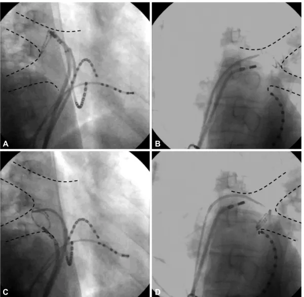
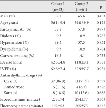
Background and Objectives: We aimed to investigate whether a large-sized Lasso catheter could increase the success rate of immediate complete pulmonary vein (PV) antral isolation and improve the outcome of catheter ablation in atrial fibrilla- tion (AF) patients. Subjects and Methods: This study included 107 consecutive patients (67 males, mean age: 57.8±9.7 years) who underwent PV mapping and ablation due to symptomatic drug-refractory AF. The first 43 patients underwent isolation of both ipsilateral PVs using the Carto-Merge 3 dimensional mapping system (group 1). The other 64 patients un- derwent isolation of both ipsilateral PVs using the same technique with a large-sized (a diameter of 30 to 35 mm) Lasso cathe- ter (group 2). When ipsilateral PVs did not show any potential after the initial circumferential ablation, we defined this as

‘immediate complete antral isolation (ICAI)’. We compared the AF recurrence rate of both groups. Results: There was no sig- nificant difference of the clinical characteristics between group 1 and group 2. All the patients were followed-up for 1 year. The ICAI rate of group 1 and group 2 was significantly different (21% vs. 78%, p<0.001), and the AF recurrence rates of group 1 and group 2 were also different (34.9% vs. 18.8%, p=0.042). Using multiple logistic regression analysis, the use of a large-sized Lasso catheter was a significant predictive factor for preventing recurrence (odds ratio: 0.489, 95% confidence interval: 0.136- 0.927). Conclusion: It is likely that a large-sized Lasso catheter plays an important role in achieving ICAI and in lowering the rate of AF recurrence. (Korean Circ J 2011;41:447-452)

KEY WORDS: Atrial fibrillation; Catheter ablation; Pulmonary vein; Tachycardia.

Received: September 10, 2010 Revision Received: December 1, 2010 Accepted: December 9, 2010

Correspondence: Yong-Seog Oh, MD, Division of Cardiology, Depart- ment of Internal Medicine, College of Medicine, The Catholic University of Korea, 505 Banpo-dong, Seocho-gu, Seoul 137-040, Korea Tel: 82-2-2258-6031, Fax: 82-2-592-3810

E-mail: [email protected]

• The authors have no financial conflicts of interest.

cc

This is an Open Access article distributed under the terms of the Cre- ative Commons Attribution Non-Commercial License (http://creativecom- mons.org/licenses/by-nc/3.0) which permits unrestricted non-commer- cial use, distribution, and reproduction in any medium, provided the origi- nal work is properly cited.

Introduction

The pulmonary vein (PV) has been the main target for ca- theter ablation in patients with atrial fibrillation (AF) ever sin- ce it was identified as the major trigger for AF.

1)2)

Different te- chniques that target the PV include direct trigger ablation,

3)

segmental ostial PV isolation

4-6)

and circumferential PV iso-

lation.

7)8)

The first two techniques are seldom used alone cur-

rently because of the high recurrence rate obtained. In cir-

cumferential PV isolation, the endpoint for ablation is the

electrical disconnection of all PV potentials from the left at-

rium (LA). The PV can be isolated individually or as ipsilat-

eral pairs, the so called ‘en bloc’ isolation, depending on the

venous anatomy, the catheter stability and the operator’s pre-

ference. In segmental ablation, to monitor complete PV iso-

lation, one or two circular mapping catheters are placed at

the ostia of the individual PV. This procedure was initially per-

formed under fluoroscopic guidance, but it is now combined

with a three dimensional (3D) electroanatomic mapping sys-

tem (CARTO or NAVx). To the best of our knowledge, the fe-

asibility and efficacy of using large-sized Lasso catheters, wh-

ich encircle the whole ipsilateral superior and inferior PVs,

in addition to using a 3D mapping system, have not yet been

reported. In the present study, we aimed to investigate wheth-

er the large-sized Lasso catheter could affect the clinical out-

(2)

comes of patients who undergo AF ablation using the Carto electroanatomic mapping system.

Subjects and Methods

Study population

This study included 107 consecutive patients (67 males, mean age: 57.8±9.7 years) who underwent PV mapping and ablation between June 2005 and July 2007. All the patients had highly symptomatic, drug refractory paroxysmal or per- sistent episodes of AF. Patients who received stent placement or bypass surgery for significant coronary artery disease were excluded. We defined persistent AF as an episode of AF that lasted for over 7 days and required cardioversion to restore si- nus rhythm. All the patients had been on treatment with at least one class I or III antiarrhythmic drug and had failed to maintain sinus rhythm before ablation. Each patient under- went transesophageal echocardiography to rule out the pres- ence of left atrial thrombi before the procedure.

The patients were recruited in a prospective, non-rando- mized manner. The first 43 patients underwent transseptal isolation of both ipsilateral PVs using the 3D electroanatomic mapping system (group 1). The remaining 64 patients under- went isolation of both ipsilateral PVs using the same techni- que by placing a large-sized (diameter 30-35 mm) circular mapping catheter at the pulmonary antrums (group 2). The initial 20 cases for each group were excluded to rule out a le- arning curve effect. Written informed consent for the proce- dure was obtained from all patients and the study protocol was approved by the institutional review board at our medi- cal center.

Electrophysiologic study and 3 dimensional electroanatomic mapping

A spiral CT scan was obtained prior to the ablation proce- dure to evaluate the PV anatomy. The patients were given oral anticoagulation for at least 2 month before the procedure.

This oral anticoagulation was discontinued 3 days before the procedure to achieve an international normalized ratio of pro- thrombin time that was less than 2.0. Amiodarone was dis- continued at least 3 weeks before AF ablation and any other antiarrhythmic drugs were suspended for 1 week prior to the procedure.

During the procedure, the patient was given intravenous sedation with midazolam and fentanyl to achieve an appropri- ate level of consciousness. Continuous, non-invasive blood pressure, heart rate and oxygen saturation monitoring were performed throughout the procedure. The surface electro- grams and bipolar endocardial electrograms were monitored and stored on a computer-based digital amplifier/recording system (Prucka CardioLab 7000 system, G.E. Medical systems, Milwaukee, WI, USA).

Three standard diagnostic catheters were positioned: a 6F hexapolar catheter at the His bundle area, a 7F decapolar catheter in the right atrium via the left femoral vein and a 7F decapolar catheter in the coronary sinus via the right jugular vein. A pigtail catheter was introduced into both pulmonary arteries via the right femoral vein, and the pulmonary veno- grams were obtained during the levo phase of both ipsilater- al pulmonary artery angiographies.

Double transseptal catheterization was performed with an 8.5F SL1 sheath (St. Jude Medical, Inc., Minnetonka, MN, USA) using the Brockenbrough technique. An initial intravenous bolus of 5,000 IU heparin was given immediately after the tr- ansseptal puncture. Repeated doses of heparin were infused every 30 minutes to maintain an activated clotting time be- tween 250 and 350 seconds.

Three-dimensional LA maps were reconstructed using a nonfluoroscopic navigation system (Carto XP, Biosense-Web- ster, Diamond Bar, CA, USA). Electroanatomic mapping was performed using an irrigated-tip catheter (Navistar Ther- mocool, Biosense-Webster, Diamond Bar, CA, USA). The Na- vistar catheter was placed inside each PV and then it was sl- owly drawn back to the LA for obtaining multiple PV points.

Following that, the Navistar explored the entire LA, includ- ing the anterior wall, the posterior wall, the septal wall, the appendage and the mitral annular area. Over 150 points were sampled in each procedure. The CT images were transferred to the Carto system equipped with the image integration software (Carto-Merge

TM

Image Integration Module). The Car- to-Merge software was used to superimpose the reconstruct- ed 3D CT images on the real time LA electroanatomic map.

Fig. 1. Three-dimensional mapping of the Carto-Merge on the right

anterior oblique view. Circumferential ablation around both ipsilat-

eral veins is shown by the red dots.

(3)

The merge technique has been previously described.

9)

Dur- ing the AF ablation, we set the fluoroscopy at 6 nGy/p with a pulse rate of 10/sec to reduce the radiation exposure.

Ablation procedure

Ablation was performed with the Navistar Thermocool ca- theter. Radiofrequency energy was applied with a power of 25 to 35 W and an irrigation rate of 17 to 40 mL/min. Radio- frequency energy was delivered in each spot for a maximum of 40 seconds or until the maximal local electrogram ampli- tude decreased by 80%. In group 1 patients, we performed circumferential antral ablation >5 mm away from the defin- ed PV ostia after mapping with CartoMerge software (Fig. 1).

After confirming that there was no anatomical gap on the 3D map, a steerable circular catheter with a diameter of either 25 mm or of variable size (Lasso, Biosense-Webster, Diamond Bar, CA, USA) was inserted into each of the PVs (Fig. 2). If the Lasso catheter showed no potential or a dissociated potential recorded in each PV, then we defined this as “immediate

complete antral isolation (ICAI)” If the residual conduction between the PV and the LA was identified, then additional segmental ablation was performed on the previous ablation line, as guided by the earliest potentials detected on the st- andard-sized Lasso catheter. To ablate as close as possible on the previous ablation line, we preferred using the variable Lasso catheter adjusted to the diameter of the PV antrum.

For group 2 patients, a large-sized Lasso catheter (30-35 mm, Biosense-Webster) was placed at the beginning of the antral ablation (Fig. 3). This could encircle the entire ipsilater- al PV in most cases. Circumferential PV isolation was guided by the large-sized Lasso catheter as well as the CartoMerge electroanatomic mapping. The isolation of each of the PVs was verified by putting the standard Lasso catheter (25 mm or variable size) into the ipsilateral superior and inferior PVs.

ICAI was achieved in 78% of the patients. In the remaining pa- tients, additional antral ablation, guided by a standard Lasso catheter, was needed in cases where the large size Lasso cath- eter did not fit correctly into the ipsilateral PV antrum.

Fig. 2. Fluoroscopic image confirming the isolation of each pulmonary vein. A variable-sized Lasso catheter was used to verify conduction block between the pulmonary vein and the left atrium. Dashed lines are schemes of the ipsilateral pulmonary vein. A: right superior pulmo- nary vein. B: left superior pulmonary vein. C: right inferior pulmonary vein. D: left inferior pulmonary vein.

C A

D

B

(4)

The roof line ablation on the LA

10)

was performed in both groups after circumferential PV isolation. For patients with persistent AF, mitral isthmus ablation was added in both gr- oups as previously described.

11)

The end point of ablation was defined as 1) the absence of PV potentials or dissociated PV potentials as documented with the standard size (25 mm) or variable size Lasso catheters within all four PVs and 2) bidi- rectional conduction block for the roof line and the mitral isthmus line.

Postablation management and follow-up

After the procedure, anticoagulation with warfarin was re- started and continued for at least 3 months. Antiarrhythmic drugs were reused in 37 (86.0%) and 43 (72.9%) patients in group 1 and 2, respectively (p=0.110). Amiodarone was in- cluded for 9 (24.3%) and 10 (26.3) patients in group 1 and 2, respectively (p=0.843). All the patients were monitored by ECG during hospitalization. After being discharged, the pa- tients underwent careful follow-up visits (one week after dis- charge followed by monthly visits) at the cardiology clinic. On each visit, patient symptoms and their surface ECG recordings were evaluated. The ambulatory 24-hour ECG monitoring was routinely examined at 3, 6 and 12 months, or whenever the patient exhibited symptoms. Any documented episode of symptomatic or asymptomatic AF that lasted for more than 30 seconds was considered to be a recurrence. AF recurrence within the first 3 months after ablation was not counted as a recurrence and those who did not show any evidence of tachycardia after 3 months of follow-up were considered as successful ablation. All the patients were followed-up for 1 year. Repeated ablation was not performed during this period.

Statistical analysis

Statistical analysis was performed using Statistical Package

for the Social Sciences (SPSS) version 12.0 (SPSS Inc., Chicago, IL, USA). Continuous variables were expressed as means±

SDs and they were compared using an unpaired Student’s t- test. The categorical variables were compared with Fisher’s exact test or the χ

2

test, where appropriate. All the compari- sons were two-sided, and p<0.05 were regarded as statistically significant. Multiple logistic regression analysis was perform- ed to identify the independent predictors of AF recurrence.

Results

Patient and procedural characteristics

The baseline characteristics are summarized in Table 1. Of the 107 study subjects, 67 (62.6%) were males (62.6%). The number of patients with diabetes and hypertension was 11 (10.3%) and 41 (38.3%), respectively. There was no signifi- cant difference for the clinical and procedural characteristics between group 1 and group 2. Non-fatal cardiac tamponade occurred in 2 patients (one in group 1 and the other in group 2, p=0.775).

Acute outcomes (immediate complete antral isolation)

The rate of successful ICAI showed a significant difference between group 1 and 2 (21% vs. 78%, respectively, p<0.001).

Follow-up for atrial fibrillation recurrence

The total AF recurrence rate of group 1 and group 2 was 34.9% and 18.8%, respectively (Table 2). The recurrence rate was not significantly different between group 1 and group 2 for paroxysmal AF (p=0.210) and persistent AF (p=0.127).

Multiple logistic regression analysis

The variables included in the multivariate analysis were LA Fig. 3. Fluoroscopic image in which a large-sized Lasso catheter (35 mm) was used. In group 2 patients, a large-sized Lasso catheter was placed at the beginning of the antral ablation. Dashed lines are schemes of ipsilateral pulmonary vein. A: right pulmonary vein. B: left pul- monary vein.

A B

(5)

size, left ventricular ejection fraction, gender, age, the type of AF and the use of a large-sized Lasso catheter. After multiple logistic regression analysis, the use of a large-sized Lasso cath- eter was significant as a predictive factor for preventing recur- rence (odds ratio: 0.489, 95% confidence interval: 0.136- 0.927).

Discussion

In this study, we observed that using a large-sized Lasso catheter was an independent predictor of freedom from AF recurrence after catheter ablation. To our knowledge, this is the first report to investigate the feasibility and efficacy of us- ing a large-sized Lasso catheter in addition to using 3D elec- troanatomic mapping.

Yamane et al.

12)

demonstrated the effectiveness of large-size Lasso catheters. The diameter of the Lasso catheter was 25-30 mm and they used it for antral isolation of individual PVs without the 3D mapping system. In contrast, we used a larger Lasso (35 mm in diameter) and performed the ‘en bloc’ ab- lation of the ipsilateral PVs.

PV antral isolation poses a difficulty in achieving a com- plete conduction block and requires a longer procedure time

as compared to segmental ostial isolation. However, AF free survival was better for antral isolation than for segmental ostial isolation.

13)14)

Ouyang et al.

7)8)

have shown the feasibili- ty of complete isolation around both ipsilateral PVs in com- bination with the 3D mapping system and the double-Lasso catheter. The reason for using the Lasso catheter in addition to the 3D mapping system is that it is easy to directly monitor the PV potentials and simultaneously observe conduction block when isolating the PV antrum. The limitation of using a large-sized Lasso catheter is that occasionally the contact of the catheter is not good enough because of the anatomic vari- ance around the PV antrum. The AF recurrence rate of our study was slightly lower than that of a previous study.

7)

The addition of the roof and mitral isthmus line might have con- tributed to the lower rate of AF relapse in our study.

In the present study, the rate of AF recurrence was lower in group 2 than in group 1 and the large-sized Lasso catheter was a significant predictor for AF recurrence using multivari- ate analysis. Complete electrical isolation of the PVs has a sig- nificant impact on the long-term efficacy of the procedure

15)

and the recovered PV conduction after the AF ablation is a do- minant factor for AF recurrence.

16)

In group 1, only 21% of the patients achieved ICAI by 3D-guided antral ablation al- one and the rest of them needed additional antral ablation using the standard Lasso catheter. In contrast, we could ach- ieve ICAI after initial antral isolation in 78% of the group 2 patients. In both groups, we tried to deliver radiofrequency energy on the previous line as close as possible, but in some cases additional ablation should have been performed inside the PVs. Although we did not check the PV reconnection in the relapsed patients by a redo procedure, we assumed that there may be some differences in the recovered PV conduc- tion between the two groups. Additional linear lesions might also have contributed to such ambiguous results.

Study limitation

First, this study has limitations because of its small sample size and non-randomized design. Second, we performed 24- hour Holter monitoring because 7-day Holter monitoring or a transtelephonic recording system was not available. Thus, we may have underestimated the asymptomatic recurrence of AF. However, this limitation would have equally affected both treatment groups. Further investigation will be required to determine whether such an approach will have practical implications.

Conclusion

The combination method of using the Carto-Merge 3D electroanatomic mapping system together with a large sized Lasso catheter for AF ablation seem to be more effective than 3D mapping only for the prevention of AF recurrence. A large sized Lasso catheter plays an important role to achieve ICAI.

Table 1. Baseline and procedural characteristics of the study population

Group 1 (n=43)

Group 2

(n=64) p

Male (%) 58.1 65.6 0.433

Age (years) 56.1±9.4 59.0±9.9 0.129

Paroxysmal AF (%) 58.1 57.8 0.973

Diabetes (%) 09.3 10.9 0.785

Hypertension (%) 49.5 37.5 0.832

Dyslipidemia (%) 09.3 10.9 0.784

Current smoking (%) 16.3 14.1 0.753

LA size (mm) 42.5±4.8 41.8±8.1 0.581

LVEF (%) 62.8±7.4 62.9±7.7 0.951

Antiarrhythmic drugs (%) Class IC

Amiodarone Sortalol

37 (86.0) 05 (11.6) 08 (18.6)

51 (79.7) 4 (6.3) 10 (15.6)

0.399 0.326 0.686 Procedure time (minute) 273±74 294±77 0.363 Fluoroscopic time (minute) 192±53 205±75 0.547 Fluoroscopy setting: 6 nGy/p, pulse rate: 10/sec. AF: atrial fibrilla- tion, LA: left atrium, LVEF: left ventricular ejection fraction Table 2. Recurrence rate observed in group 1 and group 2

Group 1 (n=43) Group 2 (n=64) p

Paroxysmal AF (%) 28.0 14.7 0.210

Persistent AF (%) 44.4 23.3 0.127

Total (%) 34.9 18.8 0.042

AF: atrial fibrillation

(6)

The reason for using the Lasso catheter in addition to the 3D mapping system is that it is easy to directly monitor the PV potentials and simultaneously observe conduction block when isolating the PV antrum.

REFERENCES

1) Haissaguerre M, Jais P, Shah DC, et al. Spontaneous initiation of at- rial fibrillation by ectopic beats originating in the pulmonary veins. N Engl J Med 1998;339:659-66.

2) Nam KB, Choi KJ, Park DW, Kim J, Rhee KS, Kim YH. Electrophy- siological characteristics of arterially-perfused canine pulmonary veins: role of the delayed afterdepolarization-induced triggered acti- vity. Korean Circ J 2005;35:643-8.

3) Jais P, Haissaguerre M, Shah DC, et al. A focal source of atrial fibrill- ation treated by discrete radiofrequency ablation. Circulation 1997;

95:572-6.

4) Haissaguerre M, Jais P, Shah DC, et al. Electrophysiological end point for catheter ablation of atrial fibrillation initiated from multiple pulmonary venous foci. Circulation 2000;101:1409-17.

5) Chen SA, Tai CT, Tsai CF, Hsieh MH, Ding YA, Chang MS. Radio- frequency catheter ablation of atrial fibrillation initiated by pulmonary vein ectopic beats. J Cardiovasc Electrophysiol 2000;11:218-27.

6) Oral H, Knight BP, Ozaydin M, et al. Segmental ostial ablation to iso- late the pulmonary veins during atrial fibrillation: feasibility and mechanistic insights. Circulation 2002;106:1256-62.

7) Ouyang F, Bansch D, Ernst S, et al. Complete isolation of left atrium surrounding the pulmonary veins: new insights from the double-Lasso technique in paroxysmal atrial fibrillation. Circulation 2004;110:

2090-6.

8) Ouyang F, Ernst S, Chun J, et al. Electrophysiological findings dur- ing ablation of persistent atrial fibrillation with electroanatomic mapp- ing and double Lasso catheter technique. Circulation 2005;112:

3038-48.

9) Dong J, Dickfeld T, Dalal D, et al. Initial experience in the use of in- tegrated electroanatomic mapping with three-dimensional MR/CT images to guide catheter ablation of atrial fibrillation. J Cardiovasc Electrophysiol 2006;17:459-66.

10) Hocini M, Jais P, Sanders P, et al. Techniques, evaluation, and conse- quences of linear block at the left atrial roof in paroxysmal atrial fi- brillation: a prospective randomized study. Circulation 2005;112:

3688-96.

11) Jais P, Hocini M, Hsu LF, et al. Technique and results of linear abla- tion at the mitral isthmus. Circulation 2004;110:2996-3002.

12) Yamane T, Date T, Kanzaki Y, et al. Segmental pulmonary vein antrum isolation using the “large-size” lasso catheter in patients with atrial fi- brillation. Circ J 2007;71:753-60.

13) Oral H, Scharf C, Chugh A, et al. Catheter ablation for paroxysmal atrial fibrillation: segmental pulmonary vein ostial ablation versus left atrial ablation. Circulation 2003;108:2355-60.

14) Arentz T, Weber R, Burkle G, et al. Small or large isolation areas ar- ound the pulmonary veins for the treatment of atrial fibrillation? Re- sults from a prospective randomized study. Circulation 2007;115:

3057-63.

15) Cheema A, Dong J, Dalal D, et al. Long-term safety and efficacy of cir- cumferential ablation with pulmonary vein isolation. J Cardiovasc Electrophysiol 2006;17:1080-5.

16) Ouyang F, Antz M, Ernst S, et al. Recovered pulmonary vein conduc-

tion as a dominant factor for recurrent atrial tachyarrhythmias after

complete circular isolation of the pulmonary veins: lessons from dou-

ble Lasso technique. Circulation 2005;111:127-35.

수치

Fig. 1. Three-dimensional mapping of the Carto-Merge on the right  anterior oblique view
Fig. 2. Fluoroscopic image confirming the isolation of each pulmonary vein. A variable-sized Lasso catheter was used to verify conduction  block between the pulmonary vein and the left atrium
Table 1. Baseline and procedural characteristics of the study  population Group 1 (n=43) Group 2(n=64) p Male (%) 58.1 65.6 0.433 Age (years) 56.1±9.4 59.0±9.9 0.129 Paroxysmal AF (%) 58.1 57.8 0.973 Diabetes (%) 09.3 10.9 0.785 Hypertension (%) 49.5 37.5

참조

관련 문서