J Vet Sci 2015, 16(4), 389-396ㆍhttp://dx.doi.org/10.4142/jvs.2015.16.4.389 JVS
Received 2 Feb. 2015, Revised 26 May. 2015, Accepted 3 Jul. 2015
*Corresponding author: Tel: +82-41-530-4873; Fax: +82-41-530-3085; E-mail: [email protected]
†
The first two authors contributed equally to this work.
‡
Present address: Department of Animal Biotechnology, College of Agricultural Life Science, Chonbuk National University, Jeonju 54896, Korea
Journal of Veterinary Scienceㆍⓒ 2015 The Korean Society of Veterinary Science. All Rights Reserved.
This is an Open Access article distributed under the terms of the Creative Commons Attribution Non-Commercial License (http://creativecommons.org/licenses/
pISSN 1229-845X eISSN 1976-555X
Anti-adipogenic effect of Artemisia annua in diet-induced-obesity mice model
Hye Kyung Baek1,†, Hyeji Shim1,†, Hyunmook Lim1, Minju Shim1, Chul-Kyu Kim2, Sang-Kyu Park2, Yong Seok Lee3, Ki-Duk Song4,‡, Sung-Jo Kim5, Sun Shin Yi1,*
Departments of 1Biomedical Laboratory Science, 2Medical Biotechnology, College of Medical Sciences, and 3Life Science and Biotechnology, College of Natural Sciences, Soonchunhyang University, Asan 31538, Korea
4Genomic Informatic Center, Han-kyong National University, Anseong 17579, Korea
5Department of Biotechnology, Hoseo University, Asan 31499, Korea
Obesity has increased continuously in western countries during the last several decades and recently become a problem in developing countries.
Currently, anti-obesity drugs originating from natural products are being investigated for their potential to overcome adverse effects associated with chemical drugs. Artemisinic acid, which was isolated from the well-known anti-malaria herb Artemisia annua (AA) L., was recently shown to possess anti-adipogenic effects in vitro. However, the anti-adipogenic effects of AA in animal models have not yet been investigated.
Therefore, we conducted daily oral administration with AA water extract in a diet-induced obesity animal model and treated 3T3-L1 cells with AA to confirm the anti-adipogenic effects in the related protein expressions. We then evaluated the physiology, adipose tissue histology and mRNA expressions of many related genes. Inhibition of adipogenesis by the AA water extract was observed in vitro. In the animal model, weight gain was significantly lower in the AA treated group, but there were no changes in food intake volume or calories. Reductions in lipid droplet size and mRNA expression associated with adipogenesis were also observed in animal epididymal fat. This study is the first to report that AA has an anti-obese effects in vivo.
Keywords: adipogenesis, animal model, Artemisia annua, diet induced obesity, obesity
Introduction
Adipose tissue plays a role in energy storage and thermoregulation, and serves as a source of various hormones including adipokines and cytokines [11,12,36]. Adipose tissue is essential to the absorption of fat-soluble vitamins [35] and for the cell membrane composition [30]. However, constant high-fat diet intake causes obesity by excessively increasing adipogenesis in the body. The increased fat accumulation produces serious negative complications such as increased insulin resistance, arteriosclerosis, cardiovascular diseases, hyperlipidemia and diabetes mellitus [18,34,41].
In western countries, many people consume high fat or high caloric diets without regular exercise. Consequently, many pharmaceutical companies have begun investigating drugs
targeting obesity. However, several promising anti-obesity drugs have been withheld because they showed unexpected side effects in humans [4,31].
Artemisia annua (AA), a well-known anti-malaria agent [23,29], was recently shown to reduce adipocyte differentiation in many in vitro studies, downregulating the level of peroxisome proliferator-activated receptor (PPAR)-, C/EBP-, C/EBP- [22]. However, its effects on adipogenesis have not yet been investigated in animal models.
In the present study, we performed daily oral administrations of AA water extract in a diet-induced obesity (DIO) animal model. AA extract was applied to 3T3-L1 cells, after which the relative protein expressions were compared among various concentrations and times from application. In addition, oil red O staining and western blot were performed in vitro, after which
Table 1. Primers Used for RT-PCR Analyses
Adipogenic gene Sequence Accession number
PPAR- [18] Forward 5’-TTG CTG AAC GTG AAG CCC ATC GAG G-3’ CG301269
Reverse 5’-GTC CTT GTA GAT CTC CTG GAG CAG-3’
C/EBP- [14] Forward 5’-GAC ATC AGC GCC TAC ATC GA-3’ NM_007678
Reverse 5’-TCG GCT GTG CTG GAA GAG-3’
C/EBP- [14] Forward 5’-ATT TCT ATG AGA AAA GAG GCG TAT GT-3’ NM_009883
Reverse 5’-AAA TGT CTT CAC TTT AAT GCT CGA A-3’
C/EBP- [14] Forward 5’-TTC CAA CCC CTT CCC TGA T-3’ NM_007679
Reverse 5’-CTG GAG GGT TTG TGT TTT CTG T-3’
the physiological data and effects on adipogenesis were observed based on the histology and mRNA expression of many related genes to evaluate the anti-obesity effects of the herb AA in an in vivo system.
Materials and Methods
AA extraction
A total of 40 g AA were boiled with 1.8 L distilled water (DW) under 1.5 bar at 80oC. After boiling for 30 min, the extract was fully cooled. The extract was filtered first by paper (185 mm;
Advantec, Japan), then by a Nalgene Rapid-Flow Battle Top Filter (0.2 m-pore membrane; Thermo Scientific, USA). Final AA extract was stored at 4°C.
Animals
Twenty-four adult C57BL/6J mice (mean = 23 g, between 21 and 25 g, 7-weeks-old) were housed at room temperature (22oC) and 60% humidity under a 12-h light: dark cycle (light cycle : dark cycle from 07:00 to 19:00). The mice were divided into four groups, two that were provided a normal chow diet (2018S; Harlan, USA) and two that were given a high fat diet (TD.06414; Harlan). Free access was allowed to water. Every day, 10 mL/1 kg/day AA extract was carefully administered with an oral sonde (0.9 × 50 mm) to half of each food group, while the same amount of DW was administered to the other half. Weight, food and water intake were recorded daily, and blood sugar level was tested once every week under non-fasting conditions. The peripheral blood was collected by cutting the tip of the mouse tail vein. Glucose levels from the peripheral blood were measured using a One Touch Ultra (LifeScan, USA) blood glucose meter with One Touch Ultra test strips (LifeScan). The experiments were performed over 4 weeks and were approved by the Institutional Animal Care and Use Committee (IACUC approval no. SCH15-0001) at Soonchunhyang University.
Tissue processing
Epididymal adipose tissues were removed before perfusion
and immersed in 4% paraformaldehyde (PFA). The animals were perfused with 0.1 M phosphate-buffered saline (PBS; pH 7.35) followed by 4% PFA in 0.1 M phosphate-buffer (PB; pH 7.35).
Cell culture
3T3-L1 cells were maintained in high-glucose (25 mM) Dulbecco’s Modified Eagle’s medium (DMEM; Gibco, USA) supplemented with 10% bovine calf serum (BCS; Hyclone, USA) and antibiotics (penicillin 100 U/mL and streptomycin 100 mg/mL) at 37oC in a 5% CO2 incubator. For adipocyte differentiation, cells were treated with differentiation inducing media (DIM) containing 1 M dexamethasone (Sigma, USA), 5 mM 3-isobutyl-1-methylxanthine (Sigma), and 4 mg/mL insulin (Sigma) in DMEM with 10% fetal bovine serum (FBS) at 2 days post-confluence. After 2 days, the cells were cultured in DMEM containing 10% FBS and insulin. Subsequently, the medium was changed every second day.
Oil red O staining
Oil red O staining was performed on adipogenic induction day 8. Briefly, cells were washed twice with PBS, then fixed with 4% paraformaldehyde for 1 h at room temperature. The cells were subsequently washed with 60% isopropanol and dried completely. Finally, cells were stained with oil red O (6 parts 0.5% oil red O powder in isopropanol and 4 parts water) for 10 min and washed with PBS.
Quantitative real-time PCR analysis
Total RNA was isolated from the epididymal adipose tissue using an Ambion PureLink RNA Mini Kit according to the manufacturer’s instructions (Ambion, USA). Quantitative real time PCR was performed with SYBR Green dye using an ABI Step One Real Time PCR instrument (Applied Biosystems, UK). For relative quantitation of gene expression, we used the comparative Ct method (2−Ct). Results were normalized to the control gene (36B4, housekeeping gene, acid ribosomal protein). The sequences of the primers and probes used are listed in Table 1 [13,17].
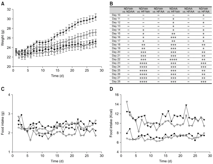
Fig. 1. Body weight gain and food intake. (A) Body weight changes. The high fat diet (HF)/Artemisia annua (AA) group gained significantly less weight than the HF/vehicle (Veh) group starting from day 14. There were no differences between the normal chow diet (ND)/Veh and ND/AA group at the beginning of the study. (B) Standard error table for body weight changes (*p < 0.05, **p < 0.01, ***p < 0.005, ****p < 0.001). (C and D) Daily food intake volume and Kcal change. There were no significant differences in food intake between groups. However, the HF/AA group’s intake was slightly less than that of the HF/Veh group.
Western blot analysis
Cell extracts were homogenized in lysis buffer (iNtRon Biotechnology, Korea), and protein concentrations were determined with a BCA kit (iNtRon Biotechnology). Lysates were separated with 10% SDS-PAGE and transferred to PVDF membranes (Bio-Rad Laboratories, USA). The membranes were probed with primary antibodies against PPAR-, fatty acid binding protein 4 (FabP4), glyceraldehyde 3-phosphate dehydrogenase (Cell Signaling Technologies, USA) and C/EBP (Abcam, UK), then incubated overnight. After further washing, membranes were incubated with HRP-conjugated secondary antibody (Vector, USA). The immunoreactive signals were detected based on their enhanced chemiluminescence and recorded in the MicroChemi 4.2 system.
Data analysis
All measurements were performed and analyzed to ensure objectivity. The intensity of the bands generated during western blotting was evaluated based on the optical density measured by transforming mean gray levels using the formula: optical density = log (256/mean gray level) with ImageJ 1.59 software (National Institutes of Health, USA). Lipid droplet size was calibrated per experiment area in the microscope. Dataf are presented as the means ± standard error (SE). Relative mRNA expression levels were automatically measured by real-time qPCR. The differences between means were analyzed using repeated two-way analysis of variance and one-way analysis of variance followed by the Bonferroni post-test and Duncan’s new multiple methods to determine differences between experimental groups.
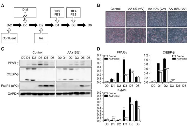
Fig. 2. Reproduction of the anti-adipogenesis effects of AA extract in vitro. (A) 3T3-L1 cells were treated with DIM and AA on day 0.
Insulin was then added on day 2, followed by 10% FBS on days 4 and 6. (B) Oil red O staining was performed on day 8. Adipose differentiation was noticeably suppressed in plates treated with AA compared to the control. (C and D) Western blotting and statistical analysis. PPAR- was significantly suppressed on day 3 in plates treated with AA. C/EBP- was the same on days 1 and 2. FabP4 was also suppressed on days 3, 5 and 8 in cells treated with AA (***p < 0.005, ****p < 0.001). Magnification: 100× (upper lane of B), 200× (lower lane of B).
Results
Physiological data
The results showed that all four groups of mice had similar weights at the start of the experiment. Daily weight measurements showed that the high fat diet (HF)/AA group weighed less than the HF/vehicle (Veh) group. This difference was statistically significant starting from day 14, and became more apparent as the experiment progressed. In both normal chow diet (ND)-fed groups, the ND/AA group weighed slightly less than the ND/Veh, but this difference was not significant (panel A and B in Fig. 1). Food intake in both ND fed groups (ND/Veh and ND/AA) did not differ significantly throughout the experiment. For the first two weeks, there were no notable differences in food intake between the HF fed groups (HF/Veh and HF/AA), but food intake in the HF/AA group became relatively lower than in the HF/Veh group after that (panel C and D in Fig. 1). There were no differences in blood sugar levels among groups. Epididymal fat tissues of the mice that received
AA (ND and HF) were lower than those of mice that received Veh (ND and HF).
Adipogenic effect of AA water extract in vitro
Adipogenesis was suppressed in the 3T3-L1 cell plate treated with AA water extract, which was the same agent administered orally to the DIO animal model during the experiment. The distinctions between the control plate and the AA treated plate became more obvious at higher concentrations of AA (panel B in Fig. 2). This anti-adipogenic effect was observed by western blotting, which was used to compare the expressions of several factors. PPAR-, transcription factor was significantly suppressed on day 3 and 5 in the plate treated with AA. Another transcription factor, C/EBP-, was also inhibited on days 1 and 2. Moreover, FabP4 which is detected in mature adipocytes, was suppressed on days 3, 5 and 8 (panel C in Fig. 2). The mean gray levels of the all bands were digitized with image J (panel D in Fig. 2).
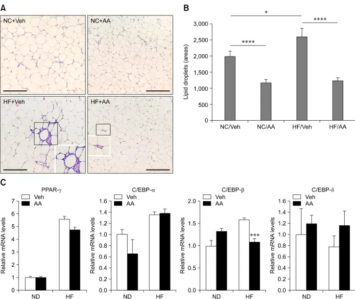
Fig. 3. Anti-adipogenesis effects of AA extract in vivo. (A and B) H&E staining of fat tissue and statistical analysis. Adipose tissue differentiation in the AA groups was more suppressed than in the Veh groups. This suppression was more evident between ND/Veh and ND/AA than between HF/Veh and HF/AA (*p < 0.05, ****p < 0.001). (C) Real time PCR. C/EBP- was more significantly suppressed in the HF/AA group than in the HF/Veh group. Levels of other transcription factors did not differ between the Veh and AA group (***p < 0.005). Scale bar = 200 m (A).
Epididymal adipose tissue hematoxylin and eosin staining The results of hematoxylin and eosin (H&E) staining demonstrated that the adipocyte sizes in mice administered AA (ND/AA, HF/AA) were smaller than those in mice administered Veh (ND/Veh, HF/Veh). The parts indicated with black squares were magnified. The sizes of the lipid droplets measured using ImageJ and were 1,981 ± 160, 1,177 ± 94, 2,594 ± 258 and 1,232 ± 91 (relative levels) in the ND/Veh, ND/AA, HF/Veh and HF/AA groups, respectively. Both AA-administered ND and HF fed groups demonstrated reduced lipid droplet sizes compared to the Veh-administered ND and HF fed groups (panel B in Fig. 3).
Transcription factor RT-PCR
PPAR-, C/EBP-, C/EBP- and C/EBP- mRNA expressions of ND/Veh, ND/AA, HF/Veh and HF/AA were measured by RT-PCR. There were no differences in PPAR-, C/EBP- and C/EBP- expressions in the AA-administered groups (ND and HF) or the Veh-administered groups (ND and HF). However, C/EBP- expression was noticeably more suppressed in HF/AA than in HF/Veh. In the ND-fed groups, this reduction of C/EBP- expression was found in the Veh-administered group, but the difference was not significant.
Discussion
The number of individuals with obesity has increased gradually in recent decades because of high calorie diets and various social stresses. Many types of anti-obesity drugs and agents have been developed, but these have numerous side effects on the cardiovascular system, gastrointestinal system and central nervous system [4,8,26]. In particular, specific serotonin reuptake inhibitors (SSRIs) and serotonin- norepinephrine reuptake inhibitors (SNRIs) have been investigated and used as appetite suppressants in medical treatments [5,16]; however, continuous administration of SSRIs and SNRIs causes regaining of weight in obesity patients, despite modifications to these agents to reduce side effects [6,7]. Therefore, there is demand for natural substances with anti-obesity effects that have low side effects and toxicity [27,32].
Many natural substances, particularly AA, have been studied for a variety of applications. AA has been used for extermination of vermin [19] and as an anti-malarial agent [29].
Recent studies have even reported that AA has inhibitory effects on cancer metastasis [38]. Additionally, the anti-adipogenic effects of AA have been investigated and the reduction of adipogenesis by AA in vitro was demonstrated [22]. However, no studies have investigated whether AA has the same effect in vivo. Therefore, we administered a daily dose of oral AA water extract to a DIO animal model in the present study. Our results confirmed the anti-adipogenic effects of AA on both the in vitro and in vivo systems. We measured food intake volume and calories daily and found no differences between the AA-treated groups (ND and HF) and the Veh-treated groups (ND and HF).
Although there were no differences in food intake rate, there was a significant difference in body weight between the AA- and the Veh-treated groups. These results indicate that AA water extract may play a primary role in inhibiting adipogenesis rather than suppressing appetite. Interestingly, body weight gain in the HF/AA group was significantly lower than that in the ND/AA group. If this outcome translates to humans, it is expected that AA could reduce body weight gain by decreasing adipogenesis in the abdomens of obesity patients without suppressing their appetite and food consumption.
Western blotting showed that the expression of PPAR-, C/EBP- and FabP4 decreased in the AA treated plate. PPAR-
is a nuclear hormone receptor that plays a major role in regulating the expression of proteins necessary for development of functional mature adipocytes [9,15]. C/EBP-
plays a synergistic role in terminal adipocyte differentiation [40], and FabP4 is detected in mature adipocytes [2,14]. Recent studies have shown that the expressions of these factors also decreased when adipogenesis was suppressed, while they increased when adipogenesis was enhanced [20,21]. Therefore, these results indicate that AA inhibits the expression of proteins
essential for adipogenesis and/or adipocyte maturation.
H&E staining revealed cell infiltration between adipocytes.
The cells may be lymphocytes or macrophages since adipocytes are known to be surrounded by immune related cells in obese animals and humans [3]. It is also well known that measurements of fat accumulation are directly proportional to adiposity [10]. Therefore, overt detection of macrophages indicates excessive adipogenesis. Macrophages, which secrete many types of adipokines and cytokines, are related to the innate inflammation response [24]. Continuous adipose tissue accumulation with macrophages maintains a chronic low inflammatory state, and pro-inflammatory factors such as interleukin-1 and interleukin-6 are continuously secreted [33,39]. In particular, interleukin-6 has a harmful effect on the hippocampus, impairing learning, cognition and memory function and leading to dementia [1,37]. Obesity-related chronic inflammation also contributes to neuropsychiatric symptoms involving depression [25,28].
Adipose tissue in HF/AA mice manifested lower cell infiltration than in HF/Veh mice, and the lipid droplet size of HF/AA was less than that of HF/Veh, indicating inhibition of pro-inflammation or inflammation in HF/AA. Additional immunohistochemistry is required to confirm the presence of macrophages; however, the results of the current study indicate that AA exerts an anti-adipogenic effect in the DIO animal model, mitigating adipose tissue accumulation and inflammation. Furthermore, a previous study demonstrated that artemisinic acid isolated from AA significantly attenuated tumor necrosis factor-α-induced secretion of interleukin 6 [22], indicating that AA has an inhibitory effect on the inflammatory state characterizing obesity. Thus, AA is likely to have beneficial effects on the brain of obese individuals, lowering the potential risk of central nervous system disease.
In conclusion, AA water extract decreased body weight gain and suppressed adipocyte differentiation in a DIO animal model, which was reflected in the in vitro results. Additionally, it is expected that AA will improve brain function, particularly on the hippocampus, suppressing the chronic inflammatory state in the DIO animal model. Our study may provide background information for further anti-obesity research.
Acknowledgments
This work was carried out with the support of the Cooperative Research Program for Agriculture Science & Technology Development (project no. PJ01104602) of the Rural Development Administration, Korea.
Conflict of Interest
There is no conflict of interest.
References
1. Aral LA, Pinar L, Göktaş G, Deveden EY, Erdoğan D.
Comparison of hippocampal interleukin-6 immunoreactivity after exhaustive exercise in both exercise-trained and untrained rats. Turk J Med Sci 2014, 44, 560-568.
2. Bag S, Ramaiah S, Anbarasu A. fabp4 is central to eight obesity associated genes: a functional gene network-based polymorphic study. J Theor Biol 2015, 364, 344-354.
3. Bai Y, Sun Q. Macrophage recruitment in obese adipose tissue. Obes Rev 2015, 16, 127-136.
4. Ballinger A. Orlistat in the treatment of obesity. Expert Opin Pharmacother 2000, 1, 841-847.
5. Bello NT, Walters AL, Verpeut JL, Cunha PP. High-fat diet-induced alterations in the feeding suppression of low-dose nisoxetine, a selective norepinephrine reuptake inhibitor. J Obes 2013, 2013, 457047.
6. Choi S, Blake V, Cole S, Fernstrom JD. Effects of chronic fenfluramine administration on hypothalamic neuropeptide mRNA expression. Brain Res 2006, 1087, 83-86.
7. Choi S, Jonak EM, Simpson L, Patil V, Fernstrom JD.
Intermittent, chronic fenfluramine administration to rats repeatedly suppresses food intake despite substantial brain serotonin reductions. Brain Res 2002, 928, 30-39.
8. Comerma-Steffensen S, Grann M, Andersen CU, Rungby J, Simonsen U. Cardiovascular effects of current and future anti-obesity drugs. Curr Vasc Pharmacol 2014, 12, 493-504.
9. Coskun H, Summerfield TLS, Kniss DA, Friedman A.
Mathematical modeling of preadipocyte fate determination.
J Theor Biol 2010, 265, 87-94.
10. Exley MA, Hand L, O’Shea D, Lynch L. Interplay between the immune system and adipose tissue in obesity. J Endocrinol 2014, 223, R41-48.
11. Fantuzzi G. Adipose tissue, adipokines, and inflammation. J Allergy Clin Immunol 2005, 115, 911-919.
12. Frühbeck G. Overview of adipose tissue and its role in obesity and metabolic disorders. Methods Mol Biol 2008, 456, 1-22.
13. Fu M, Sun T, Bookout AL, Downes M, Yu RT, Evans RM, Mangelsdorf DJ. A nuclear receptor atlas: 3T3-L1 adipogenesis. Mol Endocrinol 2005, 19, 2437-2450.
14. Garin-Shkolnik T, Rudich A, Hotamisligil GS, Rubinstein M. FABP4 attenuates PPAR and adipogenesis and is inversely correlated with PPAR in adipose tissues. Diabetes 2014, 63, 900-911.
15. Hsueh WA, Law R. The central role of fat and effect of peroxisome proliferator-activated receptor- on progression of insulin resistance and cardiovascular disease. Am J Cardiol 2003, 92 (Suppl), 3J-9J.
16. Jackson HC, Bearham MC, Hutchins LJ, Mazurkiewicz SE, Needham AM, Heal DJ. Investigation of the mechanisms underlying the hypophagic effects of the 5-HT and noradrenaline reuptake inhibitor, sibutramine, in the rat. Br J Pharmacol 1997, 121, 1613-1618.
17. Jeong HW, Lee JW, Kim WS, Choe SS, Kim KH, Park HS, Shin HJ, Lee GY, Shin D, Lee H, Lee JH, Choi EB, Lee HK, Chung H, Park SB, Park KS, Kim HS, Ro S, Kim JB. A newly identified CG301269 improves lipid and glucose metabolism without body weight gain through activation of
peroxisome proliferator-activated receptor and . Diabetes 2011, 60, 496-506.
18. Kim SK, Kim HJ, Hur KY, Choi SH, Ahn CW, Lim SK, Kim KR, Lee HC, Huh KB, Cha BS. Visceral fat th ickness measured by ultrasonography can estimate not only visceral obesity but also risks of cardiovascular and metabolic diseases. Am J Clin Nutr 2004, 79, 593-599.
19. Knudsmark Jessing K, Duke SO, Cedergreeen N. Potential ecological roles of artemisinin produced by Artemisia annua L. J Chem Ecol 2014, 40, 100-117.
20. Lai CS, Tsai ML, Badmaev V, Jimenez M, Ho CT, Pan MH.
Xanthigen suppresses preadipocyte differentiation and adipogenesis through down-regulation of PPAR and C/EBPs and modulation of SIRT-1, AMPK, and FoxO pathways. J Agric Food Chem 2012, 60, 1094-1101.
21. Lee H, Kang R, Hahn Y, Yang Y, Kim SS, Cho SH, Chung SI, Yoon Y. Antiobesity effect of baicalin involves the modulations of proadipogenic and antiadipogenic regulators of the adipogenesis pathway. Phytother Res 2009, 23, 1615-1623.
22. Lee J, Kim MH, Lee JH, Jung E, Yoo ES, Park D.
Artemisinic acid is a regulator of adipocyte differentiation and C/EBP expression. J Cell Biochem 2012, 113, 2488-2499.
23. Lee MR. Plants against malaria, part 2: Artemisia annua (Qinghaosu or the sweet wormwood). J R Coll Physicians Edinb 2002, 32, 300-305.
24. McDonald MK, Tian Y, Qureshi RA, Gormley M, Ertel A, Gao R, Aradillas Lopez E, Alexander GM, Sacan A, Fortina P, Ajit SK. Functional significance of macrophage-derived exosomes in inflammation and pain. Pain 2014, 155, 1527-1539.
25. Peterson HR, Rothschild M, Weinberg CR, Fell RD, McLeish KR, Pfeifer MA. Body fat and the activity of the autonomic nervous system. N Engl J Med 1988, 318, 1077-1083.
26. Pikkel YY. Acute bilateral glaucoma and panuveitis as a side effect of topiramate for weight loss treatment. BMJ Case Rep 2014, 2014, bcr2014203787.
27. Qi S, Zhou D. Lotus seed epicarp extract as potential antioxidant and anti-obesity additive in Chinese Cantonese sausage. Meat Sci 2013, 93, 257-262.
28. Rethorst CD, Bernstein I, Trivedi MH. Inflammation, obesity, and metabolic syndrome in depression: analysis of the 2009-2010 National Health and Nutrition Examination Survey (NHANES). J Clin Psychiatry 2014, 75, e1428-1432.
29. RITAM Artemisia annua Task Force; Willcox M, Falquet J, Ferreira JF, Gilbert B, Hsu E, de Magalhães PM, Plaizier-Vercammen J, Sharma VP, Wright CW, Yaode W.
Artemisia annua as a herbal tea for malaria. Afr J Tradit Complement Altern Med 2006, 4, 121-123.
30. Scott B, Lew J, Clandinin MT, Cinader B. Dietary fat influences electric membrane properties of neurons in cell culture. Cell Mol Neurobiol 1989, 9, 105-113.
31. Siebenhofer A, Jeitler K, Horvath K, Berghold A, Siering U, Semlitsch T. Long-term effects of weight-reducing drugs in hypertensive patients. Cochrane Database Syst Rev 2013, 3, CD007654.
32. Tan S, Gao B, Tao Y, Guo J, Su ZQ. Antiobese effects of capsaicin-chitosan microsphere (CCMS) in obese rats induced by high fat diet. J Agric Food Chem 2014, 62, 1866-1874.
33. Taupin V, Gogusev J, Descamps-Latscha B, Zavala F.
Modulation of tumor necrosis factor-, interleukin-1, interleukin-6, interleukin-8, and granulocyte/macrophage colony-stimulating factor expression in human monocytes by an endogenous anxiogenic benzodiazepine ligand, triakontatetraneuropeptide: evidence for a role of prostaglandins. Mol Pharmacol 1993, 43, 64-69.
34. Tiele OW. Fat, hypertension and arteriosclerosis in the experimental animal. Hippokrates 1974, 45, 512.
35. Torkildsen Ø, Løken-Amsrud KI, Wergeland S, Myhr KM, Holmøy T. Fat-soluble vitamins as disease modulators in multiple sclerosis. Acta Neurol Scand Suppl 2013, s196, 16-23.
36. Vereyken EJF, Bajova H, Chow S, De Graan PNE, Gruol DL. Chronic interleukin-6 alters the level of synaptic proteins in hippocampus in culture and in vivo. Eur J Neurosci 2007, 25, 3605-3616.
37. Wu B, Hu K, Li S, Zhu J, Gu L, Shen H, Hambly BD, Bao S, Di W. Dihydroartiminisin inhibits the growth and metastasis of epithelial ovarian cancer. Oncol Rep 2012, 27, 101-108.
38. Xing Z, Jordana M, Kirpalani H, Driscoll KE, Schall TJ, Gauldie J. Cytokine expression by neutrophils and macrophages in vivo: endotoxin induces tumor necrosis factor-alpha, macrophage inflammatory protein-2, interleukin-1 beta, and interleukin-6 but not RANTES or transforming growth factor-beta 1 mRNA expression in acute lung inflammation. Am J Respir Cell Mol Biol 1994, 10, 148-153.
39. Yang HN, Park JS, Woo DG, Jeon SY, Do HJ, Lim HY, Kim JH, Park KH. C/EBP- and C/EBP--mediated adipogenesis of human mesenchymal stem cells (hMSCs) using PLGA nanoparticles complexed with poly (ethyleneimmine). Biomaterials 2011, 32, 5924-5933.
40. Yoshioka S, Uemura K, Tamaya N, Tamagawa T, Miura H, Iguchi A, Nakamura J, Hotta N. Dietary fat-induced increase in blood pressure and insulin resistance in rats. J Hypertens 2000, 18, 1857-1864.