Total Transcanal Endoscopic Facial Nerve Decompression for Traumatic Facial Nerve Palsy
4
0
0
전체 글
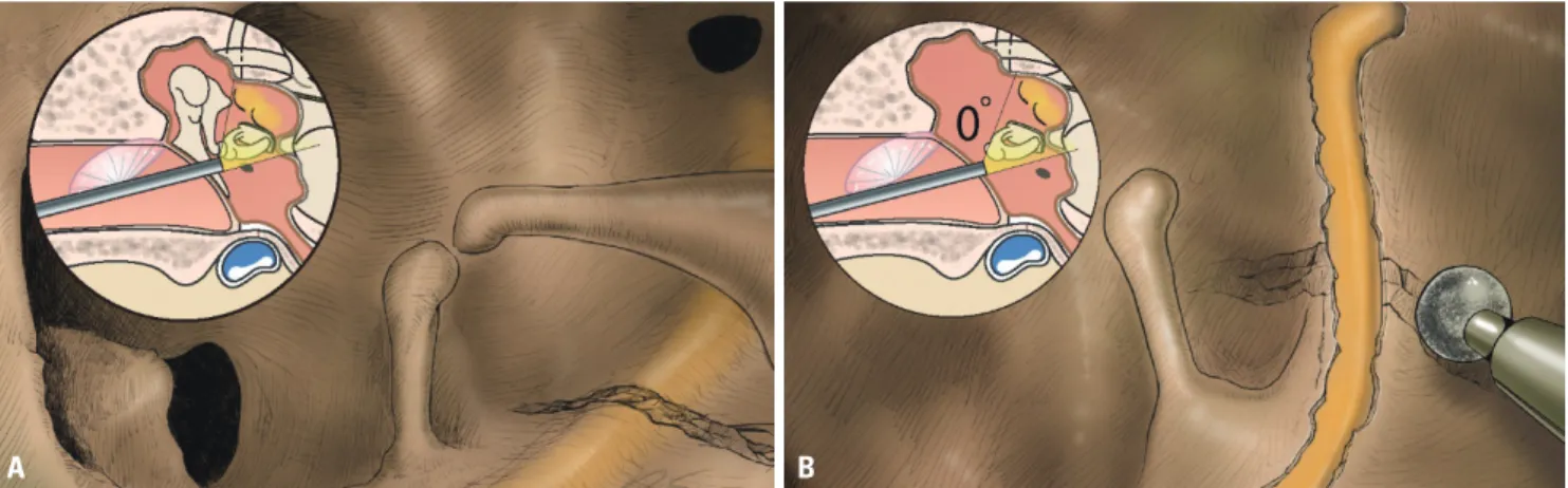
(2) Endoscopic Transcanal Facial Nerve Decompression. used as anatomic landmarks for the GG area because of the close anatomic relationship between the GG and these structures. The lateral semi-circular canal was also detected endoscopically, posteriorly, and superiorly with respect to the second genu of the facial nerve, representing the posterior limit of surgical dissection. The cog was gently removed using a micro curette to increase exposure of the GG area. The facial nerve was decompressed from the GG to the second genu. After this procedure, a gelfoam soaked with corticosteroid solution was placed in the surgical field close to the facial nerve. Partial ossicular chain reconstruction with titanium prosthesis and underlay tympanoplasty using tragal cartilage were done. The tympanomeatal flap was repositioned, and the EAC was filled with gelfoam (Fig. 1, Supplementary Video 1, only online).. nificant improvement in facial nerve function (Fig. 2D-I), having from House-Brackmann (H-B) grade IV to grade II.. Case 2 A 39-year-old male who experienced head trauma after falling down 10 days before he visited our clinic with left facial palsy and bilateral hearing impairment. HRCT scan showed fracture line crossed left tympanic segment of facial nerve, and audiological evaluation showed bilateral hearing loss (Fig. 3A and B). The patient underwent facial nerve decompression from the GG to the second genu by endoscopic transcanal approach. Then, ossiculoplasty was done. Six months after the operation, the patient showed hearing improvement at low frequencies (Fig. 3C), and facial nerve function improved from H-B grade V to III (Fig. 3D-I). This slow recovery was due to difference in degree of injury and personal vulnerability.. Patients and methods Between October and December 2016, two patients with traumatic facial nerve palsy underwent total transcanal endoscopic facial nerve decompression from the GG to the second genu. Patients agreed to the use of their medical records and images for specific purpose and informed consents were obtained.. Case 1 A 54-year-old male visited our clinic with left facial palsy and hearing impairment four weeks after a traumatic subdural hematoma and left zygomatic fracture. Otoendoscopic examination revealed left tympanic membrane perforation, and pure tone audiometry showed conductive hearing loss. High-resolution computed tomography (HRCT) of the temporal bone showed fracture line crossed left tympanic segment of facial nerve (Fig. 2A, B, and C). Intraoperatively, a fracture line across the tympanic segment of facial nerve and disarticulated incudostapedial joint were found. The patient underwent facial nerve decompression from the GG to the second genu through the endoscopic transcanal approach. Six months post-operation, there was sig-. A. DISCUSSION Management of facial nerve palsy secondary to temporal bone fractures has been an issue of dispute among otologic surgeons for years; the role, timing, and type of surgery have been debated. Middle cranial fossa approach and the transmastoid including its modifications, and the two combined together are the approaches currently used for facial nerve decompression depending on the site of injury, which is frequently deduced from the HRCT scan of the temporal bone. 1-4,9-11 Middle cranial fossa approach is used to expose the IAC and labyrinthine segment of the facial nerve while preserving hearing. The GG and tympanic portion of the nerve can also be decompressed using this approach, especially for longitudinal fractures. When this approach is combined with the transmastoid approach, the whole course of the facial nerve can be exposed and decompressed. However, the middle cranial fossa approach involves invasive procedures such as craniotomy and temporal lobe retraction.. B. Fig. 1. Schematic drawings of total transcanal endoscopic facial nerve decompression. (A) After tympanomeatal flap elevation, fracture line across the tympanic segment of facial nerve was noted. The disarticulated incudostapedial joint was also found. (B) After removal of malleus and incus, decompression of facial nerve was performed using microdrills. Using this approach, facial nerve can be exposed from geniculate ganglion to second genu.. 458. https://doi.org/10.3349/ymj.2018.59.3.457.
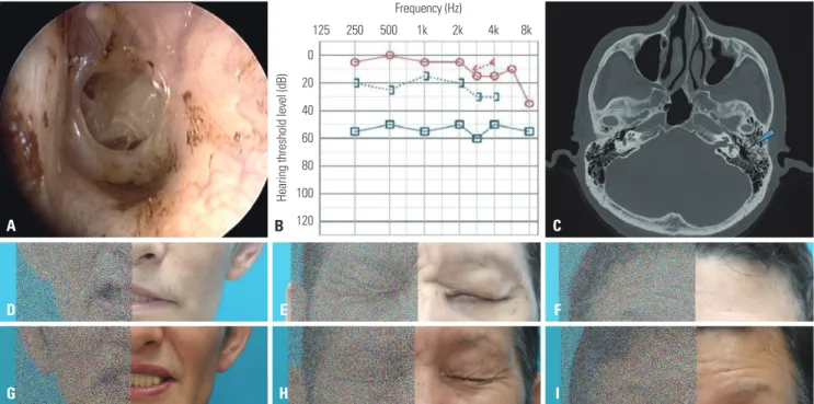
(3) Aveline Aloyce Kahinga, et al.. Transmastoid approach is appropriate when the main pathologic lesion is located on the tympanic or mastoid segment. Decompression of the GG or distal portion of the labyrinthine segment is also possible. Drawbacks of this approach,. however, include extensive drilling of the mastoid bone, long time, and alteration of normal structures. Herein, we described total transcanal endoscopic approach for facial nerve decompression as an alternative for lesions Frequency (Hz). 125. 250. 500. 1k. 2k. 4k. 8k. Hearing threshold level (dB). 0 20 40 60 80 100. B 120. A. C. D. E. F. G. H. I. Fig. 2. Otologic manifestations and facial expressions of case 1. (A and B) Traumatic tympanic membrane perforation and conductive hearing loss were noted in the left ear. (C) Pre-operative CT scan, axial view. Note the left temporal bone fracture as shown by the blue arrow. (D-F) Pre-operative and (G-I) six-month post-operative photos of the patient. There was a significant improvement in facial nerve function from House-Brackmann grade IV to II. Frequency (Hz) 250. 500. 1k. 2k. 4k. 8k. 125. 0. 0. 20. 20. Hearing threshold level (dB). Hearing threshold level (dB). 125. Frequency (Hz). 40 60 80 100. A 120. B. 250. 500. 1k. 2k. 4k. 8k. 40 60 80 100. C 120. D. E. F. G. H. I. Fig. 3. Otologic manifestations and facial expressions of case 2. (A) Pure tone audiometry showed bilateral conductive hearing loss (average ABG=30 dB). (B) Pre-operative CT scan, axial view. Note the left temporal bone fracture as shown by the blue arrow. (C) Three-month post-operative pure tone audiometry showed improvement. (D-F) Pre-operative and (G-I) five-month post-operative photos of the patient. Patient showed improvement in facial function from House-Brackmann grade V to III. https://doi.org/10.3349/ymj.2018.59.3.457. 459.
(4) Endoscopic Transcanal Facial Nerve Decompression. limited to the tympanic segment. The workhorse approaches for facial nerve decompression, are still transmastoid or middle cranial fossa craniotomies, however, endoscopic approach is less invasive compared to the other approaches, since it involves neither craniotomy nor extensive mastoid bone drilling. Despite the removal of the incus and malleus to have good exposure of the tympanic segment, which requires ossiculoplasty, this procedure is cosmetically acceptable as there are no external scars or bony depressions due to drilling. The outcome of facial nerve decompression through this approach was assessed using the H-B grading system for facial nerve function, and results were similar to those reported previously for middle cranial fossa, transmastoid, and combined approaches.2,4,9,11 In conclusion, total transcanal endoscopic facial nerve decompression is minimally invasive alternative for traumatic facial palsy whose lesion is limited to the tympanic segment.. SUPPLEMENTARY DATA Video 1. Surgical movie of total transcanal endoscopic facial nerve decompression.. ACKNOWLEDGEMENTS This research was supported by a grant of the Yonsei University College of Medicine (6-2016-0084). The authors thank Dong-Su Jang, MFA, Medical Illustrator, Medical Research Support Section, Yonsei University College of Medicine, Seoul, Korea, for his help with the illustrations.. ORCID Aveline Aloyce Kahinga In Seok Moon. 460. REFERENCES 1. Hato N, Nota J, Hakuba N, Gyo K, Yanagihara N. Facial nerve decompression surgery in patients with temporal bone trauma: analysis of 66 cases. J Trauma 2011;71:1789-92. 2. Ulug T, Ulubil SA. Management of facial paralysis in temporal bone fractures: a prospective study analyzing 11 operated fractures. Am J Otolaryngol 2005;26:230-8. 3. Nash JJ, Friedland DR, Boorsma KJ, Rhee JS. Management and outcomes of facial paralysis from intratemporal blunt trauma: a systematic review. Laryngoscope 2010;120:1397-404. 4. Darrouzet V, Duclos JY, Liguoro D, Truilhe Y, De Bonfils C, Bebear JP. Management of facial paralysis resulting from temporal bone fractures: our experience in 115 cases. Otolaryngol Head Neck Surg 2001;125:77-84. 5. Marchioni D, Alicandri-Ciufelli M, Piccinini A, Genovese E, Monzani D, Tarabichi M, et al. Surgical anatomy of transcanal endoscopic approach to the tympanic facial nerve. Laryngoscope 2011; 121:1565-73. 6. Marchioni D, Molteni G, Presutti L. Endoscopic anatomy of the middle ear. Indian J Otolaryngol Head Neck Surg 2011;63:101-13. 7. Marchioni D, Alicandri-Ciufelli M, Mattioli F, Nogeira JF, Tarabichi M, Villari D, et al. From external to internal auditory canal: surgical anatomy by an exclusive endoscopic approach. Eur Arch Otorhinolaryngol 2013;270:1267-75. 8. Marchioni D, Soloperto D, Rubini A, Nogueira JF, Badr-El-Dine M, Presutti L. Endoscopic facial nerve surgery. Otolaryngol Clin North Am 2016;49:1173-87. 9. Liu Y, Han J, Zhou X, Gao K, Luan D, Xie F, et al. Surgical management of facial paralysis resulting from temporal bone fractures. Acta Otolaryngol 2014;134:656-60. 10. Kim MW, Ryu NG, Lim BW, Kim J. Temporal lobe retraction provides better surgical exposure of the peri-geniculate ganglion for facial nerve decompression via transmastoid approach. Yonsei Med J 2016;57:1482-7. 11. Xu P, Jin A, Dai B, Li R, Li Y. Surgical timing for facial paralysis after temporal bone trauma. Am J Otolaryngol 2017;38:269-71.. https://orcid.org/0000-0001-7238-3299 https://orcid.org/0000-0002-3951-5074. https://doi.org/10.3349/ymj.2018.59.3.457.
(5)
수치
관련 문서
The index is calculated with the latest 5-year auction data of 400 selected Classic, Modern, and Contemporary Chinese painting artists from major auction houses..
1 John Owen, Justification by Faith Alone, in The Works of John Owen, ed. John Bolt, trans. Scott Clark, "Do This and Live: Christ's Active Obedience as the
Endoscopic spine surgery has become a practical, minimally invasive technique for decompression in patients with spinal disc herniation or stenosis.. However,
In this way, it is possible to shorten the time of accident restoration and maintenance by minimizing the expression error of the high-strength facial
Purpose: Calcaneal fracture is a rare fracture, which accounts for about 2% of all fractures, but is one of the most common fractures in the ankle bone.. There is
There are various legal grounds about finance bills and the budgetary system. For example, there are some nations of which the authority of the legislature is simply
Detection of Organic Vapors and Nerve Agent Simulant Vapors Based on Photoluminescent Bragg-Reflective Porous Silicon Interferometer.. Preparation of
High complication rate in locking plate fixation of lower periprosthetic distal femur fractures in patients with total knee arthroplasties.. Management and
