Presumed Necrotizing Viral Retinitis after Intravitreal Triamcinolone Injection: Case Report
4
0
0
전체 글
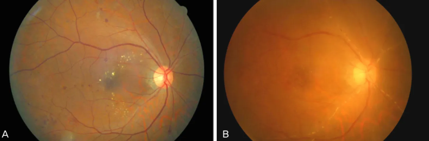
(2) Korean J Ophthalmol Vol.25, No.6, 2011. A. B. Fig. 1. Fundus photography of the right eye. (A) Before intravitreal injection of triamcinolone acetonide (IVTA), only mild diabetic changes can be seen. (B) Five months after IVTA, anterior chamber inflammation, severe arterial obstruction, and vitreous opacity developed.. A. B. Fig. 2. Montage fundus photography. (A) Five months after intravitreal triamcinolone acetonide injection, anterior chamber inflammation, severe arterial obstruction, and vitreous opacity developed. A white necrotic retinal area (black arrows) and vitreous opacity developed. Intravitreal triamcinolone is still visible in the inferior vitreous (white arrow). (B) After anti-viral treatment, the necrotic retinal area and vitreous opacity were gradually resolved. One month after antiviral medication, the white retinal necrotic area eventually disappeared.. Remnant triamcinolone particles were still visible in the inferior vitreous. (Fig. 2A, white arrow) After presumed diagnosis of viral retinitis, in particular acute retinal necrosis, laboratory work-up, including anterior chamber paracentesis for viral polymerase chain reaction (PCR), was performed. However, no viral DNA was detected by PCR, including herpes simplex virus (HSV), varicella zoster virus (VZV), cytomegalovirus (CMV), or Ebstein-Barr virus. Blood tests for human immunodeficiency virus and syphilis were negative, and the CD4 cell count was within normal limits. Intravenous acyclovir was administered for ten days, and oral valaciclovir was given for the following three weeks. Along with antiviral medications, oral aspirin and oral/topical steroids were prescribed. One week after initiation of antiviral medications, the size of the white retinal necrotic area decreased. 452. and eventually disappeared at one month (Fig. 2B). Barrier laser was performed around the necrotic retina for prevention of retinal breaks and detachment. After three months, blood samples for viral serology testing were collected. Anti-HSV, anti-CMV, and anti-VZV IgG were detected, but the IgM titers were negative. During routine six month follow-up visits, the visual acuity was limited to 20 / 100 OD due to cataract and epiretinal membrane (ERM). One year after the IVTA, vitrectomy for ERM removal combined with cataract operation was performed, and the visual acuity was restored to 20 / 40 OD.. Discussion Our case is presumed to be a form of acute retinal necrosis.
(3) M. M. M. M. F. M. 77. 69. 65. 63. 77. 30. Delyfer et al. [7]. Delyfer et al. [7] UfretVincenty et al. [11] Sekiryu et al. [8] Park et al. [9] TugalTutkum et al. [10] Shah et al. [3] Shah et al. [3]. CME from NIDDM BRVO CME from None CRVO Panuveitis Behçet disease. Exudative NIDDM AMD (with PDT) CME from NIDDM CRVO CME/ Behcet vitreitis disease. CSME from NIDDM DR Exudative None AMD (with PDT) CME from None CRVO. 2. 1. 1. 1. 3. 1. 1. 1. 1. 20. N/A. 4. 4. Retisert ×2. 8. 20. 4. 4. 4. CMV retinitis CMV retinitis. CMV retinitis CMV retinitis CMV retinitis. CMV retinitis CMV retinitis. CMV. CMV. CMV. CMV. CMV. CMV. Necrotizing HSV herpetic retinal necrosis CMV CMV retinitis. CMV retinitis ARN. Vitreous PCR. Vitreous and serum PCR. AC PCR. AC PCR. Clinical. AC PCR. AC PCR. Serum IgG. Immunocompetent. Immunocompetent N/A. 4+. 26. 27. N/A. 1/ 200. CF. NLP. 26 wk. Immunocompetent Immunocompromised Immunocompetent. 3+ N/A 20/ 200 20/ 400. Immuno2+ 23 N/A 20/ 200 competent 5 mon after Immuno- N/A N/A 20/ 40 20/ 50 Retisert competent no. 2 7 mon Immuno- (+)§ 48 20/ 40 20/ 200 competent 4 mon Immuno- Many N/A N/A LP competent 14 wk Immuno4+ N/A 20/ 200 20/ 200 competent. 3 mon. 3 mon. 3 wk. Yes. Yes. 20 /63∏ N/A. No. No. No. No. ‡. Oral. Intravitrea l, IV. Intrvitreal. Intravitrea l injection, implant IV, oral. IV, oral. Intravitreal, IV, oral. No‡. †. Systemic. Yes. HM. 20 / 32. 20 / 40. 20 / 400. 20 / 200. NLP. 1. 20. CMV. 13 wk N/A N/A 20/ 40 20/ 40 N/A No Prior CMV retinitis, clinical appearance # IV, oral 5 mon 2+ 23 20/ 40 20/ 200 20 / 40 No Han et al. 56 M CSME from NIDDM 1 4 ARN VZV Serum IgG, (present DR or response to HSV treatment case) IVTA = intravitreal triamcinolone injection; AC = anterior chamber; IOP = intraocular pressure; VA = visual acuity; CSME = clinically significant macular edema; DR = diabetic retinopathy; NIDDM = non-insulin dependent diabetes mellitus; CMV = cytomegalovirus; PDT = photodynamic therapy; ARN = acute retinal necrosis; HSV = herpes simplex virus; N/A = not available; AMD = age-related macular degeneration; BRVO = branch retinal vein occlusion; CME = cystoid macular edema; CRVO = central retinal vein occlusion; ERM = epiretinal membrane; HIV = human immunodeficiency virus; IRF = intraretinal fluid; IRU = immune recovery uveitis; IV = intravenous; PCR = polymerase chain reaction; VZV = varicella zoster virus. * Visual acuity was limited to 6 / 36 due to cataract formation. † Oral or IV antiviral treatment were administered. Exact route of drug administration was not mentioned. ‡ Vitrectomy was performed before viral retinitis was developed. § Anterior chamber inflammation was mentioned but the grade of inflammation was not available. ∏ After cataract surgery, visual acuity was improved to 20 / 63. # After cataract surgery and vitrectomy for epiretinal membrane removal, visual acuity was improved to 20 / 40.. 62 N/A CME from NIDDM BRVO 43 N/A CME from AIDS IRU. M. Aggermann 69 et al. [6]. F. M. Age Sex. Saidel et al. 75 [4] Toh et al. 62 [5]. Authors. Diagnosis confirmation. Visual acuity VA Vitrectomy Antiviral Interval to Immunocom AC Peak promised before VA affor mediretinitis cell IOP Final state IVTA ter retitreatment cation VA nitis CMV Vitreous PCR 4 mon Immuno- Trace N/A 20/ 40 20/ 80 20 /400 Yes Oral competent HSV Serum IgG and 5 mon Immuno1+ 40 6/ 18 N/A 6 / 36* No IV, oral IgM competent. Indication Systemic for IVTA diseases No. of Dose Diagnosis Cause injections (mg). Triamcinolone acetonide. Table 1. Published cases of viral retinitis after intravitreal corticosteroid implantation. JM Han, et al. Necrotizing Viral Retinitis after IVTA. 453.
(4) Korean J Ophthalmol Vol.25, No.6, 2011. (ARN) considering the prompt response to antiviral treatment, serum viral IgG results, and typical clinical presentations, although we failed to reveal the causative organism by PCR or serology. The causative association between IVTA and ARN was presumed by the visualization of triamcinolone particles at the time of ARN occurrence and no underlying systemic immune deficiency. Other possible diagnoses include CMV retinitis and intraocular lymphoma. In CMV retinitis, early lesions are located along the retinal blood vessels and sometimes on the fovea or disc. Intraocular lymphoma/leukemia could also produce whitish infiltrative subretinal lesions. However, in our case, ARN is the more likely diagnosis because a yellow-white retinal lesion was initially noticed in the peripheral retina, and occlusive vasculopathy involving all retinal arteries was observed with anterior chamber reactions, which are the pathognomonic findings of ARN. Follow-up examination showed no evidence of lymphoma or leukemia. To date, there are 12 case reports of necrotizing herpetic retinopathy, including nine of CMV retinitis and three of ARN in ten immunocompetent patients, of whom two immunocompromised patients received intravitreal corticosteroid injections (Table 1) [3-11]. The mean age of patients with necrotizing herpetic retinopathy associated with intravitreal corticosteroid injection ranged from 30 to 77 years (range, 63.75 ± 14.13 years). PCR was performed for diagnosis of pathogens in all cases except one [6], which had a lack of vitreous due to previous vitrectomy. The diagnosis was confirmed in seven cases (63.6%) by detection of viral DNA. The IOP of the infected eye according to the available IOP data in case reports was 23 to 48 mmHg (mean, 32.8 ± 10.7 mmHg). Elevated IOP is a common clinical feature of necrotizing herpetic retinopathy associated with intravitreal corticosteroid injection. All case reports mentioned inflammatory cells in the anterior chamber. The interval between intravitreal corticosteroid injection and retinitis ranged from three weeks to seven months (range, 4.0 ± 1.7 months). Local immune deficiency by intravitreal steroid injections has been suggested as the most likely pathogenesis for necrotizing herpetic retinopathy [3-10]. A history of viral retinitis, such as CMV retinitis, was not clear except in only one case of CMV retinitis. However, reactivation of a virus from prior subclinical infections may play an important role in viral retinitis after IVTA, as well as in new viral infections. Bacterial endophthalmitis, one of the most serious complications of an IVTA, presents 7.5 days after injection [12], which is earlier than that of necrotizing herpetic retinopathy (four months on average). Bacterial endophthalmitis usually develops through the inoculation of bacteria into the sterile vitreous, whereas necrotizing herpetic retinopathy can develop by reactivation or by new inoculations. Beer et al. [13] reported that triamcinolone was detected in the aqueous humor more than three months after IVTA. Moreover, triamcinolone can remain in the vitreous longer than in the aqueous humor 454. [14]. The persistent immunocompromised state of the posterior segment after IVTA appears to mimic the intraocular state of the immunocompromised patient and may be the main mechanism of viral retinitis, either by reactivation or by a new infection of a virus. In conclusion, we report a case of presumed ARN after IVTA and propose that physicians should be aware of the possibility of necrotizing herpetic retinopathy after IVTA. Heightened suspicion and prompt treatment using antiviral agents could act to preserve visual acuity.. Conflict of Interest No potential conflict of interest relevant to this article was reported.. References 1. Kramar M, Vu L, Whitson JT, He YG. The effect of intravitreal triamcinolone on intraocular pressure. Curr Med Res Opin 2007;23:1253-8. 2. Yilmaz T, Weaver CD, Gallagher MJ, et al. Intravitreal triamcinolone acetonide injection for treatment of refractory diabetic macular edema: a systematic review. Ophthalmology 2009;116:902-11. 3. Shah AM, Oster SF, Freeman WR. Viral retinitis after intravitreal triamcinolone injection in patients with predisposing medical comorbidities. Am J Ophthalmol 2010;149:433-40.e1. 4. Saidel MA, Berreen J, Margolis TP. Cytomegalovirus retinitis after intravitreous triamcinolone in an immunocompetent patient. Am J Ophthalmol 2005;140:1141-3. 5. Toh T, Borthwick JH. Acute retinal necrosis post intravitreal injection of triamcinolone acetonide. Clin Experiment Ophthalmol 2006;34:380-2. 6. Aggermann T, Stolba U, Brunner S, Binder S. Endophthalmitis with retinal necrosis following intravitreal triamcinolone acetonide injection. Ophthalmologica 2006;220:131-3. 7. Delyfer MN, Rougier MB, Hubschman JP, et al. Cytomegalovirus retinitis following intravitreal injection of triamcinolone: report of two cases. Acta Ophthalmol Scand 2007;85:681-3. 8. Sekiryu T, Iida T, Kaneko H, Saito M. Cytomegalovirus retinitis after intravitreal triamcinolone acetonide in an immunocompetent patient. Jpn J Ophthalmol 2008;52:414-6. 9. Park YS, Byeon SH. Cytomegalovirus retinitis after intravitreous triamcinolone injection in a patient with central retinal vein occlusion. Korean J Ophthalmol 2008;22:143-4. 10. Tugal-Tutkun I, Araz B, Cagatay A. CMV retinitis after intravitreal triamcinolone acetonide injection in a patient with Behcet's uveitis. Int Ophthalmol 2010;30:591-3. 11. Ufret-Vincenty RL, Singh RP, Lowder CY, Kaiser PK. Cytomegalovirus retinitis after fluocinolone acetonide (Retisert) implant. Am J Ophthalmol 2007;143:334-5. 12. Moshfeghi DM, Kaiser PK, Scott IU, et al. Acute endophthalmitis following intravitreal triamcinolone acetonide injection. Am J Ophthalmol 2003;136:791-6. 13. Beer PM, Bakri SJ, Singh RJ, et al. Intraocular concentration and pharmacokinetics of triamcinolone acetonide after a single intravitreal injection. Ophthalmology 2003;110:681-6. 14. Cheng L, Banker AS, Martin M, et al. Triamcinolone acetonide concentration of aqueous humor after decanted 20-mg intravitreal injection. Ophthalmology 2009;116:1356-9..
(5)
수치

관련 문서