The Clinical Courses of Patients with Congenital Cystic Adenomatoid Malformation Complicated by Pneumonia
Byung Woo Jhun,
1Se Jin Kim,
1Kang Kim,
1Seok Kim,
2and Ji Eun Lee
11Division of Pulmonary and Critical Care Medicine, Department of Internal Medicine, The Armed Forces Capital Hospital, Seongnam;
2Department of Thoracic Surgery, The Armed Forces Capital Hospital, Seongnam, Korea.
Received: July 7, 2014 Revised: October 2, 2014 Accepted: October 10, 2014 Corresponding author: Dr. Ji Eun Lee, Division of Pulmonary and Critical Care Medicine, Department of Internal Medicine, The Armed Forces Capital Hospital, 81 Saemaeul-ro 177beon-gil, Bundang-gu, Seongnam 463-040, Korea.
Tel: 82-31-725-6298, Fax: 82-31-706-0987 E-mail: [email protected]
∙ The authors have no financial conflicts of interest.
© Copyright:
Yonsei University College of Medicine 2015 This is an Open Access article distributed under the terms of the Creative Commons Attribution Non- Commercial License (http://creativecommons.org/
licenses/by-nc/3.0) which permits unrestricted non- commercial use, distribution, and reproduction in any medium, provided the original work is properly cited.
Purpose: We evaluated the clinical characteristics and courses of patients with con-
genital cystic adenomatoid malformation (CCAM) complicated by pneumonia.
Materials and Methods: We retrospectively reviewed the records of 19 adult pa-
tients with surgically confirmed CCAM between March 2005 and July 2013. Re-
sults: Eighteen of nineteen patients presented with acute pneumonia symptoms andsigns, and inflammatory markers were elevated. On chest computed tomography, all 18 patients had parenchymal infiltration around cystic lesions, 17 (94%) had an air-fluid level, and 2 (11%) had pleural effusion. After antibiotics treatment for a median of 22 days prior to surgery, all acute pneumonia symptoms and signs disap- peared in 17 (94%) patients at a median of 10 days. Improvements and normaliza- tion of inflammatory marker levels, occurred in 17 (94%) and 9 (50%) patients at medians of 8 and 17 days, respectively. Radiological improvement was evident in 11 (61%) patients, at a median of 18 days, of these patients, partial radiological im- provement occurred in 10 (56%) and complete radiological resolution in only 1 (6%). One patient (6%) did not improve in terms of clinical, laboratory, or radiolog- ical findings despite antibiotic treatment for 13 days. Consequently, after 17 (94%) elective and 1 (6%) emergency surgeries, all patients improved without develop- ment of complications. Conclusion: We described the clinical characteristics and courses of patients with CCAM complicated by pneumonia, and showed that sur- gery may be performed safely after clinicolaboratory improvement is attained upon antibiotic treatment, even in the absence of complete radiological resolution.
Key Words:
Antibiotic treatment, congenital cystic adenomatoid malformation, surgical resection
INTRODUCTION
Congenital cystic adenomatoid malformation (CCAM) of the lung, which was first
described as a distinct disease entity by Ch’In and Tang,
1is a rare anomaly of the
lower respiratory tract characterized by cystic adenomatous overgrowth of the ter-
minal bronchioles and airspaces.
2Approximately 80% of CCAM cases are usually
identified during the first 2 years of life, and typical manifestations include progres-
sive respiratory distress or recurrent respiratory tract infection.
evaluated. Follow-up data regarding the changes in clinical, laboratory, and radiological findings during antibiotic treat- ment before surgical resection were evaluated. Complica- tions associated with surgery and survivals were also evalu- ated. Follow-up data were last obtained on August 1, 2013.
The present study was approved by the Institutional Re- view Board of the Armed Forces Capital Hospital, which permitted review and publishing of information from the pa- tients’ records. The requirement for informed consent of the individual patients was waived given the retrospective na- ture of the study.
Patient management and treatment outcomes
Of all CCAM patients with or without pneumonia, those with CCAM complicated by pneumonia initially received antibiotic treatment after microbiological testing of peripher- al blood and sputum. During antibiotic treatment, non-surgi- cal interventions, such as catheter drainage of an abscess pocket to relieve symptoms or flexible bronchoscopy, with bronchoalveolar lavage to evaluate accurate causative agents, were performed. Follow-up chest radiographs and CT scans were usually obtained approximately every 3‒5 days initial- ly, and then at 2‒3 weeks, to evaluate the radiological re- sponse after commencement of antibiotic treatment. Patients with CCAM complicated by pneumonia subsequently un- derwent elective or emergency surgery with diagnostic and curative therapeutic intent, considering the response to anti- biotic treatment.
The response to antibiotic treatment was evaluated based on clinical, laboratory, and radiological improvements. Clin- ical improvement was defined as disappearance of the symptoms or signs noted at initial evaluation, and laboratory improvement was defined as a decrease in the WBC count, CRP level, or ESR during antibiotic treatment. Radiological improvement was defined as partial or complete disappear- ance of radiographic findings associated with pneumonia (parenchymal infiltration around the cystic lesion, air-fluid level, and pleural effusion), although multiple cystic lesions related to CCAM could persist.
Patients who initially presented with blood-tinged sputum alone, without acute pneumonia symptoms, received conser- vative treatment without antibiotic treatment, and subse- quently underwent elective surgery with diagnostic and cu- rative therapeutic intent. All non surgical interventions and determination of the timing of surgery in patients with CCAM with or without pneumonia depended on the deci- sion of the attending physician.
Adult presentation, however, is uncommon, and only a few adult cases have been reported sporadically. CCAM, in adults, usually involves the unilateral lobes of the lung and can be complicated with recurrent respiratory tract infec- tions and abscesses.
3-7CCAM has also been noted to be as- sociated with the development of malignancies such as ade- nocarcinoma
8,9and pleuropulmonary blastoma.
10Therefore, surgical resection of the affected part of the lung is the treat- ment of choice, even in asymptomatic patients, because it may prevent recurrent respiratory tract infections and malig- nant transformation.
11To date, however, no accurate data exist on the clinical characteristics, or courses and outcomes, of patients with CCAM complicated by pneumonia. Thus, in the current study, we investigated the clinical characteristics, overall re- sponses to preoperative antibiotic treatment, and the out- comes of patients treated via subsequent surgical resection in patients with CCAM complicated by pneumonia.
MATERIALS AND METHODS
Study patients and data collection
We retrospectively reviewed the medical records of consec- utive adult patients with newly diagnosed, histologically proven CCAM, confirmed by surgical resection, between March 2005 and July 2013 at the Armed Forces Capital Hos- pital, an 874-bed referral military hospital in Gyeonggi province, South Korea. Data were retrospectively collected regarding epidemiology, underlying conditions, initial pre- senting symptoms, chest radiography, contrast-enhanced chest computed tomography (CT), surgical treatment (in- cluding type of surgical approach and extent of surgical re- section), and duration from the radiological diagnosis to sur- gery. Of all such patients, those with CCAM complicated by pneumonia were evaluated.
Pneumonia was defined as the presence of a new infiltrate on chest radiography, together with at least one of the follow- ing: fever (temperature≥38.0ºC); hypothermia (temperature
<35.0ºC); a new cough with or without sputum production;
pleuritic chest pain; dyspnea, or altered breath sounds on aus-
cultation.
12Data for acute pneumonia symptoms or signs, ini-
tial inflammatory markers [such as the white blood cell
(WBC) count, C-reactive protein (CRP) level, and erythro-
cyte sedimentation rate (ESR)], microbiological tests, abnor-
mal findings of initial chest CT scans, and the initial empiri-
cal antibiotics and the duration of antibiotic treatment were
presentation. The median oxygen saturation value on room air was 94% (IQR, 88‒96%).
The WBC count and CRP and ESR levels as inflammato- ry markers were elevated in all patients, with median values of 11510/μL (IQR, 8990‒15145/μL), 11.3 mg/dL (IQR, 3.3‒19.4 mg/dL), and 45 mm/h (IQR, 29‒67 mm/h), respec- tively. As a causative agent, Streptococcus pneumoniae was positive in microbiological culture tests in seven (39%) pa- tients, comprising four bronchoalveolar lavage fluid samples
Statistical analysisAll data are presented as medians [interquartile ranges (IQR)]
for continuous variables and as numbers (percentages) for categorical variables. All statistical analyses were performed using the PASW 18.0 software (SPSS Inc., Chicago, IL, USA).
RESULTS
Baseline characteristics of patients with CCAM
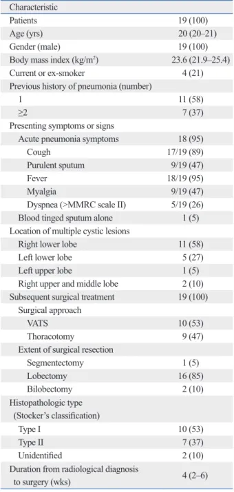
Nineteen adult patients with CCAM were identified during the study period. The clinical characteristics of the patients are shown in Table 1. The median age of these patients was 20 years (IQR, 20‒21 years) and all were males. Four (21%) patients were current or ex smokers. Seven (37%) patients had a history of recurrent pneumonia occurring at least twice. Of these 19 patients, 18 initially presented with acute pneumonia symptoms or signs including cough, purulent sputum, fever, myalgia, and/or dyspnea, and received preop- erative antibiotic treatment. The remaining patient presented with only blood-tinged sputum, without evidence of pulmo- nary infection on chest CT scan, and underwent conserva- tive treatment without prescription of antibiotics. Eventually, all 19 patients underwent surgical resection. The most com- mon location of multiple cystic lesions was the right lower lobe (n=11, 58%), followed by the left lower lobe (n=5, 27%), right upper and middle lobe (n=2, 10%), and left up- per lobe (n=1, 5%). Approximately half (n=10, 53%) of the patients underwent video-assisted thoracoscopic surgery (VATS), and most patients (n=16, 85%) underwent lobecto- my. The median duration from radiological diagnosis to sur- gery was 4 weeks (IQR, 2‒6 weeks). Upon histopathologi- cal evaluation, 10 of the 19 (53%) patients were found to have CCAM type I, and 7 (37%) CCAM type II (Stocker’s classification). We were unable to identify the CCAM type in two patients (10%).
Characteristics of the patients with CCAM complicated by pneumonia
The characteristics of the 18 patients with CCAM compli- cated by pneumonia are shown in Table 2. All 18 patients presented with pulmonary or systemic symptoms, such as cough (n=17, 94%), purulent sputum (n=9, 50%), fever (n=18, 100%), myalgia (n=9, 50%), and/or dyspnea (n=5, 28%). Seven (39%) patients had tachycardia (>100 beats/
min) and five (28%) tachypnea (>30 breaths/min) at initial
Table 1. Baseline Characteristics of Patients with CCAM Characteristic
Patients 19 (100)
Age (yrs) 20 (20‒21)
Gender (male) 19 (100)
Body mass index (kg/m2) 23.6 (21.9–25.4)
Current or ex-smoker 4 (21)
Previous history of pneumonia (number)
1 11 (58)
≥2 7 (37)
Presenting symptoms or signs
Acute pneumonia symptoms 18 (95)
Cough 17/19 (89)
Purulent sputum 9/19 (47)
Fever 18/19 (95)
Myalgia 9/19 (47)
Dyspnea (>MMRC scale II) 5/19 (26)
Blood tinged sputum alone 1 (5)
Location of multiple cystic lesions
Right lower lobe 11 (58)
Left lower lobe 5 (27)
Left upper lobe 1 (5)
Right upper and middle lobe 2 (10) Subsequent surgical treatment 19 (100)
Surgical approach
VATS 10 (53)
Thoracotomy 9 (47)
Extent of surgical resection
Segmentectomy 1 (5)
Lobectomy 16 (85)
Bilobectomy 2 (10)
Histopathologic type (Stocker’s classification)
Type I 10 (53)
Type II 7 (37)
Unidentified 2 (10)
Duration from radiological diagnosis
to surgery (wks) 4 (2–6)
CCAM, congenital cystic adenomatoid malformation; MMRC, modified medical research council; VATS, video-assisted thoracoscopic surgery.
Data are expressed as medians (interquartile range) or numbers (%).
the parenchymal infiltrate around the multiple cystic lesions, and the air-fluid interface in the right lower lobe.
Responses to treatment and outcomes in patients with CCAM complicated by pneumonia
The clinical, laboratory, and radiological responses to treat- ment and outcomes in patients with CCAM complicated by pneumonia are summarized in Table 3. At admission, all 18 patients received broad-spectrum antibiotic treat- ment; third-generation cephalosporins±macrolides (n=11, 61%), quinolone±clindamycin (n=4, 22%), piperacillin/
tazobactam±quinolone (n=2, 11%), and carbapenem (n=1, 6%). The median duration of antibiotic treatment before surgical resection was 22 days (IQR, 14‒30 days). One pa- tient received percutaneous catheter drainage during anti- biotic treatment to relieve symptoms.
Upon antibiotic treatment for a median time of 22 days (IQR, 14‒30 days) prior to surgical resection, all acute pneu- monia symptoms and abnormal signs disappeared in 17 (94%) patients, at a median time of 10 days (IQR, 8‒15 days); the median times to disappearance of cough, purulent sputum, fever, myalgia, dyspnea, tachycardia, tachypnea, and oxygen requirement were 8, 7, 5, 4, 4, 4, 3, and 5 days, respectively. Improvement or normalization of all inflamma- tory markers occurred in 17 (94%) and 9 (50%) patients at median times of 8 days (IQR, 5‒19 days) and 17 days (IQR, 13‒24 days), respectively. Radiological improvement was evident in 11 (61%) patients at a median time of 18 days (IQR, 14‒27 days); partial radiological improvements oc- curred in 10 (56%) patients and complete radiological reso- lution in only 1 (6%).
The 17 (94%) patients who exhibited clinicolaboratory improvement after preoperative antibiotic treatment under- went elective surgery to treat CCAM. However, the clinical, laboratory, and radiological findings of one patient (6%) did not improve despite 13 days of antibiotic treatment; this pa- tient subsequently underwent emergency surgery with diag- nostic and curative therapeutic intent. With 17 (94%) elec- tive and 1 (6%) emergency surgeries in patients with CCAM complicated by pneumonia, all patients survived without complications associated with surgery.
As shown in Fig. 2, we observed serial changes in clini- cal, laboratory, and radiological findings in response to pre- operative antibiotic treatment in patients with CCAM com- plicated by pneumonia. The median time of resolution of all symptoms and abnormal signs in half of the patients was ap- proximately 5 days. The times to improvement and normal- and three sputum samples.
On chest CT images, all patients had parenchymal infiltra- tion around cystic lesions, 17 (94%) had an air-fluid level, and 2 (11%) had pleural effusion. Fig. 1 shows typical CT findings in patients with CCAM complicated by pneumo- nia. The chest CT image shows parenchymal infiltration around multiple cystic lesions, and an air-fluid interface in the right lower lobe. After 27 days of broad-spectrum antibi- otic treatment, CT imaging revealed near-disappearance of
Table 2. Characteristics of Patients with CCAM Complicated with PneumoniaCharacteristics
Patients 18 (100)
Age (yrs) 21 (20–21)
Gender (male) 18 (100)
Acute pneumonia symptoms or signs 18 (100)
Cough 17/18 (94)
Purulent sputum 9/18 (50)
Fever 18/18 (100)
Myalgia 9/18 (50)
Dyspnea (>MMRC scale II) 5/18 (28) Initial vital signs
Body temperature (°C) 38.4 (38.3–39.2)
Heart rate (beats/min) 90 (74–108)
Tachycardia (>100 breaths/min) 7 (39) Tachypnea (>30 breaths/min) 5 (28)
SpO2 on room air (%) 94 (88–96)
Oxygen requirement 6 (33)
Inflammatory markers
White blood cell (/μL) 11510 (8990–15145) C-reactive protein (mg/dL) 11.3 (3.3–19.4) Erythrocyte sedimentation rate
(mm/h) 45 (29–67)
Microbiological findings Positive culture
for Streptococcus pneumoniae 7 (39) Bronchoalveolar lavage fluid 4/7
Sputum 3/7
Resistance to 3rd cephalosporin or
quinolone 0 (0)
Abnormal CT finding
Parenchymal infiltrate around the
cystic lesion 18 (100)
Air-fluid level 17 (94)
Pleural effusion 2 (11)
Need for vasopressor 0 (0)
Need for mechanical ventilation 0 (0)
CCAM, congenital cystic adenomatoid malformation; MMRC, modified medical research council; SpO2, oxygen saturation; CT, computed tomogra- phy.
Data are expressed as medians (interquartile range) or numbers (%).
Fig. 1. Chest computed tomography (CT) of a 20-year-old male with congenital cystic adenomatoid malformation complicated by pneumonia. (A) The CT im- age shows parenchymal infiltration around multiple cystic lesions with an air-fluid level in the right lower lobe. (B) A CT image obtained after 27 days of broad-spectrum antibiotic treatment reveals near-disappearance of the parenchymal infiltrate around the multiple cystic lesions, and the air-fluid interface in the right lower lobe.
A B
DISCUSSION
In the current study, we investigated the clinical characteris- tics and courses of patients with CCAM complicated by pneumonia, and showed that surgery may be performed safely without ensuing complications after clinicolaboratory improvement was attained by appropriate antibiotic treat- ment, regardless of a lack of complete radiological resolu- tion. Of the 18 study patients with CCAM complicated by pneumonia, preoperative antibiotic treatment caused all acute pneumonia symptoms and abnormal signs to disap- pear; at least one inflammatory marker improved in 17 pa- tients (94%). Partial radiological improvement occurred in 10 (56%) patients, but complete radiological resolution in only 1 (6%), However, the remaining patient (6%) exhibited no improvement in terms of clinical, laboratory, or radiolog- ical findings, despite preoperative antibiotic treatment. Nev- ertheless, 17 (94%) underwent elective and 1 (6%) emergen- cy surgeries, and all patients survived without complications.
To date, no accurate data on the clinical course or ade- quate duration of antibiotic treatment prior to subsequent surgical resection in adult patients with CCAM complicated by pneumonia are available, and a few reports have dis- cussed the treatment response and timing of subsequent sur- gical interventions.
4,5,13-15For example, Herrero, et al.
5report- ed two cases of CCAM in adults, including one patient ization of any inflammatory marker in half of the patients
were 8 and 24 days, respectively. The time to any radiologi- cal improvement in half of the patients was 20 days.
Table 3. Clinical, Laboratory, and Radiological Responses to Treatment and Outcomes in Patients with CCAM Compli- cated with Pneumonia
Characteristic
Antibiotic treatment 18 (100)
3rd cephalosporins±macrolides 11 (61)
Quinolone±clindamycin 4 (22)
Piperacillin/tazobactam±quinolone 2 (11)
Carbapenem 1 (6)
Duration of antibiotic treatment before surgery
(days) 22 (14‒30)
Percutaneous catheter drainage of abscess
in the cystic lesion 1 (6)
Changes after antibiotic treatment Improvement in any acute pneumonia
symptoms or signs 17 (94)
Cough 16/17 (94)
Purulent sputum 9/9 (100)
Defervescence 17/18 (94)
Myalgia 8/9 (89)
Dyspnea (>MMRC scale II) 4/5 (80) Tachycardia (>30 breaths/min) 6/7 (86) Tachypnea (>30 breaths/min) 5/5 (100)
Oxygen requirement 5/6 (83)
Disappearance of all acute pneumonia
symptoms and signs 17/18 (94)
Improvement in any inflammatory markers 17 (94)
White blood cell (/μL) 14/18 (78)
C-reactive protein (mg/dL) 14/14 (100) Erythrocyte sedimentation rate (mm/h) 10/10 (100) Normalization of any inflammatory marker 9/18 (50) Improvement in any radiological findings 11 (61)
Parenchymal infiltrate around the cystic
lesion 11/18 (61)
Air-fluid level 11/17 (65)
Pleural effusion 2/2 (100)
Complete radiological resolution 1/18 (6) No response in clinical, laboratory,
or radiological findings 1 (6)
Subsequent surgical resection 18 (100)
Elective surgery 17 (94)
Emergency surgery 1 (6)
Complications associated with surgical resection 0 (0)
Survival 18 (100)
CCAM, congenital cystic adenomatoid malformation; MMRC, modified medical research council.
Data are expressed as medians (interquartile range) or numbers (%). Fig. 2. Serial changes in clinical, laboratory, and radiological findings in re- sponse to antibiotic treatment before surgery.
100
80
60
40
20
0
Cumulative patients, % (n)
Hospital days
0 4 8 12 16 20 24 28
94 (17/18) 94 (17/18)
61 (11/18)
50 (9/18)
6 (1/18)
*
Disappearance of all acute pneumonia symptoms and abnormal signs Improvement in any inflammatory marker Normalization of any inflammatory marker Any radiologic improvement
Complete radiologic resolution
*
involvement of the lower lobes (n=16, 85%) and right lower lobes (n=10, 53%), and 2 (1%) patients had involvement of the upper and middle lobes. Studies similar to ours regard- ing lower lobe or right side predominance have been report- ed;
21,22however, they involved short case series. Thus, further studies concerning the epidemiological tendencies are re- quired.
The current study had a number of limitations. First, there were epidemiological biases for age and gender in the pa- tient group because this study was conducted at a military hospital, and all enrolled patients were young males, which may limit the applicability of our data to general populations, although most of the reported adult CCAM patients were not elderly individuals. Second, no long-term follow up of the study patients was performed. Because young Korean males are obliged to serve in the military for 2 years, the median postoperative follow-up period was less than 2 years at our hospital, which may have been insufficient to evaluate treatment outcomes. Thus, to perform a more accurate anal- ysis, further studies of the response to medical and surgical treatment in adult patients with CCAM complicated by pneumonia are needed.
In conclusion, we described in the current study, the clini- cal characteristics and courses of patients with CCAM com- plicated by pneumonia, and showed that surgery may be performed safely without ensuing complications after clini- colaboratory improvements were achieved by appropriate antibiotic treatment, even in the absence of complete radio- logical resolution.
ACKNOWLEDGEMENTS
Kang Kim and Seok Kim collected data and critically re- viewed the study proposal. Byung Woo Jhun, Se Jin Kim, and Ji Eun Lee made substantial contributions to analysis and interpretation of data, and conception and design of study.
REFERENCES
1. Ch’In KY, Tang MY. Congenital adenomatoid malformation of one lobe of a lung with general anasarca. Arch Pathol (Chic) 1949;
48:221-9.
2. Laberge JM, Flageole H, Pugash D, Khalife S, Blair G, Filiatrault D, et al. Outcome of the prenatally diagnosed congenital cystic adenomatoid lung malformation: a Canadian experience. Fetal Di-
presenting with recurrent pneumonia. This 46-year-old fe- male with recurrent pneumonia was initially treated with broad-spectrum antibiotics and clinically improved, but her lung consolidation did not resolve completely. After 3 months, when a follow-up chest CT scan showed almost complete resolution of the consolidation, the patient subse- quently underwent lobectomy. Huang, et al.
14reported a 51-year-old male with recurrent lower respiratory tract in- fection symptoms and intermittent febrile episodes for more than 10 years who underwent planned surgical resection us- ing a VATS approach because the symptoms did not re- spond to antibiotic treatment. However, these previous stud- ies have some limitations; they were small case series or not focused on analysis of the response to, or duration of, medi- cal treatment and the timing of surgical intervention.
Thus, the data in Fig. 2 contain useful information on the times required for improvements in clinical, laboratory, and radiological findings in response to antibiotic treatment in patients with CCAM complicated by pneumonia. The medi- an times to disappearance of all acute pneumonia symptoms and abnormal signs, decrease in the level of any inflamma- tory marker, and radiological improvement in half of the pa- tients were 5, 8, and 20 days, respectively. Patients with complicated pneumonia (such as those with an abscess) usu- ally show clinical improvement with decreased fever within 3 to 4 days after antibiotic treatment
16,17and Hirshberg, et al.
18showed that factors predictive of the mortality of 77 patients with a lung abscess indicated the importance of surgical in- tervention; however, the adequate duration of antibiotic treat- ment and appropriate timing of subsequent surgical resection remain controversial. Therefore, our data may have clinical significance and facilitate to estimate the response to medi- cal treatment and determinate adequate duration of antibiot- ic treatment and appropriate timing of subsequent surgical intervention in patients with CCAM complicated by pneu- monia.
During our study period, 19 patients with CCAM with or
without pneumonia were identified; interestingly, more than
half (n=10, 53%) of these patients underwent right lower lo-
bectomy, 5 (27%) underwent left lower lobectomy, 2 (10%)
underwent bilobectomy (right upper and middle lobe), 1
(5%) underwent left upper lobectomy, and 1 (5%) under-
went right lower segmentectomy. Surgical resection was
done by VATS in 10 (53%) patients and thoracotomy in 9
(47%) patients. It is generally known that CCAM has no
predilection for a particular location or side and usually in-
volves a single lobe.
19,20However, most of our patients had
for community-acquired pneumonia: a randomized trial in low- risk patients. Ann Intern Med 2005;142:165-72.
13. Harada K, Noguchi T, Miura T, Kawano Y, Kashima K, Kawahara K. Successful treatment of an adult patient with pulmonary ab- scess secondary to congenital cystic adenomatoid malformation.
Jpn J Thorac Cardiovasc Surg 2005;53:580-2.
14. Huang HJ, Talbot AR, Liu KC, Chen CP, Fang HY. Infected cystic adenomatoid malformation in an adult. Ann Thorac Surg 2004;78:
337-9.
15. Dahabreh J, Zisis C, Vassiliou M, Arnogiannaki N. Congenital cystic adenomatoid malformation in an adult presenting as lung abscess. Eur J Cardiothorac Surg 2000;18:720-3.
16. Schweppe HI, Knowles JH, Kane L. Lung abscess. An analysis of the Massachusets General Hospital cases from 1943 through 1956.
N Engl J Med 1961;265:1039-43.
17. Pohlson EC, McNamara JJ, Char C, Kurata L. Lung abscess: a changing pattern of the disease. Am J Surg 1985;150:97-101.
18. Hirshberg B, Sklair-Levi M, Nir-Paz R, Ben-Sira L, Krivoruk V, Kramer MR. Factors predicting mortality of patients with lung ab- scess. Chest 1999;115:746-50.
19. Oh BJ, Lee JS, Kim JS, Lim CM, Koh Y. Congenital cystic adeno- matoid malformation of the lung in adults: clinical and CT evalua- tion of seven patients. Respirology 2006;11:496-501.
20. Cloutier MM, Schaeffer DA, Hight D. Congenital cystic adeno- matoid malformation. Chest 1993;103:761-4.
21. Luján M, Bosque M, Mirapeix RM, Marco MT, Asensio O, Do- mingo C. Late-onset congenital cystic adenomatoid malformation of the lung. Embryology, clinical symptomatology, diagnostic procedures, therapeutic approach and clinical follow-up. Respira- tion 2002;69:148-54.
22. Sapin E, Lejeune V, Barbet JP, Carricaburu E, Lewin F, Baron JM, et al. Congenital adenomatoid disease of the lung: prenatal diag- nosis and perinatal management. Pediatr Surg Int 1997;12:126-9.
agn Ther 2001;16:178-86.
3. Collins AM, Ridgway PF, Killeen RP, Dodd JD, Tolan M. Con- genital cystic adenomatoid malformation of the lung: hazards of delayed diagnosis. Respirology 2009;14:1058-60.
4. Khan NU, Jones MT, Greaves M. Case report: congenital cystic adenomatoid malformation of an entire lung in a 33-year-old man:
a case report and review of the literature. Br J Radiol 2008;81:
e276-8.
5. Herrero Y, Pinilla I, Torres I, Nistal M, Pardo M, Gómez N. Cystic adenomatoid malformation of the lung presenting in adulthood.
Ann Thorac Surg 2005;79:326-9.
6. Kumar KJ, Anilkumar MG, Shivamurthy YL, M Kumar P. Con- genital cystic adenomatoid malformation presenting as lung ab- scess in a child. Tuberk Toraks 2012;60:389-92.
7. Ikegame S, Nagamatsu Y, Nagata N, Kazumi Y, Mitarai S, Iwasa- ki Y, et al. Congenital cystic adenomatoid malformation in adult- hood complicated by Mycobacterium celatum infection. Intern Med 2012;51:2203-7.
8. Kaslovsky RA, Purdy S, Dangman BC, McKenna BJ, Brien T, Ilves R. Bronchioloalveolar carcinoma in a child with congenital cystic adenomatoid malformation. Chest 1997;112:548-51.
9. Benouaich V, Marcheix B, Begueret H, Brouchet L, Velly JF, Jou- gon J. Malignancy of congenital cystic adenomatoid malformation of lung in aged. Asian Cardiovasc Thorac Ann 2009;17:634-6.
10. Nur S, Badr R, Sandoval C, Brudniki A, Yeh A. Syndromic pre- sentation of a pleuropulmonary blastoma associated with congeni- tal cystic adenomatoid malformation. A case report. J Pediatr Surg 2007;42:1772-5.
11. Nagata K, Masumoto K, Tesiba R, Esumi G, Tsukimori K, Norio W, et al. Outcome and treatment in an antenatally diagnosed con- genital cystic adenomatoid malformation of the lung. Pediatr Surg Int 2009;25:753-7.
12. Carratalà J, Fernández-Sabé N, Ortega L, Castellsagué X, Rosón B, Dorca J, et al. Outpatient care compared with hospitalization