Skeletal maturation evaluation using mandibular third molar development in adolescents
10
0
0
전체 글
(2) Vol. 39, No. 2, 2009. Korean J Orthod. strong relationship between cervical vertebrae maturity and hand-wrist maturity. Hellsing11 persisted with a statistically significant correlation between the length of cervical vertebrae and height during puberty. Hassel and Farman12 suggested six stages of classification of cervical vertebrae based on the shape of the second to the fourth vertebrae on lateral cephalograph. Dental maturity is also used for evaluating adolescent growth. In many previous studies, relationships between dental maturity and skeletal maturity have been reported. Lauterstein,13 Engström et al14 and Sierra7 have suggested a strong relationship between dental maturity and skeletal maturity. In contrast, Garn and Lewis and Garn,15 Green16 and Demirjian et al6 have insisted on the presence of a weak relationship between dental maturity and skeletal maturity. The third molar offers a unique point over other teeth because its development tends to continue over a long 17 period and until a later age. Since Banks investigated the calcification time of the third molar in adolescent patients, many studies have been carried out to accurately estimate third molar development, but the results were controversial. The continuation of third molar development during adolescence provides a different point of reference from the other teeth. If it is verified that there is a positive correlation between third molar development and general growth, it could be possible to use the third molar as a growth indicator in pubertal patients. The aims of this study were to estimate dental maturity using the Demirjian Index for the mandibular third molar, to investigate the relationships between dental maturity and skeletal maturity among growing patients and to evaluate the clinical value of the third molar as a growth evaluation index.. MATERIAL AND METHODS Materials The samples were derived from panoramic, lateral cephalometric and hand-wrist radiographs of 270 female subjects registered as patients at the orthodontic department of the dental hospital at Yonsei University. The age range of the sample was from 9.9 to 19.5 years, and the mean age was 13.7 years. All samples. Skeletal maturation evaluation using mandibular third molar. were female to eliminate any sexual differences. The selection criteria were as follows: 1. Well-nourished and free of any known serious disease. 2. Normal growth and dental development; no missing teeth or supernumerary teeth. 3. No congenital oral or maxillofacial anomalies such as a cleft lip and palate.. Methods Dental maturity evaluation on panoramic radiograph In this study, we used the left lower third molar as a sample because estimation errors occur more frequently in calculating the maturation of the upper molar than the lower molar. Sometimes the upper third molar root is overlapped with anatomic structures such as the palate, the inferior border of the zygomatic arch or the maxillary sinus septum. Therefore, it is difficult to observe the root. We decided to use the mandibular left third molar as our sample based on their study. The cases in which left and right third molar development remarkably differed or in which developmental anomalies were observed were excluded. Tooth calcification was rated according to the method described by Demirjian et al18 in which one of eight stages of calcification (A to H) was assigned to the third-molar tooth (Table 1). Cervical vertebrae maturity evaluation on lateral cephalogram Cervical vertebrae maturation indicators (CVMIs) were induced by dividing the second, third and fourth vertebrae into six groups depending on their maturation patterns on lateral cephalograph using the classification of Hassel and Farman12 (Table 2). Hand-wrist maturity evaluation on hand-wrist radiograph Skeletal Maturity Indicators (SMIs) were assigned by selecting six anatomical sites from the first, third and fifth phalanges based on the Skeletal Maturation 9 Assessment (SMA) developed by Fishman (Fig 1). Only the samples with an SMI Level of 6 or higher were included in this study, because the number of. 121.
(3) Cho SM, Hwang CJ. 대치교정지 39권 2호, 2009년. Table 1. Dental calcification stages using Demirjian Index Stage A: Calcification of single occlusal points without fusion of different calcifications. Stage B: Fusion of mineralization points; the contour of the occlusal surface is recognizable. Stage C: Enamel formation has been completed at the occlusal surface, and dentine formation has commenced. The pulp chamber is curved, and no pulp horns are visible. Stage D: Crown formation has been completed to the level of the cementoenamel junction. Root formation has commenced. The pulp horns are beginning to differentiate, but the walls of the pulp chamber remain curved. Stage E: The root length remains shorter than the crown height. The walls of the pulp chamber are straight, and the pulp horns have become more differentiated than in the previous stage. In molars the radicular bifurcation has commenced to calcify. Stage F: The walls of the pulp chamber now form an isosceles triangle, and the root length is equal to or greater than the crown height. In molars the bifurcation has developed sufficiently to give the roots a distinct form. Stage G: The walls of the root canal are now parallel, but the apical end is partially open. In molars only the distal root is rated. Stage H: The root apex is completely closed (distal root in molars). The periodontal membrane surrounding the root and apex is uniform in width throughout.. Table 2. Cervical vertebrae maturation indicators (CVMI) 1. Initiation ㆍ Very significant amount of adolescent growth expected ㆍ C2, C3 and C4 inferior vertebral body borders are flat ㆍ Superior vertebral borders are tapered posterior to anterior 2. Acceleration ㆍ Significant amount of adolescent growth expected ㆍ Concavities developing in lower borders of C2 and C3 ㆍ Lower border of C4 vertebral body is flat ㆍ C3 and C4 are more rectangular in shape 3. Transition ㆍ Moderate amount of adolescent growth expected ㆍ Distinct concavities in lower borders of C2 and C3 ㆍ C4 developing concavity in lower border of body ㆍ C3 and C4 are rectangular in shape 4. Deceleration ㆍ Small amount of adolescent growth expected ㆍ Distinct concavities in lower borders of C2, C3 and C4 ㆍ C3 and C4 are nearly square in shape 5. Maturation ㆍ Insignificant amount of adolescent growth expected ㆍ Accentuated concavities of inferior vertebral body borders of C2, C3 and C4 ㆍ C3 and C4 are square in shape 6. Completion ㆍ Adolescent growth is completed ㆍ Deep concavities are present for inferior vertebral body borders of C2, C3 and C4 ㆍ C3 and C4 heights are greater than widths. 122.
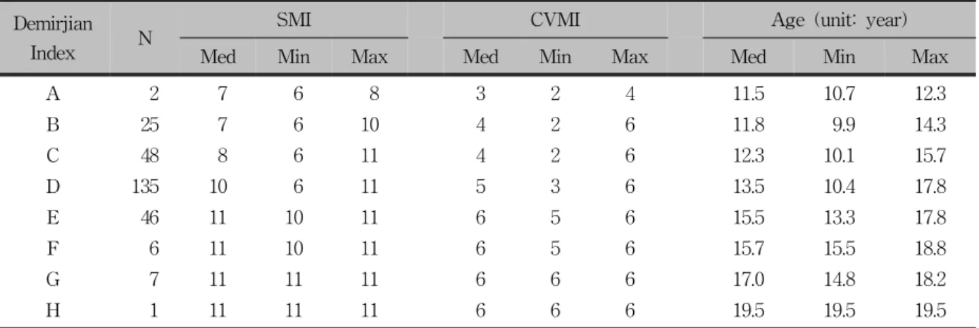
(4) Vol. 39, No. 2, 2009. Korean J Orthod. Skeletal maturation evaluation using mandibular third molar. samples with SMI Levels of 5 or lower was minor.. confirmed. The first radiograph was taken within six months after menarche, and the next radiographs were taken at six-month intervals.. Correlation between Demirjian Index and age at menarche Correlations between the Demirjian Index and the age at menarche were examined in 224 out of the 270 female subjects whose ages at menarche were. Correlation between Demirjian Index and malocclusion type Every female in this study was classified into skeletal Class I, II, or III malocclusion to measure the Demirjian Index in an attempt to evaluate the presence of difference in development for each class.. Statistical analysis (1) The median value, minimum value and maximum value of SMI, CVMI and chronologic age were calculated at each stage of the Demirjian Index. (2) The Pearson correlation analysis was used to verify the relationship among the SMI, the CVMI and the Demirjian Index. (3) The Spearman rank order correlation analysis was used to assess the relationship between age at menarche and third molar development. (4) To evaluate the variations of third molar development among malocclusion classes, median, minimum, and maximum values of the Demirjian Index at every SMI stage for each malocclusion class were determined followed by a Kruskal-Wallis Test for the verification of variations. Fig 1. Eleven skeletal maturity indicators.. Table 3. Median, minimum and maximum values of skeletal maturation indicator (SMI), cervical vertebrae maturation indicator (CVMI) and chronological age in relation to Demirjian Index Demirjian Index. N. SMI. CVMI. Age (unit: year). Med. Min. Max. Med. Min. Max. Med. Min. Max. 3. 2. 4. 11.5. 10.7. 12.3. A. 2. 7. 6. 8. B. 25. 7. 6. 10. 4. 2. 6. 11.8. 9.9. 14.3. C. 48. 8. 6. 11. 4. 2. 6. 12.3. 10.1. 15.7. D. 135. 10. 6. 11. 5. 3. 6. 13.5. 10.4. 17.8. E. 46. 11. 10. 11. 6. 5. 6. 15.5. 13.3. 17.8. F. 6. 11. 10. 11. 6. 5. 6. 15.7. 15.5. 18.8. G. 7. 11. 11. 11. 6. 6. 6. 17.0. 14.8. 18.2. H. 1. 11. 11. 11. 6. 6. 6. 19.5. 19.5. 19.5. N, Number of subjects; Med, median; Min, minimum; Max, maximum.. 123.
(5) Cho SM, Hwang CJ. 대치교정지 39권 2호, 2009년. RESULTS Changes in chronological age along with the development of the third molar The median values were SMI Stage 10 and CVMI Stage 5 at Demirjian Stage D, and the mean chronologic age was 13.5 years. All the samples were at SMI Stage 11 and CVMI Stage 6 at Demirjian Stage G and H where root formation is almost completed (Table 3).. Distribution of SMI and CVMI by Demirjian Index Deciding on the levels of SMI where a high proportion is occupied by Demirjian Stages from A to C is challenging because the number of samples in the stages is insignificant and has a diverse distribution.. SMI Level 10 occupies nearly half (43.7%) of Demirjian Stage D. SMI Levels 10 and 11 are characterized by their concentration in Demirjian Stages beginning with E. Demirjian Stages G and H were all distinguished by SMI Level 11 (Table 4). In spite of the difference in ratio, Demirjian Stages from A to D displayed various distributions in CVMI Levels from 2 to 6. Demirjian Stages beginning with E were characterized by their concentration of CVMI Levels 5 and 6, which corresponded to the pattern of SMI distribution. Demirjian Stages G and H were all distinguished by CVMI Level 6 (Table 5).. Intercorrelations of SMI, CVMI and the Demirjian Index Upon examination of the intercorrelations between SMI and the Demirjian Index, and CVMI and the Demirjian Index, each showed a statistically significant. Table 4. Distribution of SMI by Demirjian Index [unit: number (%)] SMI. Demirjian Index. Total. A. B. C. D. E. F. G. H. 6. 1 (50.0). 9 (36.0). 8 (16.7). 4 (3.0). 0. 0. 0. 0. 22. 7. 0. 7 (28.0). 13 (27.0). 12 (8.9). 0. 0. 0. 0. 32. 8. 1 (50.0). 2 (8.0). 4 (8.3). 13 (9.6). 0. 0. 0. 0. 20. 9. 0. 0. 8 (16.7). 17 (12.6). 0. 0. 0. 0. 25. 10. 0. 7 (28.0). 9 (18.8). 59 (43.7). 7 (15.2). 1 (16.7). 0. 0. 83. 11. 0. 0. 6 (12.5). 30 (22.2). 39 (84.8). 5 (83.3). 7 (100). 1 (100). 88. Total. 2 (100). 25 (100). 48 (100). 135 (100). 46 (100). 6 (100). 7 (100). 1 (100). 270. N, Number of subjects. Table 5. Distribution of cervical vertebrae maturation indicator (CVMI) by Demirjian Index [unit: number (%)] CVMI. Demirjian Index. Total. A. B. C. D. E. F. G. H. 2. 1 (50.0). 5 (20.0). 3 (6.2). 0. 0. 0. 0. 0. 9. 3. 0. 2 (8.0). 11 (22.9). 7 (5.2). 0. 0. 0. 0. 20. 4. 1 (50.0). 11 (44.0). 14 (29.2). 25 (18.5). 0. 0. 0. 0. 51. 5. 0. 6 (24.0). 14 (29.2). 56 (41.5). 9 (19.6). 1 (16.7). 0. 0. 86. 6. 0. 1 (4.0). 6 (12.5). 47 (34.8). 37 (80.4). 5 (83.3). 7 (100). 1 (100). 104. Total. 2 (100). 25 (100). 48 (100). 135 (100). 46 (100). 6 (100). 7 (100). 1 (100). 270. N, Number of subjects.. 124.
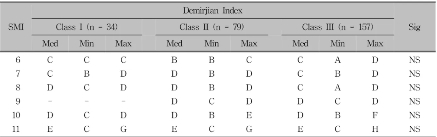
(6) Vol. 39, No. 2, 2009. Korean J Orthod. Skeletal maturation evaluation using mandibular third molar. correlation, with a slightly higher correlation existing between SMI and the Demirjian Index. The Spearman. rank-order correlation coefficient between SMI and CVMI was 0.83, which was also statistically significant (p < 0.001) (Table 6).. Table 6. Correlation coefficients of skeletal maturation indicator (SMI), cervical vertebrae maturation indicator (CVMI), Demirjian Index. Correlation between Demirjian Index and age at menarche. Demirjian. SMI. CVMI. SMI. 1.00. 0.83. 0.64. *. CVMI. 0.83. 1.00. 0.59. *. 0.64. 0.59. 1.00. *. Demirjian index. Index. The Demirjian Index and age at menarche showed a weak (although statistically significant) correlation (r = 0.26). The period between the age at menarche and the age when the radiograph was taken was divided into six-month intervals (Table 7).. Significance. *. p < 0.001.. Table 7. Correlation between Demirjian Index and menarche age (Number of subjects according to the difference between menarche and the timing when the radiograph was taken) Menarche. Demirjian Index. < 0.5 yr. < 1 yr. < 1.5 yr. < 2 yr. < 2.5 yr. < 3 yr. > 3 yr. A. 0. 1. 0. 0. 0. 0. 1. B. 4. 2. 3. 1. 0. 1. 10. C. 11. 5. 4. 3. 2. 1. 11. D. 17. 20. 18. 14. 17. 11. 22. E. 1. 0. 3. 1. 4. 6. 20. F. 0. 0. 0. 0. 0. 2. 4. G. 0. 0. 0. 0. 0. 0. 4. H. 0. 0. 0. 0. 0. 0. 0. r. Sig. 0.26. *. *. yr, Years; r, correlation coefficient; Sig, significance; p < 0.001.. Table 8. Comparison of Demirjian Index among Class I, II and III malocclusions Demirjian Index SMI. Class I (n = 34) Med. Min. Class II (n = 79). Max. Med. Min. Class III (n = 157). Max. Med. Min. Max. Sig. 6. C. C. C. B. B. C. C. A. D. NS. 7. C. B. D. D. B. D. C. B. D. NS. 8. D. C. D. D. B. D. C. A. D. NS. 9. -. -. -. D. C. D. D. C. D. NS. 10. D. C. D. D. B. E. D. B. F. NS. 11. E. C. G. E. C. G. E. C. H. NS. n, Number of subjects; Med, median; Min, minimum; Max, maximum; Sig, significance; NS, not significant.. 125.
(7) Cho SM, Hwang CJ. Correlation between the Demirjian Index and malocclusion type Every sample was classified into malocclusion classes I, II, or III to compare the median, minimum and maximum value of the Demirjian Index for the SMI of each class. There was no statistically significant difference among the malocclusion classes (Table 8).. DISCUSSION Many growth evaluation methods for precise prediction have been suggested.5-7 Dental maturity, in particular, has the advantage of easy evaluation during regular dental treatment. The relationship between dental maturity and bone maturity, however, shows diverse opinions among different studies.5-7,13-16 Third molars were excluded from most of the studies due to their assumed developmental variations. The aim of this study was to investigate the relationship between dental maturity and skeletal maturity using the development of the third molar in the mandible and also to evaluate the clinical value of the third molar. Dental maturity can generally be determined by the stage of tooth eruption or the stage of tooth formation. The first disadvantage of this method is in determining its exact timing because it happens quickly. In addition, tooth eruption can be altered by local factors, systemic diseases and nutritional habits; the reliability of the method is questionable. Therefore, dental maturity in this study was determined by evaluating the stages of tooth formation, with an emphasis on Demirjian’s method. The root of the third molar tends to be less divergent and more fused, making it harder to evaluate its development according to root length. The Demirjian Index evaluates the evident changes in the shape of the tooth without letting the tooth length affect the reliability of evaluation. Recent studies have verified that Demirjian’s classification system shows the least intra-examiner and inter-examiner errors and a high correlation with biological age.19,20 Therefore, Demirjian’s classification system was utilized in this study to assess third molar development. Bolaños et al21 reported that the crown formation of. 126. 대치교정지 39권 2호, 2009년. the third molar was complete at the age of 14, and the root formation terminated at an average of 18.5 years based on their studies in a Spanish population. More22 over, Kullman et al indicated that the root formation began at about 15.1 years and was completed at an average age of 19.3 years. The crown formation was also completed by 13.5 years in this study, which resembled the ranges of previous studies. The distribution of SMI was investigated at each Demirjian Stage. It is difficult to characterize the specific pattern of distribution due to its diversity in Demirjian Stages below D. All the samples were within SMI 6 to 8 at Demirjian Stage A. But this result was due to the insufficient sample size. If sample size becomes larger, the result will be more various just as at Stage B to D. On the contrary, the distribution of SMI was consistent at Demirjian Stage E to H even though sample size was small. Even if sample size becomes larger, this consistent pattern will be maintained. Only SMI Levels 10 and 11 were presented in Demirjian Stage E and F, and SMI Level 11 alone was seen in Demirjian Stage G and H. In the same way, the distribution of CVMI was investigated at each Demirjian stage, with similar patterns observed to those seen for SMI; manifold distribution up to Stage D, the presentation of only CVMI Levels 5 and 6 in Stages E and F, and the appearance of CVMI Level 6 in Stages G and H. Overall, the samples in Demirjian Stages E or above, the stage for the end of crown formation of the third molar and the onset of root formation, are predicted to occur at the higher levels of SMI Level 10 and CVMI Level 5. There was a statistically significant correlation between the development of the third molar (Demirjian Index) and SMI (r = 0.64), and the Index and CVMI (r = 0.59), with a slightly higher correlation found between SMI and the Demirjian Index. SMI and CVMI also had a high correlation (r = 0.83) that corresponded with the results from Hassel and Farman's 12 study. The findings also correspond to those of Demisch and Wartmann23 (r = 0.73) and Engström et al14 (r = 0.71), who reported a strong correlation between third molar formation and skeletal maturity. However, Lewis and Garn,15 Demirjian et al,6 and Krailassiri et al24 indicated that the relationship be-.
(8) Vol. 39, No. 2, 2009. Korean J Orthod. tween the development of the third molar and bone maturity was poor. Moorrees et al25 indicated that excessively subdivided stages not only make it difficult to divide the development into precise stages, but also adversely affect the evaluation due to the insufficient amount of time to progress to the following stage. Thus, the division of tooth development into only a few stages is desirable for a meaningful comparison. Different samples may also affect the result of correlations between third molars and bone maturity. Demisch and Wartmann23 stated that among white children, the correlation between third molars and bone maturity extended to 0.8, while Thai kids showed only a 0.3 correlation using the same method.24 Mincer et al26 studied 823 U.S. citizens between the ages of 14.1 and 24.9 years. No significant difference in the development of the third molar was observed with regard to race. However, Gorgani et al27 examined 229 black and 221 white U.S. citizens between 6 and 14 years of age, and determined that third-molar crown mineralization was completed about a year earlier in black Americans than in whites. The comparison of thirdmolar mineralization among Germans, Japanese and South Africans conducted by Olze et al19 using Demirjian’s classification system showed that the development completed first in South Africans, then in Germans, and finally in the Japanese participants. 28 Nanda suggested that there was a correlation between age at menarche and dental maturity (r = 0.59). Demirjian et al6 maintained that dental development was independent and that dental maturity had a low correlation with skeletal and sexual maturity due to the predominant accuracy of the dental development index 29 over others. Garn et al stated that although a correlation was present between dental development and skeletal and sexual maturity in general, the development of the third molar was independent, resulting in a weak correlation with skeletal and sexual maturity. In this study, 224 out of 270 females with a verified age at menarche showed little correlation between age at menarche and the development of third molars, which is in agreement with Garn et al’s study29 (r = 0.26). Although it appeared to be higher than that seen in previous reports, the correlation coefficient is not enough to be meaningful for the correlation between. Skeletal maturation evaluation using mandibular third molar. age at menarche and the development of the third molar. In this study, no statistically significant difference in the development of the third molar was present among Class I, II or III malocclusions. Nanda30 indicated that subjects with a skeletal open bite presented with an earlier onset of the adolescent growth spurt in the maturation of the facial bones than did those with a deep bite, and so did the dental maturation. Janson et al31 investigated the influence of facial type on dental development in subjects of the same chronological age. They showed that subjects with long faces tended to have an advanced dental maturation in comparison with short faces, which was expressed by a mean difference in dental age of six months. Our results showed no significant difference in third molar development among Class I, II or III malocclusions. The type of malocclusion should not affect the development of the third molar. The revealed correlation between lower third molar development and skeletal maturity in this study will allow clinicians to use the mandibular third molar as an adjunctive tool to adolescent growth assessment in combination with cervical vertebrae and hand-wrist maturity evaluations. Individual variations should be taken into consideration when using the developmental stage of the third molar in growth evaluations because third molars are known for their many variations based on previous studies. This cross-sectional study has limitations on evaluating the results because the females in this study were in the pubertal growth period and mainly concentrated in Demirjian Stages C, D and E. Further longitudinal studies with a larger sample size are recommended for more accurate results.. CONCLUSION The aims of this study were to evaluate a correlation between dental maturity and skeletal maturity of cervical vertebrae and hand-wrist and to estimate the clinical predictive value of the third molar. For this study, the samples were derived from panoramic, lateral cephalometric and hand-wrist radiographs of 270 female adolescents. Dental maturity (Demirjian Index) and skeletal maturity (skeletal maturation indicators) and. 127.
(9) Cho SM, Hwang CJ. 대치교정지 39권 2호, 2009년. cervical vertebrae maturation indicators were estimated from these radiographs. The results were as follows: 1. There was a significant correlation (r = 0.64) between the SMI and the Demirjian Index, and a similar correlation (r = 0.59) was observed between the CVMI and the Demirjian Index (p < 0.001). 2. If the Demirjian Index was above Stage E, SMI was above Stage 10 and CVMI was above Stage 5. 3. There was a weak correlation (r = 0.26) between menarche and the Demirjian Index (p < 0.001). 4. There was no significant difference in the Demirjian Index among Class I, II or III malocclusions.. 연구 결과 Demirjian index와 SMI, CVMI 간의 상관관계에서 SMI와 Demirjian index (r = 0.64), CVMI와 Demirjian index (r = 0.59)는 통계적으로 유의한 양의 상관관계를 보였다 (p < 0.001). 그러나 초경 연령과 Demirjian index간의 상관관 계(r = 0.26)는 낮았으며 (p < 0.001), Demirjian index를 통 해 평가한 제3대구치의 치아성숙도는 I, II, III급 부정교합 간 에 차이가 없는 것으로 나타났다. 그리고 제3대구치 치관이 완성되고 치근이 형성되기 시작하는 Demirjian index E단계 이상이면 SMI 10단계, CVMI 5단계 이상에 속하였다. 하악 제3대구치의 발육단계를 이용한 치아성숙도 평가는 경추 및 수완부 골성숙도와 조합하여 사용한다면 사춘기 성장 평가 에 있어 하나의 보조적인 수단으로 활용될 수 있을 것이다. 주요 단어: 성장 평가, 성숙, 성장 예측, 성장과 발육. Based on the results of this study, a dental maturity evaluation using the mandibular third molar would be an adjunctive tool to adolescent growth assessment in combination with cervical vertebrae and hand-wrist maturity evaluations. In particular, since it has been proven that a Demirjian Index score above Stage E means that the SMI Stage is above 10 and the CVMI Stage is above 5, judging the completion of growth may be possible when the beginning of root formation of the third molar is seen on a radiograph. Individual variations, however, should be taken into consideration to use the developmental stage of the third molar in growth evaluations because third molars are known for their many variations from previous studies. - 국문초록 -. 하악 제3대구치의 성숙도를 이용한 성장 평가 조선미aㆍ황충주b. 성장기 환자의 교정 치료에서 바람직한 치료 결과를 얻기 위해서는 성장을 정확하게 예측하는 것이 중요하다. 성장 과 정은 개인마다 상당한 차이를 보이기 때문에 성장 평가를 위 해서는 연대 연령이 아닌 생리적인 연령을 사용해야 한다. 본 연구의 목적은 사춘기 동안 발육하는 하악 제3대구치에 서 치아성숙도를 측정하고 성장 평가 지표로서의 그 가치를 평가하고자 하는 데 있다. 연세대학교 치과대학병원 교정과 에 내원한 성장기 여자 환자 270명을 대상으로 제3대구치의 치아 성숙도(Demirjian index), 경추의 골 성숙도(CVMI), 수 완부 골 성숙도(SMI), 초경 연령 등을 평가하여 분석하였다.. 128. REFERENCES 1. Lee JH, Kang YG, Lee KS, Nam JH. Maturation of cervical vertebrae in relation to menarche. Korean J Orthod 2009;39: 28-35. 2. Lee KH, Hwang YI, Kim YJ, Park YH, Baek SH, Cha KS. Skeletal maturation associated with the fourth cervical vertebra and menarcheal timing. Korean J Orthod 2008;38:52-9. 3. Bishara SE, Jamison JE, Peterson LC, DeKock WH. Longitudinal changes in standing height and mandibular parameters between the ages of 8 and 17 years. Am J Orthod 1981;80:115-35. 4. Fishman LS. Maturational patterns and prediction during adolescence. Angle Orthod 1987;57:178-93. 5. Chertkow S, Fatti P. The relationship between tooth mineralization and early radiographic evidence of the ulnar sesamoid. Angle Orthod 1979;49:282-8. 6. Demirjian A, Buschang PH, Tanguay R, Patterson DK. Interrelationships among measures of somatic, skeletal, dental, and sexual maturity. Am J Orthod 1985;88:433-8. 7. Sierra AM. Assessment of dental and skeletal maturity. A new approach. Angle Orthod 1987;57:194-208. 8. Hägg U, Taranger J. Maturation indicators and the pubertal growth spurt. Am J Orthod 1982;82:299-309. 9. Fishman LS. Radiographic evaluation of skeletal maturation. A clinically oriented method based on hand-wrist films. Angle Orthod 1982;52:88-112. 10. Lamparski DG. Skeletal age assessment utilizing cervical vertebrae [master's thesis]. Pittsburgh (PA): University of Pittsburgh; 1972. 11. Hellsing E. Cervical vertebral dimensions in 8-, 11-, and 15-year-old children. Acta Odontol Scand 1991;49:207-13. 12. Hassel B, Farman AG. Skeletal maturation evaluation using cervical vertebrae. Am J Orthod Dentofacial Orthop 1995; 107:58-66. 13. Lauterstein AM. A cross-sectional study in dental development and skeletal age. J Am Dent Assoc 1961;62:161-7. 14. Engström C, Engström H, Sagne S. Lower third molar devel-.
(10) Vol. 39, No. 2, 2009. Korean J Orthod. 15. 16.. 17. 18. 19.. 20.. 21.. 22.. opment in relation to skeletal maturity and chronological age. Angle Orthod 1983;53:97-106. Lewis AB, Garn SM. The relationship between tooth formation and other maturational factors. Angle Orthod 1960;30:70-7. Green LJ. The interrelationships among height, weight and chronological, dental and skeletal ages. Angle Orthod 1961;31: 189-93. Banks HV. Incidence of third molar development. Angle Orthod 1934;4:223-33. Demirjian A, Goldstein H, Tanner JM. A new system of dental age assessment. Hum Biol 1973;45:211-27. Olze A, Bilang D, Schmidt S, Wernecke KD, Geserick G, Schmeling A. Validation of common classification systems for assessing the mineralization of third molars. Int J Legal Med 2005;119:22-6. Arany S, Iino M, Yoshioka N. Radiographic survey of third molar development in relation to chronological age among Japanese juveniles. J Forensic Sci 2004;49:534-8. Bolaños MV, Moussa H, Manrique MC, Bolaños MJ. Radiographic evaluation of third molar development in Spanish children and young people. Forensic Sci Int 2003;133:212-9. Kullman L, Johanson G, Akesson L. Root development of the lower third molar and its relation to chronological age. Swedish Dent J 1992;16:161-7.. Skeletal maturation evaluation using mandibular third molar. 23. Demisch A, Wartmann P. Calcification of the mandibular third molar and its relation to skeletal and chronological age in children. Child Dev 1956;27:459-73. 24. Krailassiri S, Anuwongnukroh N, Dechkunakorn S. Relationships between dental calcification stages and skeletal maturity indicators in Thai individuals. Angle Orthod 2002;72:155-66. 25. Moorrees CF, Fanning EA, Hunt EE Jr. Age variation of formation stages for ten permanent teeth. J Dent Res 1963: 1490-502. 26. Mincer HH, Harris EF, Berryman HE. The A.B.F.O. study of third molar development and its use as an estimator of chronological age. J Forensic Sci 1993;38:379-90. 27. Gorgani N, Sullivan RE, DuBois L. A radiographic investigation of third-molar development. ASDC J Dent Child 1990; 57:106-10. 28. Nanda RS. Eruption of human teeth. Am J Orthod 1960:46; 363-78. 29. Garn SM, Lewis AB, Bonné B. Third molar formation and its development course. Angle Orthod 1962;32:270-9. 30. Nanda SK. Patterns of vertical growth in the face. Am J Orthod Dentofacial Orthop 1988;93:103-16. 31. Janson GR, Martins DR, Tavano O, Dainesi EA. Dental maturation in subjects with extreme vertical facial types. Eur J Orthod 1998;20:73-8.. 129.
(11)
수치
![Table 5. Distribution of cervical vertebrae maturation indicator (CVMI) by Demirjian Index [unit: number (%)]](https://thumb-ap.123doks.com/thumbv2/123dokinfo/5467397.658952/5.892.99.800.616.831/table-distribution-cervical-vertebrae-maturation-indicator-demirjian-index.webp)
관련 문서
