지유 에탄올추출물의 생체외 항산화 활성
임태진*
상지대학교 동물생명자원학부 동물생명공학전공
In vitro Antioxidant Activity of Sanguisorbae Radix Ethanol Extracts
Tae-Jin Rhim*
Department of Animal Biotechnology in Division of Animal and Life Resources, Sangji University, Wonju 220-702, Korea
Abstract - The objective of this study was to investigate the antioxidative capacity of ethanol extracts from Sanguisorbae officinalis L. root (Sanguisorbae radix) in vitro. The concentration of Sanguisorbae radix extract at which DPPH radical scavenging activity was inhibited by 50% was 0.33 mg/mL, which was similar to IC50 of α-tocopherol (0.40 mg/mL), as compared to 100% by pyrogallol as a reference. Total antioxidant status was examined by total antioxidant capacity against ABTS radical reactions. Total antioxidant capacities of Sanguisorbae radix extract were significantly (p<0.05) higher than those of α-tocopherol. Superoxide scavenging activities of Sanguisorbae radix extract were significantly (p<0.05) higher than those of catechin. Oxygen radical absorbance capacities of Sanguisorbae radix extract were significantly (p<0.05) higher than those of ascorbic acid. Cupric reducing antioxidant capacities of Sanguisorbae radix extract were significantly (p<0.05) higher than those of α-tocopherol. Sanguisorbae radix extract prevented supercoiled DNA strand breakage induced by hydroxyl radical and peroxyl radical. Total phenolic contents of Sanguisorbae radix extract at concentrations of 0.5 and 5 mg/mL were 0.50 and 3.33 mM gallic acid equivalents, respectively. Sanguisorbae radix extract at concentration of 0.01, 0.1 and 0.5 mg/mL inhibited 0.2 mM tert-butyl hydroperoxide-induced cytotoxicity by 33.8, 79.1 and 96.9%, respectively, in HepG2 cell culture system. Thus, strong antioxidant and cytotoxicity-ihnibiting effects of Sanguisorbae radix extract seem to be due to, at least in part, the prevention from free radicals-induced oxidation as well as high levels in total phenolic contents.
Key words -Sanguisorbae officinalis L., DPPH radical, ABTS radical, Superoxide, ORAC, CUPRAC, DNA breakage, Total phenolic content, Cytotoxicity
*교신저자(E-mail) :[email protected]
서 언
Superoxide anion, hydroxyl radical, hydrogen peroxide 등의 반응산소종(reactive oxygen species, ROS)은 활성산소라고도 불리우며, 단백질 불활성화, 지질 과산화, DNA 변성 등을 초래하여 세포의 기능장애를 유발 하고, 암을 비롯하여 동맥경화증, 당뇨병, 파킨슨 질병 등 수많은 질병을 일으키는 것으로 보고되고 있다(Halliwell et al., 1992; Thannickal and Fanburg, 2000). 따라서, 최근에는 이러한 ROS를 제거해줌으로써 생체 내에서 산화 성 스트레스로 인하여 생성되는 산화물질들을 방어하는 항
산화제의 활용이 증가하고 있고, 특히 식물계에 널리 분포 되어 있는 천연 항산화제의 중요성과 이에 관한 연구가 증 가하고 있다.
오이풀(Sanguisorbae officinalis L.)은 장미과에 속하 는 다년생 초본으로, 우리나라에 널리 자생하고 있으며, 그 동속 식물의 뿌리를 지유(Sanguisorbae radix)라고 한 다. 민간에서는 화상, 내출혈 치료제로 이용되어져 왔고 (Cheng and Cao, 1992), pomolic acids, sanguiins, triterpenoids, triterpene glycosides 등의 성분들이 지 유로부터 분리되어 보고된 바 있다(Cheng and Cao, 1992; Yokozawa et al., 2000; Mimaki et al., 2001;
Liu et al., 2005). 지유 추출물은 높은 항암 활성을 나타 Original Research Article
내었으며(Goun et al., 2002; An et al., 2004b), 항바이 러스 및 항균 활성도 보고된 바 있다(Kim et al., 2001;
An et al., 2004b). 흰쥐를 이용한 in vivo 실험에서 지유 추출물 투여는 간 보호(Jeong et al., 2003)와 뇌세포 보 호(Nguyen et al., 2008) 효과를 나타내었다. 지유 추출 물의 항산화 효과는 DPPH 소거활성 억제(An et al., 2005), superoxide 소거활성 억제(An et al., 2004a) 및 신장 피질세포 미토콘드리아의 지질과산화 억제(Yokozawa et al., 2002) 등이 보고된 바 있다.
이와 같이, 지유의 항암, 항균, 간 및 뇌세포 보호 기능 등에 관해 보고되고 있으나, 항산화 활성에 관한 연구결과 는 많이 보고되지 않고 있다. 따라서, 본 연구에서는 지유 추출물의 free radical 소거활성과 세포독성 억제효과들을 조사하였다.
재료 및 방법
시약
실험에 사용된 ammonium acetate, ascorbic acid, 2,2'-azino-bis(3-ethylbenzothiazoline-6-sulfonic acid) diammonium salt(ABTS), 2,2'-azobis(2-methyl propionamidine) dihydrochloride(AAPH), tert-butyl hydroperoxide(t-BHP), catechin, cupric chloride, 2,2- diphenyl-1-picrylhydrazyl(DPPH), ferrous sulfate, fluorescein sodium salt, Folin-Ciocalteu phenol reagent, gallic acid, hydrogen peroxide, 6-hydroxy- 2,5,7,8-tetramethylchroma-2-carboxylic acid(Trolox), neocuproine, β-nicotinamide adenine dinucleotide (NADH), nitroblue tetrazolium(NBT), phenazine methosulfate(PMS), pyrogallol, RPMI-1640 medium, sesamol, sodium acetate, sodium bicarbonate, thiazolyl blue tetrazolium bromide(MTT), α-tocopherol, Tris (hydroxymethyl) aminomethane는 Sigma-Aldrich Co.
(St. Louis, MO, USA)로부터 구입하였고, dimethyl sulfoxide(DMSO), penicillin/streptomycin은 Bio Basic Inc.(Ontario, Canada)로 부터 구입하였다. pBR322 DNA는 KOSCHEM(Seoul, Korea)으로부터 구입하였고, GelRed nucleic acid gel stain은 Biotium(Hayward, CA, USA)으로부터 구입하였으며, fetal bovine serum (FBS)은 Lonza(Walkersville, MD, USA)로부터 구입하 여 사용하였다.
지유 추출물
지유는 경상북도 경산시에서 구입하여 본 연구의 시료로 사용하였다. 건조된 시료 500 g을 마쇄하여, 수직으로 환 류냉각관을 부착시킨 round flask에 넣고 450 mL의 95%
에탄올을 첨가하여 혼합한 후, heating mantal(E105, Minsung Scientific Co., Seoul, Korea)로 80℃에서 4시 간 강열 환류 추출하였다. 이 과정을 3회 반복하여 얻은 추 출액을 Whatman No.2 여과지로 여과하여 불순물을 제거 하였다. 여과된 용액은 감압농축기(Eyela N-1 NW, Tokyo Rikakikai Co., Tokyo, Japan)를 사용하여 45℃에서 감 압 농축시켰으며, 동결건조 후 62 g의 추출물을 회수하였 다. 지유 추출물은 분석시까지 -80℃에서 보관하였다.
DPPH radical 소거활성 측정
DPPH radical 소거활성은 Malterud et al.(1993)의 방 법에 따라 측정하였다. DPPH 용액(45 μg/mLmethanol) 을 추출물과 혼합한 다음, GENESYS 10S spectro- photometer(Thermo Scientific, Waltham, MA, USA) 를 사용하여 515 nm에서 흡광도의 감소를 30초 간격으로 5분간 측정하였다. DPPH radical 소거활성은 pyrogallol 용액(125 μg/mLDMSO)의 흡광도 감소를 100%로 기준하 여 표기하였다. 시료 농도와 DPPH radical 소거활성 간의 회귀분석에 의해 50%의 DPPH radical 소거활성에 필요한 지유 추출물 및 α-tocopherol의 농도(IC50)를 측정하였다.
또한, 양성 대조군으로 α-tocopherol을 사용하여 DPPH radical 소거활성을 비교 조사하였다.
총항산화능 측정
총항산화능은 Trolox equivalent antioxidant capacity (TEAC) 방법을 수정한 Erel(2004)의 방법에 따라 측정하 였다. 추출물에 0.35 M acetate 완충용액과 0.89 mM ABTS 용액 및 0.44 mM hydrogen peroxide 용액 등을 첨가하고, 혼합한 뒤 5분 후에 660 nm에서 흡광도를 측정 하였다. Trolox를 표준시약으로 사용하여 표준곡선을 작 성하였고, 총항산화능은 mM Trolox equivalent로 표기하 였다. 또한, 양성 대조군으로 α-tocopherol을 사용하여 총항산화능을 비교 조사하였다.
Superoxide 소거활성 측정
Superoxide 소거활성은 Liu et al.(1997)의 방법에 따
라 측정하였다. 추출물에 62 μM NBT와 98 μM NADH를 함유한 20 mM Tris 용액(pH 8.0)을 혼합한 다음, 20 mM Tris 용액과 33 μM PMS를 각각 첨가하였다. 즉, 비효소 적으로 PMS/NADH로 유발된 superoxide는 NBT를 자주 색의 formazan으로 환원시키며, 생성된 formazan을 측 정하기 위해 560 nm에서 10 분 동안 반응물의 흡광도를 측정하였다. 추출물의 superoxide 소거활성(%)은 [(흡광
도추출물무첨가-흡광도추출물)/흡광도추출물무첨가]×100의 공식으
로 계산하였다. 또한, 양성 대조군으로 catechin을 사용하 여 superoxide 소거활성을 비교 조사하였다.
Oxygen radical absorbance capacity (ORAC) 측정 ORAC는 Huang et al.(2002)의 방법에 따라 추출물에 6×10⁻⁵ mM fluorescein 용액을 첨가하고, 37℃에서 10분 간 가열한 다음, 19 mM AAPH 용액을 첨가한 뒤, GEMINI XS fluorescence microplate reader(Molecular Devices, Sunnyvale, CA, USA)를 사용하여 excitation 파장 485 nm와 emission 파장 530 nm에서 2분 간격으로 60분간 형광도를 측정하였다. 표준시약으로 Trolox를 사 용하였으며, 표준시약과 추출물의 area under the curve(AUC)를 측정하였다. ORAC는 표준시약 농도와 AUC 간의 회귀곡선을 이용하여 μM Trolox equivalent로 표기하였다. 또한, 양성 대조군으로 ascorbic acid를 사용 하여 ORAC를 비교 조사하였다.
Cupric reducing antioxidant capacity (CUPRAC) 측정
CUPRAC는 구리이온 환원력을 나타내며, Apak et al.(2004)의 방법에 따라 측정하였다. 추출물에 2.44 mM cupric chloride 용액과 1.83 mM neocuproine 및 0.24 M ammonium acetate 완충용액(pH 7.0) 등을 첨가하고 혼합한 뒤, 실온에서 1시간 방치한 후 450 nm에서 흡광도 를 측정하였다. Trolox를 표준시약으로 사용하여 표준곡 선을 작성하였고, CUPRAC 활성은 mM Trolox equivalent로 표기하였다. 또한, 양성 대조군으로 α -tocopherol을 사용하여 CUPRAC를 비교 조사하였다.
Supercoiled DNA strand 절단
① Hydroxyl radical에 의한 DNA strand의 절단은 Hiramoto et al.(1996)의 방법에 따라 측정하였다.
Supercoiled pBR322 DNA 0.2 μg에 추출물을 넣고, 0.1 mM hydrogen peroxide 용액과 0.1 mM ferrous sulfate 용액과 함께 37℃에서 1시간 배양하였다. 양성 대조군으로 sesamol을 사용하였다.
② Peroxyl radical에 의한 DNA strand의 절단은 Hu et al.(2000)의 방법에 따라 측정하였다. Supercoiled pBR322 DNA 0.2 μg에 추출물을 넣고, 5 mM AAPH와 함께 37℃에서 2시간 배양하였다. 양성 대조군으로 Trolox를 사용하였다.
배양이 끝난 다음, gel loading buffer를 첨가하고, 0.01% GelRed nucleic acid stain이 포함된 0.8%
agarose에서 전기영동을 실시하였다. 자외선 하에서 사진 을 촬영한 후, DNA band의 density는 Image J 1.44 program(NIH, Bethesda, MD, USA)을 사용하여 측정하 였으며, 지유 추출물의 supercoiled DNA strand의 절단 억제효과는 supercoiled DNA strand의 retention percent를 측정함으로써 조사하였다. Retention percent 는 (Asample/Anative)×100의 공식으로 계산하였으며, Asample
은 radical과 추출물 처리시의 supercoiled DNA strand 의 양을 나타내며, Anative는 radical과 추출물 무처리시의 supercoiled DNA strand의 양을 나타낸다.
총페놀 함량 측정
추출물내 총페놀 함량은 Singleton et al.(1999)의 방법 에 따라 추출물에 0.08 N Folin-Ciocalteu 시약을 첨가하 고, 실온에서 6분간 방치한 다음, 3% sodium bicarbonate 용액을 첨가하고 90분간 방치한 뒤, 760 nm에서 흡광도를 측정함으로써 결정하였다. 표준시약으로 gallic acid를 사 용하여 표준곡선을 작성하였고, 총페놀 함량은 mM gallic acid equivalent로 표기하였다.
세포독성 억제효과 측정
지유 추출물의 세포독성 억제효과를 조사하기 위해, t-BHP로 유도된 HepG2 세포 배양실험에서 추출물 첨가 에 의한 세포 생존율을 측정하였다. HepG2 세포(KCLB No. 88065)는 한국세포주은행으로부터 구입하였다. HepG2 세포를 10% FBS와 100 U/mL penicillin 및 100 μg/mL streptomycine이 포함된 RPMI-1640 배지를 사용하여 5×10⁴ cells/well이 되도록 24-well plate(SPL, Pocheon, Korea)에 분주하고, 37℃, 5% CO2 incubator (NAPCO
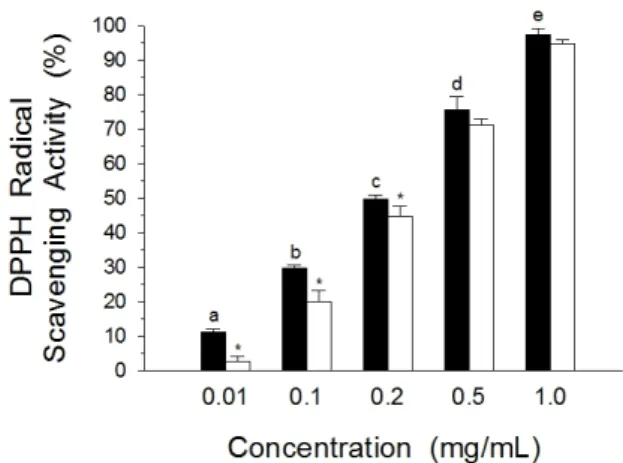
Fig. 1. DPPH radical scavenging activity of Sanguisorbae radix extracts. Data results were expressed as % radical scavenging activity relative to 100% radical scavenging activity of pyrogallol solution as a reference. Each bar represents the mean±SD of quadraplicate determinations.
■: extract, □: α-tocopherol (positive control). abcdeValues with different letters are significantly different at p<0.05.
*p<0.05 when compared with the extract within same group.
6001, Precision Scientific In., Chicago, IL, USA)에서 배양하였다. 100% confluent에 도달한 후, t-BHP(0.2 mM) 또는 지유 추출물이 포함된 배지로 교체하고 5시간 동안 배양하였다. 배양액을 제거하고, 세포 단층에 Mosmann(1983)의 방법에 따라 MTT 시약(5 mg/mL)을 첨가하고, 37℃, 5% CO2 incubator에서 3시간 배양한 후, 0.04 M HCl 용액을 첨가한 뒤, 570 nm에서 흡광도를 측 정함으로써 MTT 값을 측정하였다. 세포 생존율은 대조군 의 MTT 값을 100%로 기준하여, 처리군의 MTT 값으로 표 기하였다.
통계 분석
추출물 농도별 항산화 활성은 일원 분산분석을 사용하여 조사하였고, 농도별 평균값의 차이는 Duncan's multiple range test(Steel and Torrie, 1980)를 사용하여 p<0.05 에서 유의성을 조사하였다. 또한, 추출물과 양성대조군의 항산화 효능 비교는 대응표본 T-검정을 사용하여 p<0.05 에서 유의성을 조사하였다.
결과 및 고찰
DPPH radical 소거활성
지유 추출물의 농도별 DPPH radical 소거활성은 Fig. 1 에 나타나 있다. Pyrogallol의 억제율을 100%로 기준하였 을 때, 지유 추출물의 0.01 mg/mL 농도에서 DPPH radical 소거활성은 11.3%이었고, 추출물 농도가 증가할수 록 소거활성도 증가하여, 0.1, 0.2, 0.5 및 1 mg/mL 농도 의 추출물은 각각 29.7, 49.7, 75.6 및 97.4%의 소거활성 을 나타내었다. 양성대조군으로 사용한 α-tocopherol의 DPPH radical 소거활성은 0.01, 0.1 및 0.2 mg/mL 농도 에서 각각 2.5, 20.0 및 44.8%로 측정되어, 지유 추출물의 DPPH radical 소거활성이 α-tocopherol에 비해 유의적 으로(p<0.05) 높게 나타났다. 반면에, 0.5 및 1 mg/mL의 농도에서 α-tocopherol의 DPPH radical 소거활성은 각 각 71.1 및 94.6%로 측정되어, 지유 추출물과 α -tocopherol 간의 소거활성에는 유의적인(p>0.05) 차이 가 관찰되지 않았다. 또한, 시료 농도와 DPPH radical 소 거활성 간의 회귀분석 결과(지유 추출물의 경우, Y=
23.11+81.84X; α-tocopherol의 경우, Y=14.71+88.10X:
Y는 radical 억제%이며, X는 시료 농도), 지유 추출물 및
α-tocopherol의 IC50은 각각 0.33 및 0.40 mg/mL로 측 정되어, 지유 추출물의 DPPH radical 소거활성이 α -tocopherol과 유사함을 알 수 있었다. 본 연구에서 관찰 된 지유 추출물의 뛰어난 DPPH radical 소거활성은 기존 에 발표된 연구결과(An et al., 2004a; An et al., 2005) 와도 일치하고 있다.
총항산화능
지유 추출물의 농도별 총항산화능은 Fig. 2에 나타나 있 다. 지유 추출물 0.01 mg/mL 농도의 총항산화능은 0.14 mM Trolox equivalent이었으며, 추출물 농도가 증가함에 따라 총항산화능도 비례적으로 증가하여, 0.1, 0.5 및 1 mg/mL 농도에서는 각각 0.40, 2.60 및 3.01 mM Trolox equivalent를 나타내었다. 반면에, 양성대조군으로 사용 한 α-tocopherol의 총항산화능은 0.01, 0.1, 0.5 및 1 mg/mL 농도에서 각각 0.11, 0.29, 0.83 및 1.49 mM Trolox equivalent로 측정되었다. 모든 농도에서 지유 추 출물의 총항산화능이 α-tocopherol에 비해 유의적으로 (p<0.05) 높게 관찰되어, 지유 추출물의 ABTS radical 소 거활성이 α-tocopherol에 비해 탁월함을 알 수 있었다.
Fig. 2. Total antioxidant capacity of Sanguisorbae radix extracts. Data results were expressed as in terms of mM Trolox equivalent. Each bar represents the mean±SD of quadraplicate determinations. ■: extract, □: α-tocopherol (positive control). abcdValues with different letters are significantly different at p<0.05. *p<0.05 when compared with the extract within same group.
Fig. 3. Superoxide radical scavenging activity of Sang- uisorbae radix extracts. Data results were expressed as % inhibition of the activity. Each bar represents the mean ± SD of quadraplicate determinations. ■: extract, □:
catechin (positive control). abcdeValues with different letters are significantly different at p<0.05. *p<0.05 when compared with the extract within same group.
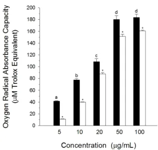
Fig. 4. Oxygen radical absorbance capacity of Sanguisorbae radix extracts. Data results were expressed as in terms of μM Trolox equivalent. Each bar represents the mean±SD of triplicate determinations. ■: extract, □: ascorbic acid (positive control). abcdValues with different letters are significantly different at p<0.05. *p<0.05 when compared with the extract within same group.
Superoxide 소거활성
지유 추출물의 농도별 superoxide 소거활성은 Fig. 3에 나타나 있다. 지유 추출물 0.01 mg/mL 농도의 superoxide 소거활성은 5.8%이었으며, 추출물 농도가 증가함에 따라 소거활성도 증가하여, 0.1, 0.2, 0.5 및 1 mg/mL 농도의
지유 추출물의 superoxide 소거활성은 각각 39.6, 59.6, 75.5 및 85.5%로 나타났다. An et al.(2004a)도 지유 추 출물의 superoxide dismutase(SOD) 유사활성이 농도의 존적으로 증가하여, 1 mg/mL의 농도에서 65.4%의 높은 활성을 나타내었다고 보고한 바 있다. 반면에, 양성대조군 으로 사용한 catechin의 superoxide 소거활성은 0.01, 0.1, 0.2, 0.5 및 1 mg/mL 농도에서 각각 1.8, 22.3, 38.5, 60.8 및 80.4%로 측정되었다. 1 mg/mL을 제외한 모든 농도에서, 지유 추출물의 소거활성이 catechin에 비 해 유의적으로(p<0.05) 높게 관찰되어, 지유의 superoxide 소거활성이 탁월함을 알 수 있었다. 또한, 본 연구결과와 유사하게, 효소적 방법을 이용한 superoxide 소거활성 연 구에서도 5 μg(dry weight)/mL 농도의 지유 추출물이 28.6%의 superoxide 소거활성을 나타내었다고(Masaki et al., 1995) 보고된 바 있다.
ORAC
ORAC assay는 AUC를 측정함으로써, free redical 손 상에 대한 억제 시간과 억제율을 모두 반영하는 항산화능 측정 방법이다. ORAC는 Trolox를 표준물질로 사용하여
Fig. 5. Cupric reducing antioxidant capacity of Sanguisorbae radix extracts. Data results were expressed as in terms of mM Trolox equivalent. Each bar represents the mean±SD of triplicate determinations. ■: extract, □: α-tocopherol (positive control). abcdeValues with different letters are significantly different at p<0.05. *p<0.05 when compared with the extract within same group.
(A)
(B)
Fig. 6. Electrophoresis of pBR322 DNA treated with hydroxyl radical and peroxyl radical in the presence of Sanguisorbae radix extracts. (A) hydroxyl radical was generated by 0.1 mM hydrogen peroxide and 0.1 mM ferrous sulfate; (B) peroxyl radical was generated by 5 mM AAPH. Lane 1, DNA alone; Lane 2, DNA+radical;
Lane 3, DNA+radical+0.001 mg/mL extracts; Lane 4, DNA+radical+0.01 mg/mL extracts; Lane 5, DNA+
radical+0.05 mg/mL extracts; Lane 6, DNA+radical+0.1 mg/mL extracts; Lane 7, DNA+radical+0.1 mg/mL sesamol(A) or 0.01 mg/mL Trolox(B). N, nicked open circular form; L, linear form; S, supercoiled form.
AAPH에 의해 생성된 peroxyl radical에 대한 소거활성을 나타낸다. 지유 추출물의 농도별 ORAC는 Fig. 4에 나타나 있다. 지유 추출물 5 μg/mL 농도의 ORAC는 41.6 μM Trolox equivalent이었으며, 추출물 농도가 증가함에 따 라 ORAC도 증가하여, 10, 20, 50 및 100 μg/mL 농도의 지유 추출물의 ORAC는 각각 77.4, 108.4, 180.1 및 183.6 μM Trolox equivalent로 나타났다. 반면에, 양성대조군 으로 사용한 ascorbic acid의 ORAC는 5, 10, 20, 50 및 100 μg/mL 농도에서 각각 11.0, 39.9, 87.8, 151.1 및 160.6 μM Trolox equivalent로 측정되었다. 모든 농도에 서, 지유 추출물의 ORAC가 ascorbic acid에 비해 유의적 으로(p<0.05) 높게 관찰되었다. 본 연구에서 관찰된 지유 추출물의 뛰어난 peroxyl radical 소거활성은 기존에 발표 된 연구결과(Liao et al., 2008)와도 일치하고 있다.
CUPRAC
지유 추출물의 농도별 CUPRAC는 Fig. 5에 나타나 있 다. 지유 추출물 0.05 mg/mL 농도의 CUPRAC는 0.44 mM Trolox equivalent이었으며, 추출물 농도가 증가함에 따라 CUPRAC도 비례적으로 증가하여, 0.1, 0.2, 0.5 및
1 mg/mL 농도에서는 각각 0.77, 1.39, 3.19 및 4.72 mM Trolox equivalent를 나타내었다. 반면에, 양성대조군으 로 사용한 α-tocopherol의 CUPRAC는 0.05, 0.1, 0.2, 0.5 및 1 mg/mL 농도에서 각각 0.13, 0.25, 0.48, 1.08 및 1.67 mM Trolox equivalent로 측정되었다. 모든 농도 에서 지유 추출물의 CUPRAC이 α-tocopherol에 비해 유 의적으로(p<0.05) 높게 관찰되어, 지유 추출물의 구리이온 환원력이 α-tocopherol에 비해 탁월함을 알 수 있었다.
Supercoiled DNA strand 절단 억제효과
DNA 전기영동을 이용하여 지유 추출물의 농도별 hydroxyl 및 peroxyl radical에 의해 유도된 DNA strand 절단에 미 치는 효과는 Fig. 6과 Table 1에 나타나 있다. Fenton 시 약인 hydrogen peroxide와 ferrous sulfate 처리에 의해 생성되는 hydroxyl radical 존재하에, pBR322 DNA의 supercoiled form은 nicked open circular form 및 linear form으로 전환되었다(lane 1 vs 2, Fig. 6A).
Hydroxyl radical 존재하에 plasmid DNA를 지유 추출물
Table 1. Retention percent of supercoiled DNA strand with the addition of Sanguisorbae radix extracts in hydroxyl radical- and peroxyl radical-induced pBR322 plasmid DNA breakage
Retention percent
Hydroxyl radical Peroxyl radical Extract(mg/mL)
0.01 45.5±5.4a 78.6±6.7a 0.05 74.2±8.3b 86.1±10.4a,b
0.1 84.9±7.7b,c 93.4±2.6b Positive Control1) 96.0±2.9c 87.8±6.6a,b Supercoiled pBR322 DNA were treated with 0.1 mM hydrogen peroxide, 0.1 mM ferrous sulfate (for hydroxyl radical generation) or 5 mM AAPH (for peroxyl radical generation) in the presence of Sanguisorbae radix extracts or positive control. Retention percent of supercoiled DNA strand was calculated as described in Materials and Methods. The values are means±SD of triplicate determinations.
1)0.1 mg/mL sesamol for hydroxyl radical- and 0.01 mg/mL Trolox for peroxyl radical-induced supercoiled DNA strand breakage.
abcValues in the same column with different superscripts are significantly different (p<0.05).
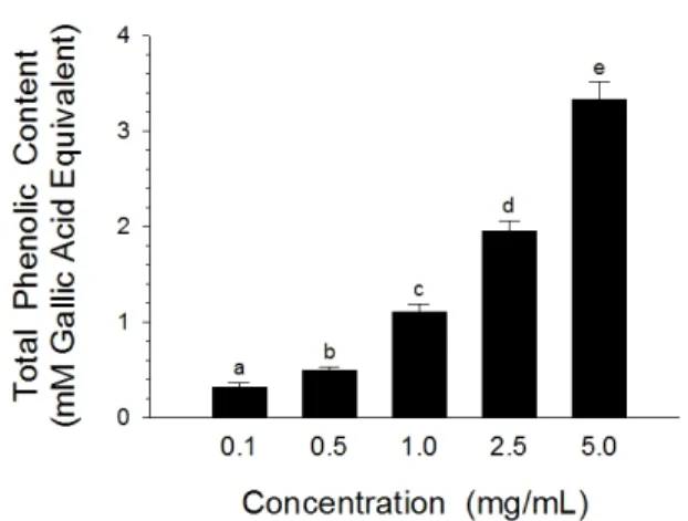
Fig. 7. Total phenolic content of Sanguisorbae radix extracts. Data results were expressed as in terms of mM gallic acid equivalent. Each bar represents the mean±SD of quadraplicate determinations. abcdeValues with different letters are significantly different at p<0.05.
과 함께 배양하였을 때, 0.001 mg/mL 농도의 추출물 첨가 는 supercoiled DNA strand의 절단 억제에 영향을 주지 않았다(lane 2 vs 3, Fig. 6A). 그러나, 추출물 농도가 증 가함에 따라 nicked open circular form은 감소한 반면 supercoiled form은 증가하여, 지유 추출물은 농도 의존 적으로 hydroxyl radical에 의해 유도된 single-strand 절단을 억제하였다. 특히, 0.1 mg/mL 농도의 지유 추출물 첨가시 hydroxyl radical에 대한 supercoiled DNA strand의 retention은 84.9%로 나타나, 동일한 농도의 양 성대조군인 sesamol에 의한 retention percent(96.0%)와 차이가 없었다(p>0.05).
AAPH 처리에 의해 생성되는 peroxyl radical 존재하 에, pBR322 DNA의 supercoiled form은 nicked open circular form 및 linear form으로 전환되었다(lane 1 vs 2, Fig. 6B). Peroxyl radical 존재하에 plasmid DNA를 지유 추출물과 함께 배양하였을 때, 0.001 mg/mL 농도의 추출물 첨가는 supercoiled DNA strand의 절단 억제에 영향을 주지 않았다(lane 2 vs 3, Fig. 6B). 그러나, 0.01 mg/mL 농도의 지유 추출물 첨가시 peroxyl radical에 대
한 supercoiled DNA strand의 retention percent는 78.6%로 나타나, 동일한 농도에서 양성대조군인 Trolox의 retention percent(87.8%)와 차이가 없었고(p>0.05), hydroxyl radical에 대한 supercoiled DNA strand의 retention percent(45.5%)에 비해 높게 나타났다(lane 4, Fig. 6A vs lane 4, Fig. 6B). 따라서, 지유 추출물이 hydroxyl radical에 비해 peroxyl radical에 대한 supercoiled DNA strand의 절단 억제효과가 탁월함을 알 수 있었다.
총페놀 함량
지유 추출물의 농도별 총페놀 함량은 Fig. 7에 나타나 있다. 지유 추출물 0.1 mg/mL 농도의 총페놀 함량은 0.32 mM gallic acid equivalent이었으며, 추출물 농도가 증가 함에 따라 총페놀 함량도 비례적으로 증가하여, 0.5, 1, 2.5 및 5 mg/mL 농도에서 각각 0.50, 1.10, 1.95 및 3.33 mM gallic acid equivalent로 나타났다. 항산화활성이 높 은 개머루덩굴과 만병초의 총페놀 함량은 1 mg/mL 농도 에서 각각 1.12와 1.25 mM gallic acid equivalent로 보고 된 바 있어(Rhim and Choi, 2010,2011), 지유 추출물의 총페놀 함량이 높음을 알 수 있었다.
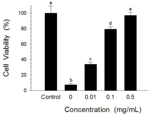
세포독성 억제효과
지유 추출물의 농도별 세포독성 억제효과는 Fig. 8에 나
Fig. 8. The effect of Sanguisorbae radix extracts on HepG2 cell viability. HepG2 cells were cultured for 5 h in the presence of 0.2 mM t-BHP and Sanguisorbae radix extracts. Cell viability was determined using MTT method.
Each bar represents the mean±SD of triplicate deter- minations. abcdValues with different letters are signi- ficantly different at p<0.05.
타나 있다. 세포 생존율은 대조군(t-BHP 무첨가군)의 MTT 값을 100%로 기준하여 표기하였다. HepG2 세포에 0.2 mM 농도의 t-BHP를 첨가한 뒤 5시간 배양한 결과, 세포 생존율은 7.5%로 나타나, t-BHP가 HepG2 세포독성 을 유발하였음을 알 수 있었다. HepG2 세포배양에서 0.2 mM 농도의 t-BHP 존재하에 지유 추출물 0.01 mg/mL 농 도의 첨가는 33.8%의 생존율을 나타내어, 지유 추출물이 세포독성에 의한 손상을 유의적으로(p<0.05) 억제시켰음 을 알 수 있었다. 추출물 농도가 증가함에 따라 세포 생존 율도 비례적으로 증가하여, 0.1 및 0.5 mg/mL 농도에서 각각 79.1 및 96.9%로 나타났다. 특히, 0.5 mg/mL 농도 의 추출물 첨가군과 대조군(t-BHP 무첨가군) 간에 세포 생존율의 유의적(p>0.05) 차이는 없었다. 흰쥐의 대뇌피질 신경세포를 이용한 연구에서 지유 추출물은 hydrogen peroxide처리에 의한 뇌세포 사멸과 ROS 생성을 억제시 켜 뇌세포 보호효과를 나타내었으며(Nguyen et al., 2008), 마우스 dermal fibroblast 세포를 이용한 연구에 서 지유 추출물은 superoxide에 의해 유발된 세포 손상을 억제시켰고(Masaki et al., 1995), HepG2 세포를 이용한 연구에서 지유 추출물에서 분리한 성분인 (+)-gallo- catechin은 acetylcholinesterase 억제제인 tacrine으로 유발된 세포독성을 농도 의존적으로 억제시켜 간세포 보호
효과를 나타내었다고(An et al., 2005) 보고된 바 있어, 본 연구에서 관찰된 지유 추출물의 세포독성 억제효과는 기존 연구결과와도 일치하고 있다.
따라서, 본 연구 결과들은 지유 추출물의 강력한 항산화 효과와 세포독성 억제효과를 나타내 보이고 있으며, 이러 한 효능들은 적어도 free radical의 산화 억제와 높은 환원 력 및 총페놀 함량에 기인하는 것으로 사료된다. 또한, 향 후 지유 추출물의 항산화 성분 분석과 동정 및 항산화 기전 연구와 더불어 지유 추출물의 천연 항산화제로의 개발 가 능성을 시사하고 있다.
적 요
본 연구에서는 지유 에탄올추출물의 항산화 효과를 조사 하였다. Pyrogallol의 억제율을 100%로 기준하였을 때, DPPH 라디칼을 50% 억제시키는데 필요한 지유 추출물의 농도는 0.33 mg/mL으로 α-tocopherol의 IC50(0.40 mg/mL)과 유사하게 나타났다. 지유 추출물의 총항산화능 은 α-tocopherol에 비해 높게 나타났다. 지유 추출물의 superoxide 소거활성은 catechin에 비해 높게 나타났다.
지유 추출물의 peroxyl 라디칼 소거활성은 ascorbic acid 에 비해 높게 나타났다. 지유 추출물의 구리이온 환원력은 α-tocopherol에 비해 높게 나타났다. 지유 추출물은 hydroxyl 라디칼 및 peroxyl 라디칼로 유발된 supercoiled DNA strand의 절단을 억제시켰다. 지유 추출물 0.5 및 5 mg/mL의 총페놀 함량은 각각 0.50 및 3.33 mM gallic acid와 동등한 수준이었다. 또한, HepG2 세포주를 이용한 세포배양에서 지유 추출물 0.01, 0.1 및 0.5 mg/mL 농도 의 첨가는 0.2 mM t-BHP로 유도된 세포독성을 각각 33.8, 79.1 및 96.9% 감소시켰다. 따라서, 본 연구 결과들 은 지유 추출물의 강력한 항산화 효과와 세포독성 억제효 과를 나타내며, 이러한 효능은 적어도 자유라디칼의 산화 억제와 높은 총페놀 함량에 기인하는 것으로 사료된다.
사 사
이 논문은 2011년도 상지대학교 교수 연구년제 지원에 의한 것임.
인용문헌
An, B.J., J.T. Lee, S.A. Lee, J.H. Kwak, J.M. Park, J.Y. Lee and J.H. Son. 2004a. Antioxidant effects and application as natural ingredients of Korean Sanguisorbae officinalis L. J.
Korean Soc. Appl. Biol. Chem. 47:244-250.
An, B.J., S.A. Lee, J.H. Son, J.H. Kwak, J.M. Park and J.Y. Lee.
2004b. Cytotoxic and antibacterial activities of Sanguisorbae officinalis L. J. Korean Soc. Appl. Biol. Chem. 47:141-145.
An, R.B., Y.H. Tian, H. Oh and Y.C. Kim. 2005. In vitro free radical scavenging and hepatoprotective compound from Sanguisorbae radix. Nat. Prod. Sci. 11:119-122.
Apak, R., K. Güçlü, M. Özyürek and S.E. Karademir. 2004.
Novel total antioxidant capacity index for dietary polyphenols and vitamins C and E, using their cupric ion reducing capability in the presence of neocuproine: CUPRAC method.
J. Agric. Food Chem. 52:7970-7981.
Cheng, D.L. and X.P. Cao. 1992. Pomolic acid derivatives from the root of Sanguisorba officinalis. Phytochemistry 31:1317- 1320.
Erel, O. 2004. A novel automated direct measurement method for total antioxidant capacity using a new generation, more stable ABTS radical cation. Clin. Biochem. 37:277-285.
Goun, E.A., V.M. Petrichenko, S.U. Solodnikov, T.V. Suhinina, M.A. Kline, G. Cunningham, C. Nguyen and H. Miles. 2002.
Anticancer and antithrombin activity of Russian plants. J.
Ethnopharmacol. 81:337-342.
Jeong, C.S., I.O. Suh, J.E. Hyun and E.B. Lee. 2003. Screening of hepatoprotective activity of medicinal plant extracts on carbon tetrachloride-induced hepatotoxicity in rats. Nat.
Prod. Sci. 9:87-90.
Halliwell, B., J.M.C. Gutteridge and C.E. Cross. 1992. Free radicals, antioxidants, and human disease:where are we now?
J. Lab Clin. Med. 119:598-620.
Hiramoto, K., N. Ojima, K.I. Sako and K. Kikugawa. 1996.
Effect of plant phenolics on the formation of the spin-adduct of hydroxyl radical and the DNA strand breaking by hydroxyl radical. Biol. Pharm. Bull. 19:558-563.
Hu, C., Y. Zhang and D.D. Kitts. 2000. Evaluation of anti- oxidant and prooxidant activities of bamboo Phyllostachys nigra var. Henonis leaf extract in vitro. J. Agric. Food Chem.
48:3170-3176.
Huang, D., B. Ou, M. Hampsch-Woodill, J.A. Flanagan and R.
L. Prior. 2002. High-throughput assay of oxygen radical absorbance capacity (ORAC) using a multichannel liquid
handling system coupled with a microplate fluorescence reader in 96-well format. J. Agric. Food Chem. 50:4437-4444.
Kim, T.G., S.Y. Kang, K.K. Jung, J.H. Kang, E. Lee, H.M. Han and S.H. Kim. 2001. Antiviral activities of extracts isolated from Terminalis chebula Retz., Sanguisorba officinalis L., Rubus coreanus Miq. and Rheum palmatum L. against hepatitis B virus. Phytother. Res. 15:718-720.
Liao, H., L.K. Banbury and D.N. Leach. 2008. Antioxidant activity of 45 Chinese herbs and the relationship with their TCM characteristics. Evid Based Complement Alternat.
Med. 5:429-434.
Liu, F., V.E.C. Ooi and S.T. Chang. 1997. Free radical scaveng- ing activities of mushroom polysaccharide extracts. Life Sci.
60:763-771.
Liu, X., Y. Cui, Q. Yu and B. Yu. 2005. Triterpenoids from Sanguisorba officinalis. Phytochemistry 66:1671-1679.
Malterud, K.E., T.L. Farbrot, A.E. Huse and R.B. Sund. 1993.
Antioxidant and radical scavenging effects of anthraquinones and anthrones. Pharmacology 47:77-85.
Masaki, H., S. Sakaki, T. Atsumi and H. Sakurai. 1995. Active- oxygen scavenging activity of plant extracts. Biol. Pharm.
Bull. 18:162-166.
Mimaki, Y., M. Fukushima, A. Yokosuka, Y. Sashida, S.
Furuya and H. Sakagami. 2001. Triterpene glycosides from the roots of Sanguisorba officinalis. Phytochemistry 57:773- 779.
Mosmann, T. 1983. Rapid colorimetric assay for cellular growth and survival: application to proliferation and cytotoxicity assays. J. Immunol. Methods 65:55-63.
Nguyen, T.T.H., S.O. Cho, J.Y. Ban, J.Y. Kim, H.S. Ju, S.B.
Koh, K.S. Song and Y.H. Seong. 2008. Neuroprotective effect of Sanguisorbae radix against oxidative stress-induced brain damage: in vitro and in vivo. Biol. Pharm. Bull.
31:2028-2035.
Rhim, T.J. and M.Y. Choi. 2010. The antioxidative effects of Ampelopsis brevipedunculata extracts. Korean J. Plant Res.
23:445-450 (in Korean).
Rhim, T.J. and M.Y. Choi. 2011. The antioxidative effects of Rhododendron brachycarpum extracts. Korean J. Plant Res.
24:456-460 (in Korean).
Singleton, V.L., R. Orthofer and R.M. Lamuela-Raventós.
1999. Analysis of total phenols and other oxidation substrates and antioxidants by means of Folin-Ciocalteu reagent. Methods Enzymol. 299:152-178.
Steel, R.G.D. and J.H. Torrie. 1980. Principles and Procedures
of Statistics, 2nd ed, McGraw-Hill, New York, USA, pp.
186-187.
Thannickal, V.J. and B.L. Fanburg. 2000. Reactive oxygen species in cell signaling. American J. Physiol. Lung Cell Mol. Physiol. 279:L1005-L1028.
Yokozawa, T., C.P. Chen, D.Y. Rhyu, T. Tanaka, J.C. Park and K. Kitani. 2002. Potential of sanguiin H-6 against oxidative
damage in renal mitochondria and apoptosis mediated by peroxynitrite in vivo. Nephron 92:133-141.
Yokozawa, T., C.P. Chen, T. Tanaka and K. Kitani. 2000. A study on the nitric oxide production-suppressing activity of Sanguisorbae Radix components. Biol. Pharm. Bull. 23:
717-722.
(Received 6 July 2012 ; Revised 13 September 2012 ; Accepted 29 October 2012)