Fructus Sophorae Enhances the Production of Prostaglandin E
2and Tumor Necrosis Factor-α through Activation of MAPKs and PI3K/AKT Signaling Pathways in Murine Macrophages
Young-Soon Kang
1, Min Ho Han
1,2, Moon Hee Lee
1,2, Su Hyun Hong
1, Heungsik Park
3, Jae-Chul Jung
3, Jeongrai Lee
3, Eun-Woo Lee
2,4, Kyung Hwa Kang
5, Cheol Min Kim
6,7, Byung-Woo Kim
2,4and
Yung Hyun Choi
1,2*
1
Department of Biochemistry, Dongeui University College of Oriental Medicine, Busan 614-052, Korea
2
Anti-Aging Research Center & Blue-Bio Industry RIC, Dongeui University, Busan 614-714, Korea
3
NOVAREX Co., Ltd. Life Science Institute, Chungbuk 363-885, Korea
4
Department of Life Science and Biotechnology, College of Life Sciences, Dongeui University, Busan 614-714, Korea
5
Department of Pathology, Dongeui University College of Oriental Medicine, Busan 614-052, Korea
6
Research Center for Anti-Aging Technology Development, Busan 609-735, Korea
7
Department of Biochemistry, Busan National University College of Medicine, Yangsan 626-870, Korea
Received October 4, 2013 /Revised October 25, 2013 /Accepted November 12, 2013Fructus Sophorae, the dried ripe fruit of Styphnolobium japonicum (L.), is an herbal ingredient used in traditional Oriental medicine. This study was carried out to investigate the effects of Fructus Sophorae extracts (FSE) on immune modulation in a murine RAW 264.7 macrophage model. As immune re- sponse parameters, the production of prostaglandin E
2(PGE
2) and tumor necrotic factor-α (TNF-α) were evaluated. Our data revealed that FSE increased the macrophage activation and the production of PGE
2and TNF-α, which was consistently correlated with upregulation of cyclooxygenase-2 (COX-2) and TNF-α expression at both transcriptional and translational levels. On comparative cytokine protein array, FSE significantly increased several cytokines, which was associated with phosphorylation of mi- togen-activated protein kinases (MAPKs), including extracellular signal-regulated kinase (ERK), p38 MAPK and c-Jun N-terminal kinase (JNK), and Akt in RAW 264.7 cells. However, each inhibitor of these molecules attenuated the FSE-induced PGE
2production. These results indicate that FSE activated macrophages through the activation of MAPKs and phosphatidylinositol-3-kinase (PI3K)/Akt signal- ing pathways in RAW 264.7 macrophages. These findings suggest that FSE may provide a promising source of an immunoenhancing agent.
Key words : Fructus Sophorae, immune modulation, macrophages, PGE
2, cytokines
*Corresponding author
*Tel : +82-51-850-7413, Fax : +82-51-853-4036
*E-mail : [email protected]
This is an Open-Access article distributed under the terms of the Creative Commons Attribution Non-Commercial License (http://creativecommons.org/licenses/by-nc/3.0) which permits unrestricted non-commercial use, distribution, and reproduction in any medium, provided the original work is properly cited.
Journal of Life Science 2013 Vol. 23. No. 11. 1397~1403 DOI : http://dx.doi.org/10.5352/JLS.2013.23.11.1397
서 론
면역 반응은 생체 자기 방어기작 중의 하나로 감염성 질환 으로부터의 보호와, 외부물질과 자신을 생리적으로 인식하여 체내에 유입된 유해 물질의 중화 또는 제거하는 현상이다. 생 체의 항상성 유지를 위하여, 면역에 관여하는 기관들이 면역 계를 구성하며 이들이 서로 협동하고 집합적으로 작용하여 면역반응을 일으키며, 면역반응의 이상으로 인한 면역시스템 에 불균형은 각종 난치성 질환의 발병과 진행의 원인이 된다 [7, 26].
체내에서 대식세포(macrophage)는 innate immunity와 adaptive immunity 모두에 관여하고 있는데, adaptive im- munity 작용으로 다른 면역 세포나 림프구를 활성화 시킬 수 있는 능력을 가지고 있다. 따라서 대식세포는 생체 내에서 염 증 및 면역반응을 조절하는 대표적인 세포로서 감염이나 암세 포 등으로부터 일차적인 보호기능을 갖고 있다. 대식세포는 자극을 받지 않은 상태에서는 전혀 phagocytic 하지 않으나 감염이나 외상으로 인한 자극을 받을 경우 활성화된다. 이렇 게 활성화 된 대식세포는 세포 내 여러 신호전달체계 및 유전 자 발현 변화를 통해서 다양한 cytokine과 prostaglandin E
2(PGE
2), nitric oxide (NO) 등의 분비와 함께 면역세포로서의
여러 특징을 나타내고 활발한 phagocytosis 작용을 시작한다
[4, 20]. 염증 반응으로 생성되는 PGE
2와 NO는 주로 대식세포
나 비만세포(mast cell)에 의해 분비되며, 염증반응, 면역반응
및 암 발생에도 깊은 관계가 있는 것으로 알려져 있다. 대식세
포로부터 분비되는 대표적인 염증성 cytokine인 tumor ne-
crotic factor-α (TNF-α)는 염증반응조절에 관여하는 것으로 잘
알려져 있으며, TNF-α의 증가로 인한 대식세포의 phag- ocytosis 작용이 과도하게 자극이 되면 염증반응과 산화적 스 트레스를 증가시키게 되지만, 적절한 수준의 발현은 암세포의 살상 및 전이의 억제 작용 등이 있는 것으로 밝혀져 있다[1,5].
한편 콩과의 회화나무(Styphnolobium japonicum L.) 열매인 괴각(槐角)은 고지혈증, 동맥경화증 및 지방간 치료의 목적으 로 오랜 동안 사용되어 온 한약재의 일종이지만, 이에 대한 구체적인 과학적인 근거는 매우 부족하다. 최근 연구들에 의 하면 괴각 추출물은 골다공증, 골 재흡수 및 골 소실 억제 효능 [10, 12, 25], 항염증[11], 항암활성[18], 폐경기 증후군 감소[19], 혈액응고 방지[13] 및 지혈 작용[6] 등 다양한 약리학적 효능이 있는 것으로 밝혀지고 있는데, 이러한 효능은 괴각에 함유된 다양한 종류의 flavonoid 계열 물질에 기인한 것으로 보고되 어지고 있다[2, 3, 14-16]. 본 연구에서는 면역 활성 증가 효능을 가지는 천연물의 스크린 과정에서 괴각 추출물을 후보물질로 선정하였으며, 이에 대한 몇 가지 근거를 제시하고자 한다.
재료 및 방법
시약 및 항체
세포배양을 위한 Dulbecco's modified eagle's medium (DMEM), bovine serum (FBS), penicillin/streptomycin은 WELGENE (Daegu, Republic of Korea)에서 구입하였고, Escherichia coli 유래 lipopolysaccaride (LPS), 3-(4,5-Dimethyl- thiazol-2-yl)-2,5-diphenyl tetrazoliumbromide (MTT), Griess reagent, sodium nitrite는 Sigma-Aldrich Chemical Co. (St.
Lousi, MO, USA)에서 구입하였다. TNF-α 측정용 enzyme- linked immunosorbent assay (ELISA) kit는 R&D Systems (Minneapolis, MN, USA)에서, PGE
2ELISA kit와 cyclo- oxygenase-2 (COX-2) 항체는 Cayman Chemical (Ann Arbor, MI, USA)에서, extracellular signal-regulated kinase (ERK) 및 p38 mitogen-activated protein kinase (MAPK) 항체는 Santa Cruz Biotechnology Inc. (Santa Cruz, CA, USA)에서 구입하 였다. c-Jun N-terminal kinase (JNK), phospho-JNK, phos- pho-ERK, phosphor-p38 MAPK 항체는 Cell Signaling (Beverly, MA, USA)에서 구입하였다.
세포 배양, 괴각 추출물의 처리 및 MTT assay
Murine macrophage cell line인 RAW 264.7 세포는 American Type Culture Collection (Manassas, VA, USA)에 서 구입하여, 10% FBS와 penicillin/streptomycin 100 unit/ml 이 함유된 DMEM 배지를 사용하여 37℃, 5% CO
2incubator에 서 배양하였으며, 2-3일에 한 번씩 계대 배양을 시행하였다.
본 연구에 사용된 20% 정도의 isoflavone이 함유되어 있는 괴 각 추출물(Fructus Sophorae extracts, FSE)은 ㈜렉스진 바이 오텍 생명과학연구소에서 제공받았으며, 이는 건조된 괴각을
60% ehthanol을 이용하여 추출한 것으로 증류수에 희석하여 적정 농도로 희석하여 배양 배지에 처리하였다. RAW 264.7 대식세포의 증식에 미치는 FSE의 영향을 조사하기 위하여 24 시간 동안 적정 농도의 FSE를 처리한 후 MTT assay를 실시하 였다.
PGE
2및 TNF-α 생성능 측정
PGE
2및 TNF-α의 생성에 미치는 FSE의 영향을 조사하기 위하여 RAW 264.7 대식세포에 적정 농도의 FSE를 24시간 동 안 처리한 후, 상등액을 회수하여 PGE
2및 TNF-α ELISA kit를 이용하여 측정하였다.
RNA의 분리 및 reverse transcriptase polymerase chain reaction (RT-PCR) 분석
FSE가 함유된 배지에서 배양된 세포를 대상으로 Tryzol®
Reagent (Invitrogen Co., Carlsbad, CA, USA)를 이용하여 RNA를 추출하였으며, 해당되는 유전자들의 primer (COX-2:
5' CAA AGG CCT CCA TTG ACC AGA 3' and 5' TGG ACG AGG TTT TTC CAC CAG 3'; TNF-α: 5' CCC CTC AGC AAA CCA CCA AGT 3' and 5' CTT GGG CAG ATT GAC CTC AGC 3')와 ONE-STEP RT-PCR PreMix kit (iNtRON Biotechnology, Republic of Korea)를 이용하여 PCR을 수행하 였다. 이후 증폭된 DNA 산물을 1.5% agarose gel를 사용하여 100 volt에서 20분간 전기 영동하여 ethidium bromide (EtBr, Sigma-Aldrich)로 염색 후 UV 하에서 관찰하였다. 아울러 PCR 반응의 대조군으로 glyceraldehyde-3-phosphate de- hydrogenase (GAPDH: 5' GTC ATC ATC TCC GCC CCT TCT GC 3' and 5' GAT GCC TGC TTC ACC ACC TTC TTG 3')를 사용하였다.
단백질의 분리와 Western blot 분석
FSE 처리에 따른 번역 수준에서의 유전자 발현 변화의 관찰 을 위하여 준비된 세포들을 250 mM NaCl, 25 mM Tris-HCl (pH 7.5), 10 mM EDTA, 1% NP-40와 0.1 mM phenyl-methyl- sulfonylfluoride, protease inhibitor 등이 함유된 lysis buffer 를 사용하여 용해시켰다. 분리된 단백질들의 농도를 측정한 후, Western blot analysis를 위해 동량의 단백질들을 sodium dodecyl sulfate (SDS)–polyacrylamide gel electrophoresis 를 이용하여 분리하고 nitrocellulose membrane (Schleicher
& Schuell, Keene, NH, USA)으로 전이시켰다. 각각의 mem- brane을 적정 항체 및 enhanced chemiluminescence (ECL, Amersham Corp. Arlington Heights, IL, USA) 용액을 이용하 여 단백질들의 발현 변화를 조사하였다.
Cytokine array 분석
다양한 종류의 cytokine 생성에 미치는 FSE의 영향을 조사
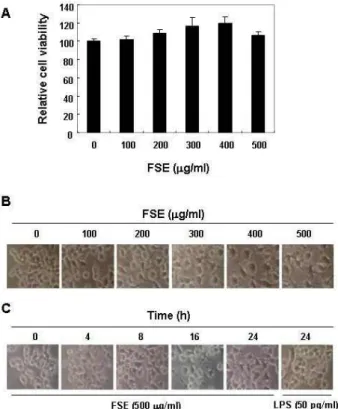
Fig. 1. Effects of FSE on cell viability and morphology in RAW 264.7 macrophages. (A) After RAW 264.7 cells were seed- ed, the cells were treated with the indicated concen- trations of FSE for 24 h, and then cell viability was meas- ured by MTT assay. The data is expressed as the mean
± SD of three independent experiments. (B and C) RAW 264.7 macrophages were treated with various concen- trations of FSE for 24 h (B), or treated with 500 μg/ml FSE or 50 pg/ml LPS for the indicated times (C). The morphological changes of RAW 264.7 macrophages were imaged using an inverted microscope (original magnifi- cation, x200).
하기 위하여 24시간 동안 500 mg/ml의 FSE 또는 100 ng/ml 의 LPS가 처리된 배지에서 배양된 세포의 단백질을 분리하였 다. 분리된 단백질을 정량 후, 급성기 단백질(acute phase pro- tein), cytokine 및 chemokine 등 40종(BLC, C5a, G-CSF, GM- CSF, I-309, Eotaxin, sICAM-1, IFN-γ, IL-1α, IL- 1β, IL-1ra, IL-2, IL-3, IL-4, IL-5, IL-6, IL-7, IL-10, IL-12p70, IL-13, IL-16, IL-17, IL-23, IL-27, IP-10, I-TAC, KC, M-CSF, JE, MCP-5, MIG, MIP- 1α, MIP-1β, MIP-2, RANTES, SDF-1, TARC, TIMP-1, TNF-α 및 TREM-1) 생성 변화를 Proteome profiler array kit (Mouse Cytokine Array Panel A, R & D Systems)을 이용하여 분석하였다[28, 30].
통계처리
모든 실험 결과는 평균 ± 표준편차로 표시하였고 SigmaPlot (Systat Software Inc., San Jose, CA, USA)을 이용하여 Student t-test를 이용하여 통계적 유의성을 얻었다.
결과 및 고찰
FSE 처리에 따른 RAW 264.7 대식세포의 활성화
FSE에 의한 RAW 264.7 대식세포 활성화 여부의 조사를 위한 실험 조건 설정을 위하여 다양한 농도의 FSE가 함유된 배지에서 배양된 RAW 264.7 세포를 대상으로 MTT assay를 실시하였다. Fig. 1의 결과에서 알 수 있듯이, FSE의 농도 400 μ g/ml까지 농도 의존적으로 RAW 267.4 세포의 증식이 증가 하였으며, 500 μg/ml 처리군에서도 106%로 대조군에 비하여 다소 증가하였다. 이러한 FSE 처리에 의한 증식 촉진이 대식세 포 활성화와 연관성이 있는지를 조사하기 위하여 LPS 처리군 을 양성 대조군으로 설정하여 형태적 변화를 관찰하였다. Fig.
1A 및 B에 나타낸 바와 같이, FSE 처리 농도 및 시간의 증가에 따라 전형적인 대식세포 활성형태인 LPS 처리군과 유사한 형 태적 변화를 관찰할 수 있었으며, 이러한 결과는 FSE가 RAW 264.7 대식세포의 활성을 촉진시켰다는 간접적인 증가가 될 수 있을 것으로 생각된다.
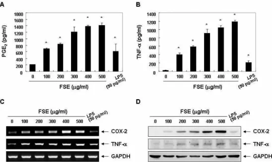
RAW 264.7 대식세포에서 FSE 처리에 따른 PEG2 및 TNF-α의 생성 증가
대식세포가 다양한 조직에 존재하고 염증성 자극을 포함한 인자들에 대한 생체방어 기능을 한다는 점에서 이러한 인자들 에 의하여 활성화된 대식세포는 PEG
2와 같은 염증성 매개인 자나 TNF-α를 포함하는 cytokine의 생성을 촉진한다[8, 22].
따라서 상기에서 FSE 처리에 따른 RAW 264.7 세포의 형태적 변화가 대식세포 활성화와 직접 연관성이 있는지를 조사하기 위하여 PEG
2및 TNF-α의 생성에 미치는 FSE의 영향을 조사하 였다. Fig. 2A 및 Fig. 3A의 결과에서 알 수 있듯이 LPS 처리군 과 비교하여 FSE의 처리에 따라 RAW 264.7 대식세포의 PEG
2의 생성이 유의적으로 증가하였음을 알 수 있었으며, TNF-α의
생성 또한 FSE의 처리 농도 의존적으로 증가되었다. 그러나
흥미롭게도 동일 농도로 고정된 조건에서 TNF-α의 생성은
FSE 처리 8시간 전후에 최고 생성능을 보여, FSE의 처리에
따라 PEG
2에 비하여 TNF-α의 생성이 앞서 이루어지고 있음을
알 수 있었다. 이러한 FSE 처리에 따른 생성능의 차이가 이들
인자들의 생성에 관여하는 유전자 발현의 차이에 기인할 것으
로 추측되어 FSE의 처리에 따른 COX-2 및 TNF-α 유전자의
발현 변화를 전사 및 번역 수준에서 비교하였다. RT-PCR 및
Western blot 분석 결과에서 알 수 있듯이, 이들 유전자들의
발현 경향성은 PEG
2및 TNF-α 생성의 정도와 유사하게 관찰
되었다(Fig. 3, 4). 따라서 FSE 처리에 의한 PEG
2및 TNF-α의
생성은 이들 산물을 조절하는 유전자의 전사 수준에서 조절되
고 있음을 알 수 있었다.
Fig. 2. Induction of PGE2 and TNF-α production in RAW 264.7 macrophages after exposure to FSE. (A and B) Following 24 h treatment with various concentrations of FSE or 50 pg/ml LPS, the supernatants were prepared, and then the amounts of PGE2(A) and TNF-α (B) were measured by ELISA. The data are shown as means ± SD of three independent experiments (*,
p
<0.05 vs. untreated control). (C and D) The levels of COX-2 and TNF-α mRNA (C) and protein (D) were assessed by RT-PCR and Western blot analysis after 24 h treatment. GAPDH and actin were used as internal controls for the RT-PCR and Western blot assays, respectively.Fig. 3. Increased release of PGE2 and TNF-α by FSE treatment in RAW 264.7 macrophages. (A and B) Cells were treated with 500 μg/ml FSE for the indicated times, and the supernatants were prepared, and then the amounts of PGE2(A) and TNF-α (B) were measured by ELISA. The data are shown as means ± SD of three independent experiments (*,
p
<0.05 vs. untreated control). (C and D) The levels of COX-2 and TNF-α mRNA (C) and protein (D) were assessed by RT-PCR and Western blot analysis after treatment for the indicated times. GAPDH and actin were used as internal controls for the RT-PCR and Western blot assays, respectively.RAW 264.7 대식세포에서 다양한 cytokine의 발현에 미 치는 FSE의 영향
한편 다양한 종류의 cytokine이 대식세포의 활성화에 관여
하며, cytokine의 종류에 따라 다양한 면역반응을 매개함으로
서 대식세포의 운명도 달라진다[27, 29]. 그러나 대식세포의
활성과 면역체계에 연관된 cytokine 신호체계 기전은 여전히
Fig. 4. Effect of FSE on multiple cytokines expression in RAW 264.7 macrophages. RAW 264.7 cells were incubated with 500 μg/ml FSE or 100 ng/ml LPS for 24 h, and then the proteome profiler array system was used to screen for activation of different acute phase proteins, cytokines, and chemokines involved in the activation of RAW264.7 macrophages by FSE treatment.
Fig. 5. Activation of MAPKs and PI3K/Akt signaling pathways by FSE treatment in RAW 264.7 macrophages. (A) Cells were treated with 500 μg/ml FSE for the indicated times, and the cells were sampled, lysed, and 30~50 μg of protein was separated by electrophoresis on SDS-polyacrylamide gels. Western blot analyses were then performed using the indicated antibodies and an ECL detection system. (B) The cells were pretreated with the indicated inhibitors (PD98059, 50 μM; SP600125, 20 μM; LY294002, 20 μM; SB203580, 20 μM; and PDTC, 20 μM) for 1 h and then treated with 500 μg/ml FSE for 24 h. The supernatants were prepared, and then the amounts of PGE2were measured by ELISA. The data are shown as means ± SD of three independent experiments (*,
p
<0.05 vs. untreated control;#,p
<0.05 vs. FSE-treated group).많은 부분이 알려져 있지 않다. 본 연구에서는 FSE가 함유된 배지에서 배양된 RAW 264.7 세포에서 분비되는 cytokine에 대한 기초 정보를 확립하기 위하여 cytokine array를 실시하였
으며, 이를 LPS 처리군과 비교하였다. Fig. 4의 결과에서 알 수 있듯이 LPS 처리군과 동일하게 유의적으로 증가된 cycto- kine은 9종(KC, G-CCSF, IL-10, IL-1ra, TIMP-1, TNF-α, JE, MIP-2 및 RANTES)이었으나, 2종(MIP-1α 및 MIP-1β)은 다소 감소되었으며, LPS 처리군에서 증가된 cyctokine은 7종 (GM-CSF, IL-2, IL-6, IL-10, IL-1α, IL-1β 및 IL-27)으로 다소 차이를 보여 주었다. 이 결과는 세포 내 존재하는 cytokine의 상대적인 증가량을 보여 주는 결과로 이들의 세포 외 분비 및 역할에 대한 추가적인 연구가 진행되어야겠지만, 향후 FSE 의 면역 조절 관련 기능적 기전 연구 수행의 기초 자료로서 활용될 수 있을 것이라 생각한다.
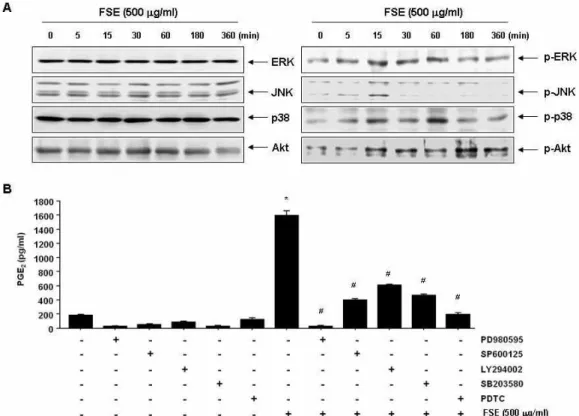
FSE 처리에 따른 MAPKs 및 PI3K/Akt 신호전달계의 역할
대식세포의 활성화에는 다양한 세포 내 신호전달계가 관여 하고 있음은 여러 선행 연구에서 확인되었으며, 이들 신호계 의 제어 현상에 대한 이해는 대식세포 활성 촉진 물질의 발굴 및 그들의 세포 내 역할에 대한 많은 정보를 제공하여 준다.
이러한 세포 내 신호전달계 중, MAPKs는 대식세포 활성을
포함한 세포 활성물질의 생성 및 다양한 생물학적기능을 조절 하며[21, 23], 세포의 증식 및 성장에 관여한다고 알려진 phos- phatidylinositol-3-kinase (PI3K)/Akt 경로 역시 대식세포의 활성에 핵심적인 역할을 하는 것으로 알려져 있다[9, 17]. 따라 서 FSE 처리에 따른 RAW 264.7 대식세포 활성에 MAPKs 및 PI3K/Akt 신호전달계의 관련성 여부를 조사하였다. Fig. 5A 의 결과에서 알 수 있듯이, 조사된 단백질들의 인산화 촉진에 따른 시간적 차이는 있지만, MAPKs에 속한 3가지 인자(ERK, JNK 및 p38 MAPK)와 Akt 단백질 모두가 활성화되었음을 알 수 있었다. 이들 중 어떤 인자들이 FSE 처리에 의한 RAW 264.7 대식세포 활성에 더 크게 관여하는 지를 다양한 선택적 저해제를 이용하여 PEG
2생성능을 비교함으로서 조사하였다.
이를 위하여 대식세포 활성에 핵심적인 역할을 하는 것으로 잘 알려진 nuclear factor (NF)-κB의 저해제인 pyrrolidine di- thiocarbamate (PDTC) [24]를 양성 대조군으로 사용하였다.
Fig. 5B의 결과에서 알 수 있듯이, PDTC의 경우처럼 본 연구에 사용된 모든 신호전달 저해제가 모두가 FSE에 의한 PEG
2의 생성을 억제하였으나, ERK 저해제인 PD98059가 가장 큰 저해 효과를 보였으며, p38 MAP 저해제인 SB203580, JNK 저해제 인 SP600125 및 PI3K 저해제인 LY294002 또한 유의적인 저해 효과를 보여 주었다. 이러한 결과는 FSE 처리에 따른 RAW 264.7 대식세포의 활성에는 MAPKs 및 PI3K/Akt 신호계를 포함한 NF-κB 연관 신호 전달계의 활성이 동시에 관여하고 있음을 의미하는 것이다.
감사의 글
본 연구는 산업통상자원부 및 한국산업기술진흥원의 산업 기술연구기반구축사업 일환으로 수행하였음(항노화산업 지 원을 위한 인프라구축 및 제품개발, N0000697).
References
1. Blatteis, C. M., Li, S., Li, Z., Perlik, V. and Feleder, C. 2004.
Signaling the brain in systemic inflammation: the role of complement.
Front Biosci
9, 915-931.2. Chang, L., Ren, Y., Cao, L., Sun, Y., Sun, Q., Sheng, N., Yuan, L., Zhi, X. and Zhang, L. 2012. Simultaneous determi- nation and pharmacokinetic study of six flavonoids from
Fructus Sophorae
extract in rat plasma by LC-MS/MS.J Chromatogr B Analyt Technol Biomed Life Sci
904, 59-64.3. Chang, L., Zhang, X. X., Ren, Y. P., Cao, L., Zhi, X. R. and Zhang, L. T. 2013. Simultaneous quantification of six major flavonoids from
Fructus sophorae
by LC-ESI-MS/MS and statistical analysis.Indian J Pharm Sci
75, 330-338.4. Erwig, L. P. and Henson, P. M. 2007. Immunological con- sequences of apoptotic cell phagocytosis.
Am J Pathol
171, 2-8.5. Fullerton, J. N., O'Brien, A. J. and Gilroy, D. W. 2013.
Pathways mediating resolution of inflammation: when enough is too much.
J Pathol
231, 8-20.6. Gan, T., Liu, Y. D., Wang, Y. and Yang, J. 2010. Traditional Chinese Medicine herbs for stopping bleeding from haemorrhoids.
Cochrane Database Syst Rev
6, CD006791.7. Ghazal, P., Dickinson, P. and Smith, C. L. 2013. Early life response to infection.
Curr Opin Infect Dis
26, 213-218.8. Guslandi, M. 1998. Nitric oxide and inflammatory bowel diseases.
Eur J Clin Invest
28, 904-907.9. Ha, T., Liu, L., Kelley, J., Kao, R., Williams, D. and Li, C.
2011. Toll-like receptors: new players in myocardial ische- mia/reperfusion injury.
Antioxid Redox Signal
15, 1875-1893.10. Joo, S. S., Kang, H. C., Lee, M. W., Choi, Y. W. and Lee, D. I. 2003. Inhibition of IL-1beta and IL-6 in osteoblast-like cell by isoflavones extracted from
Sophorae fructus
.Arch Pharm Res
26, 1029-1035.11. Joo, S. S., Kwon, S. H., Hwang, K. W. and Lee, D. I. 2005.
Improvement of menopausal signs by isoflavones derived from
Sophorae fructus
in ovariectomized female rats and the antioxidant potentials in BV2 cells.Arch Pharm Res
28, 566-572.12. Joo, S. S., Won, T. J., Kang, H. C. and Lee, D. I. 2004.
Isoflavones extracted from
Sophorae fructus
upregulate IGF-I and TGF-beta and inhibit osteoclastogenesis in rat bone marrow cells.Arch Pharm Res
27, 99-105.13. Kim, H. Y., Eo, E. Y., Park, H., Kim, Y. C., Park, S., Shin, H. J. and Kim, K. 2010. Medicinal herbal extracts of
Sophorae
radix,Acanthopanacis
cortex,Sanguisorbae
radix andTorilis
fructus inhibit coronavirus replicationin vitro
.Antivir Ther
15, 697-709.14. Kite, G. C., Rowe, E. R., Lewis, G. P. and Veitch, N. C. 2011.
Acylated flavonol tri- and tetraglycosides in the flavonoid metabolome of
Cladrastis kentukea
(Leguminosae).Phyto- chemistry
72, 372-384.15. Kite, G. C., Stoneham, C. A. and Veitch, N. C. 2007. Flavonol tetraglycosides and other constituents from leaves of
Styphnolobium japonicum
(Leguminosae) and related taxa.Phytochemistry
68, 1407-1416.16. Kite, G. C., Veitch, N. C., Boalch, M. E., Lewis, G. P., Leon, C. J. and Simmonds, M. S. 2009. Flavonol tetraglycosides from fruits of
Styphnolobium japonicum
(Leguminosae) and the authentication of Fructus Sophorae and Flos Sophorae.Phytochemistry
70, 785-794.17. Krakauer, T. 2012. PI3K/Akt/mTOR, a pathway less recog- nized for staphylococcal superantigen-induced toxicity.
Toxins (Basel)
4, 1343-1366.18. Lau, F. Y., Chui, C. H., Gambari, R., Kok, S. H., Kan, K.
L., Cheng, G. Y., Wong, R. S., Teo, I. T., Cheng, C. H., Wan, T. S., Chan, A. S. and Tang, J. C. 2005. Antiproliferative and apoptosis-inducing activity of
Brucea javanica
extract on hu- man carcinoma cells.Int J Mol Med
16, 1157-1162.19. Lee, J., Kim, K. W., Kim, H. K., Chae, S. W., Jung, J. C., Kwon, S. H. and Rheu, C. H. 2010. The effect of Rexflavone (Sophorae fructus extract) on menopausal symptoms in postmenopausal women: a randomized double-blind place- bo controlled clinical trial.
Arch Pharm Res
33, 523-530.초록:대식세포에서 MAPKs 및 PI3K/AKT 신호전달계 활성을 통한 괴각 추출물의 prostaglandin E
2및 tumor necrosis factor-α 생성의 촉진
강영순
1․한민호
1,2․이문희
1,2․홍수현
1․박흥식
3․정재철
3․이정래
3․이은우
2,4․강경화
5․김철민
6,7․김병우
2,4․ 최영현
1,2*
(
1동의대학교 한의과대학 생화학교실,
2동의대학교 항노화연구소 및 블루바이오소재개발센터,
3노바렉스 생 명과학연구소,
4동의대학교 자연과학대학 생명응용학과,
5동의대학교 한의과대학 생리학교실,
6항노화기술 개발사업단,
7부산대학교 의과대학 생화학교실)
괴각(Fructus Sophorae)은 회화나무(Styphnolobium japonicum L.)의 열매를 건조한 것으로 전통 한의학에서 널 리 사용되는 약재 중의 하나이다. 본 연구에서는 murine RAW 264.7 대식세포 모델을 이용하여 괴각 추출물 (Fructus Sophorae extracts, FSE)이 면역 조절능에 미치는 영향을 조사하였다. 이를 위한 대식세포 활성과 연관된 면역 반응 parameter로서 prostaglandin E
2(PGE
2)와 tumor necrotic factor-α (TNF-α)의 생성에 미치는 FSE의 영 향을 조사하였다. 본 연구의 결과에 의하면 FSE는 대식세포의 활성을 유도하였고, PGE
2및 TNF-α의 생성을 촉진 하였으며, 이는 cyclooxygenase-2 (COX-2)와 TNF-α 유전자의 전사 및 번역 수준에서의 활성화와 연관성이 있었 다. 또한 FSE 처리에 의하여 다양한 종류의 cytokine 발현의 증가를 cytokine array 분석을 통하여 확인하였으며, RAW 264.7 대식세포의 활성화에는 mitogen-activated protein kinases (MAPKs) 및 phosphatidylinositol-3-kinase (PI3K)/Akt 경로 활성화가 연관되어 있음을 알 수 있었다. 본 연구의 결과는 괴각 추출물이 면역 증강제로서의 개발 가능성이 매우 높음을 시사한다.
20. Li, Y., Lee, P. Y. and Reeves, W. H. 2010. Monocyte and macrophage abnormalities in systemic lupus erythematosus.
Arch Immunol Ther Exp (Warsz)
58, 355-364.21. Rao, K. M. 2001. MAP kinase activation in macrophages.
J Leukoc Biol
69, 3-10.22. Ritchlin, C. T., Haas-Smith, S. A., Li, P., Hicks, D. G. and Schwarz, E. M. 2003. Mechanisms of TNF-alpha- and RANKL-mediated osteoclastogenesis and bone resorption in psoriatic arthritis.
J Clin Invest
111, 821-831.23. Schorey, J. S. and Cooper, A. M. 2003. Macrophage signal- ling upon mycobacterial infection: the MAP kinases lead the way.
Cell Microbiol
5, 133-142.24. Schreck, R., Meier, B., Männel, D. N., Dröge, W. and Baeuerle, P. A. 1992. Dithiocarbamates as potent inhibitors of nuclear factor kappa B activation in intact cells.
J Exp Med
175, 1181-1194.25. Shim, J. G., Yeom, S. H., Kim, H. J., Choi, Y. W., Lee, D.
I., Song, K. Y., Kwon, S. H. and Lee, M. W. 2005. Bone loss preventing effect of Sophorae Fructus on ovariectomized rats.
Arch Pharm Res
28, 106-110.26. Sirisinha, S. 2011. Insight into the mechanisms regulating immune homeostasis in health and disease.
Asian Pac J Allergy Immunol
29, 1-14.27. Varin, A. and Gordon, S. 2009. Alternative activation of macrophages: Immune function and cellular biology.
Immunobiology
214, 630-641.28. Wang, Q. S., Cui, Y. L., Wang, Y. F. and Chi, W. 2011. Effects of compounds from Bi-Qi Capsule on the expression of in- flammatory mediators in lipopolysaccharide-stimulated RAW 264.7 macrophages.
J Ethnopharmacol
136, 480-487.29. Whyte, C. S., Bishop, E. T., Rucker, l. D., Gaspar-Pereira, S., Barker, R. N., Allen, J. E., Rees, A. J. and Wilson, H.
M. 2012. Suppressor of cytokine signaling (SOCS)1 is a key determinant of differential macrophage activation and function.
J Leukoc Biol
90, 845-854.30. Yano, S., Umeda, D., Maeda, N., Fujimura, Y., Yamada, K.
and Tachibana, H. 2006. Dietary apigenin suppresses IgE and inflammatory cytokines production in C57BL/6N mice.