Received: August 22, 2017 Revised: October 7, 2017 Accepted: October 20, 2017
TRAUMA AND INJURY
Correspondence to
Sung Youl Hyun, M.D.
Department of Trauma Surgery, Gachon University Gil Medical Center, 21 Nam- dong-daero 774beon-gil, Namdong-gu, Incheon 21565, Korea
Tel: +82-32-460-3015 Fax: +82-32-460-3019 E-mail: sungyoul@gilhospital.com
http://www.jtraumainj.org Copyright © 2017 The Korean Society of Trauma
Prognosis of Pulmonary Function in Patients with Multiple Rib Fractures
Hee Beom Park, M.D.
1, Sung Youl Hyun, M.D.
2, Jin Joo Kim, M.D.
1, Yeon Sik Jang, M.D.
1Departments of
1Emergency Medicine,
2Trauma Surgery, Gachon University Gil Medical Center, Incheon, Korea
Purpose: Rib fracture is the most common complication of blunt thoracic trauma.
We investigated the effect of rib fracture on pulmonary function in the conservatively treated patients.
Methods: From January 2000 to February 2017, we reviewed the records of 72 pa- tients with rib fracture and pulmonary function tests were performed. According to the number of rib fractures, patients were classified into two groups: less than six fractured ribs (group A) and more than six fractured ribs (group B). The groups were compared concerning demographics, underlying diseases, associated thoracic inju- ries, surgery, mechanical ventilator times, days spent in the intensive care unit and pulmonary function test.
Results: There were no statistically significant differences in the demographic data between the two groups. Mean hospitalization was 13.5 days in group A and 27.0 days in group B (p<0.001). There was no statistically significant difference between the two groups in the pulmonary function test.
Conclusions: We conclude that pulmonary function is restored by conservative treat- ment in patients with rib fractures even if the number of rib fractures increases. In patients with multiple rib fractures, studies comparing open rib fixation and conser- vative treatment of long term pulmonary function are required.
Keywords: Rib fractures; Pulmonary function tests; Thoracic injuries; Outcome
INTRODUCTION
Thoracic traumas comprise 10-15% of all traumas and are the cause of death in 25%
of all trauma-related fatalities [1]. The most common type of thoracic trauma is decel-
erating injuries caused by collisions like traffic accidents. Trauma related to accidents
like falls and slip down has also increased [2]. Rib fractures are the most common
bony injuries in thoracic trauma and are diagnosed in approximately 50% of patients admitted to the hospital following thoracic trauma. Rib fractures can be markers of potential internal injury. The principal diagnostic goal with clinically suspected rib fractures is the detection of significant associated complications, such as hemopneu- mothorax, pulmonary contusion, intra-abdominal injury, or major vascular injury. The pain of rib fractures can greatly interfere with ventilation, cause splinting and atel- ectasis, and prolong the time for weaning from ventilator support. Patients with multiple fractured ribs will often have difficulty coughing or adequately clearing secretions, and should be considered for 24 to 48 hours of observa- tion unit admission [3]. The presence of three or more rib fractures has been associated with increased mortality and duration of care in intensive care units (ICUs) and hospitals [4]. Increasing number of rib fractures correlates directly with increasing pulmonary morbidity (atelectasis, pneumonia) and mortality [5-7].
This morbidity and mortality associated with rib frac- tures is caused by three main problems: hypoventilation due to pain, impaired gas exchange in damaged lung un- derlying the fractures, and altered breathing mechanics.
An injury severe enough to fracture ribs will invariably cause a substantial contusion to the underlying lung. The damaged lung is poorly compliant and will not take part in gas exchange [8]. Pulmonary contusion usually resolves without causing permanent complications [9]. However, it may also have long-term adverse effects on pulmonary function. Fibrosis of the lungs can occur, resulting in dys- pnea (shortness of breath), low blood oxygenation, and reduced functional residual capacity after the injury [10].
Contusion can also permanently reduce the compliance of the lungs [11].
There are not many studies on the effect of rib fracture on pulmonary function. We investigated the effect of rib fracture on pulmonary function in the conservatively treated patients.
METHODS
Study design and population
From January 2000 to February 2017, we retrospectively
reviewed the records of patients with rib fracture treated in our university hospital emergency medical center.
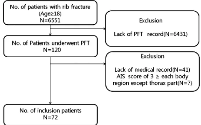
The center has approximately 90,000 patient visits an- nually. Of the reviewed patients, 120 had undergone pulmonary function tests 6-23 months following dis- charge. The included were over 18 years of age and had experience blunt trauma-related rib fracture. Exclusion criteria were lack of necessary medical records, absence of pulmonary function testing, abbreviated injury scale score≥3 for each body region, except the thorax, and history of mechanical ventilation or ICU stay due to damage to the abdomen and other organs. Finally, 72 patients were enrolled (Fig. 1).
Indications for admission were patients who those with more than three rib fractures, clinical symptoms includ- ing dyspnea and chest pain, and significant associated complications like hemopneumothorax and pulmonary contusion.
Data collection
Electronic medical records and radiographs of each pa- tient were retrospectively reviewed by one emergency medicine doctor. Patients were classified into two groups based on the extent of rib fracture: less than six ribs (group A) and more than six ribs (group B). Variables included number of rib fractures, age, sex, underlying diseases, as- sociated thoracic injuries, surgery, mechanical ventilator days, ICU days, and pulmonary function test.
Fig. 1. Flow chart of study subjection selection. PFT: pulmonary func-
tion test, AIS: abbreviated injury scale.
Outcomes
The primary outcome was hospitalization in days includ- ing ICU days. Secondary outcome included the pulmo- nary function of patients with rib fractures. Pulmonary function tests were performed following the American Thoracic Society/European Respiratory Society guide- lines by technicians experienced in pulmonary function testing. Spirometry was performed according to current recommendations [12,13]. Restrictive pattern was defined as a measured forced vital capacity (FVC) <80% of the predicted value for the patient. Obstructive pattern was defined as a measured forced expiratory volume in 1 sec- ond (FEV1)/FVC and FEV1<80% of the predicted value for the patient.
Statistical analyses
Univariate analyses involved the chi-squared test for the analysis of categorical variables and the Wilcoxon rank- sum test for the analysis of continuous variables. Mul- tivariate analyses used logistic regression and multiple regression. Dependent variables in logistic regression were mechanical ventilator care, FEV1, FVC, and ICU admission days. All statistical analyses were performed us- ing STATA version 13 (Stata Corp., College Station, TX, USA). The significance level was 0.05 (p-value). Quantita- tive data are presented as the mean±standard deviation.
RESULTS
Of the 72 patients 11 (15%) were women and 61 (85%) were men. The mean age was 51.2±12.0 (range 19 to 83 years). There were 22 (30%) patients in group A and 50 (70%) in group B. There were no statistically significant differences in sex ratio, age, and underlying lung disease between the two groups.
Group B patients were more likely to have been treated using closed thoracostomy than group A (50% vs. 90%, p=0.001). There was no statistically significant difference between the group A and B concerning rib fixation (n=0, 0% vs. n=5, 10.0%). The mean hospitalization was 13.5 days in group A and 27.0 days in group B (p<0.001). ICU admission was one (4.5%) in group A and 37 (74.0%) in group B. The incidence of ICU admission and ventilator
application was significantly higher in group B (both p<0.001). During the hospital stay, pneumonia and acute respiratory distress syndrome (ARDS) were observed in two groups. In group A, there were 0 cases of pneumonia, ARDS among 22 patients (0%). Group B also had com- plications in 0 out of 50 patients (0%). FVC was <80% in 6 patients (27.3%) in group A and 24 patients (48%) in group B. FEV1 was <80% in 8 patients (36.4%) in group A and 21 patients (42%) in group B. The FEV1/FVC was
<80% in six (27.3%) group A patients and six (12.0%) group B patients. There was no statistically significant dif- ference between the two groups in the pulmonary func- tion test. In the total population, FEV1/FVC was ≥80%
in 60 (83.3%) patients and FEV1 was ≥80% in 43 (59.7%) patients (Table 1).
The mean duration of mechanical ventilation was 0 days in group A and 4 days in group B. The mean ICU stay was 0.1 days in group A and 6.3 days in group B (p<0.001).
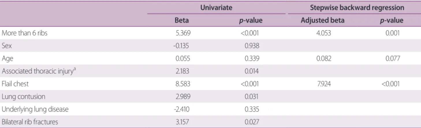
Univariate analysis revealed that factors affecting ICU admission were ≥6 rib fractures (β=5.369, p<0.001) and flail chest (β=8.583, p<0.001). In multivariate analysis, factors affecting ICU admission were also ≥6 rib fractures (adjusted β=4.053, p=0.001) and flail chest (adjusted β=7.924, p<0.001) (Table 2).
No factor affected pulmonary function (Table 3).
DISCUSSION
A recent study highlighted the detrimental effects of in-
creasing number of rib fractures and age. The authors
demonstrated that patients older than 65 years with more
than four fractures had higher morbidity and mortality
[14]. Another study reported that patients older than 45
years of age with more than four rib fractures are at risk of
prolonged ICU stays, ventilator days, and overall hospital
days [15]. Increasing number of rib fractures has been
correlated directly with increasing pulmonary morbidity
and mortality [5]. In this study, we also demonstrated
that patients with more than six rib fractures exhibit pro-
longed ICU stays, ventilator days, and overall hospital
days. Mechanical ventilator apply was performed in 31
patients. The reason for the intubation was as follows.
Table 1. Characteristics of patients with rib fractures
Rib fracture Total (n=72) Group A (n=22) Group B (n=50)
p-valueMale 61 (84.7) 21 (95.5) 40 (80.0) 0.186
Age (year) 51.2±12.0 50.5±9.2 51.5±13.1 0.731
Past medical disease COPD or asthma 6 (8.5) 2 (9.5) 4 (8.0%) 1.000
Associated thoracic injury 0.039
Pneumothorax 6 (8.3) 4 (18.2) 2 (4.0)
Hemothorax 12 (16.7) 6 (27.3) 6 (12.0)
Hemopneumothorax 52 (72.2) 11 (50.0) 41 (82.0)
Flailchest 11 (15.3) 1 (4.5) 10 (20.0) 0.186
Lungcontusion 43 (59.7) 7 (31.8) 36 (72.0) 0.003
Closed thoracostomy 56 (77.8) 11 (50.0) 45 (90.0) 0.001
Rib fixation 5 (6.9) 0 (0.0) 5 (10.0) 0.301
Hospital days 22.0 (15.0, 35.0) 13.5 (9.0, 23.0) 27.0 (20.0, 43.0) <0.001
ICU admission 38 (52.8) 1 (4.5) 37 (74.0) <0.001
Mechanical ventilator apply 31 (43.1) 1 (4.5) 30 (60.0) <0.001
FVC
<80 30 (41.7) 6 (27.3) 24 (48.0) 0.166
≥80 42 (58.3) 16 (72.7) 26 (52.0)
FEV1
<80 29 (40.3) 8 (36.4) 21 (42.0) 0.851
≥80 43 (59.7) 14 (63.6) 29 (58.0)
FEV1/FVC
<80 12 (16.7) 6 (27.3) 6 (12.0) 0.208
≥80 60 (83.3) 16 (72.7) 44 (88.0)
Values are presented as mean±standard deviation, median (25 percentile, 75 percentile), or number (%).
COPD: chronic obstructive pulmonary disease, ICU: intensive care unit, FVC: forced vital capacity, FEV1: forced expiratory volume in the first second.
Table 2. Factors related to ICU days
Univariate Stepwise backward regression
Beta
p-valueAdjusted beta
p-valueMore than 6 ribs 5.369 <0.001 4.053 0.001
Sex -0.135 0.938
Age 0.055 0.339 0.082 0.077
Associated thoracic injury
a2.183 0.014
Flail chest 8.583 <0.001 7.924 <0.001
Lung contusion 2.989 0.031
Underlying lung disease -2.410 0.335
Bilateral rib fractures 3.157 0.027
ICV: intensive care uint.
a
Associated thoracic injury: pneumothorax, hemothorax, hemopneumothorax.
11 were due to flail chest and 19 were due to lung con- tusion. The other was performed because of diaphragm injury. Table 1 shows that flail chest and lung contusion more occurred in group B. Compared with group A, lung contusion and flail chest occurred frequently in group B, Mechanical ventilation application was performed more than group A. The mean duration of mechanical ventila- tion was 4 days in group B. Lung contusion was the most common reason for longer duration of mechanical venti- lation when patients were applied for more than 4 days (13 patients, 5.8 days). The next was flail chest (5 patients, 5.8 days).
Flagel et al. [5] found that when six or more ribs are fractured, pulmonary morbidity and flail chest are in- creased. It would have a significant effect on the pulmo- nary function outcome. So we divided them into two groups based on six. In this study, increasing number of rib fractures was not associated with pulmonary function previous study that reported improvement in pulmonary function tests after 6 to 18 months [16]. Other authors reported that in a cohort of 13 survivors of severe thoracic trauma with lung contusion, substantial physiological recovery was evident with good pulmonary function tests [17]. Surgical fixation of ribs can be carried out as a first- line therapeutic option for bilateral rib fractures or flail segments without significant complications if the risk fac- tors associated with mortality are carefully considered [18].
The authors also opined that, with a view of restoring pul-
monary function as well as chest wall configuration, early operative stabilization of the ribs can be more helpful than conventional treatment for patients with multiple rib fractures. Early restoration of the chest wall integrity and respiratory functions prevents restrictive impairment of pulmonary functions, as proven by the total lung capac- ity values surgically treated patients presented 6 months postoperatively, which were all significantly higher than 85% of the predicted value [19]. The indications for fixa- tion of ribs remain controversial mostly because of a lack of adequate studies comparing operative and non-opera- tive treatment. In this study, the majority of patients with multiple rib fractures were treated without rib fixation.
Only five patients underwent rib fixation, and fixation was performed by suture fixation. In a previous study, rib fixation was performed using a metal plate. In plate fixation, firm fixation might not be achieved, creating a risk of dislocation. Complications, such as osteomyelitis, are related to fixation to the bone marrow through the ribs. The patients enrolled in this study tended to recover pulmonary function without rib fixation. The collective findings highlight the need for more studies on the surgi- cal reduction of rib fracture.
The primary limitation for this study is related to se- lection bias, because this study was conducted in a single center and had limited sample size. The small number of patients makes it difficult to prove that variables affect morbidity and pulmonary function. Another significant Table 3. Factors related to FEV1 and FEV1/FVC
FEV1 FEV1/FVC
Univariate Stepwise backward regression Univariate Stepwise backward regression OR (95% CI) Adjusted OR (95% CI) OR (95% CI) Adjusted OR (95% CI)
More than six ribs 0.79 (0.27-2.19) 2.75 (0.76-10.04) 3.19 (0.75-14.51)
Sex 0.34 (0.07-1.21) 0.35 (0.07-1.26) 0.33 (0.02-1.94)
Age 0.99 (0.95-1.03) 0.94 (0.88-0.99) 0.92 (0.86-0.98)
Associated thoracic injury
a0.63 (0.29-1.22) 0.83 (0.28-1.83)
Flail chest 0.50 (0.13-1.85) 2.20 (0.36-42.49)
Lung contusion 0.52 (0.19-1.37) 1.61 (0.45-5.73)
Underlying lung disease 0.62 (0.11-3.61) 0.91 (0.13-18.39)
Bilateral rib fractures 0.37 (0.13-0.99) 0.70 (0.20-2.62)
FEV1: forced expiratory volume in the first second, FVC: forced vital capacity, OR: odds ratio, CI: confidence interval.
a