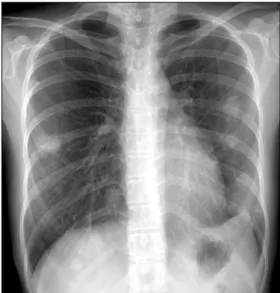
A Case of Balsalazide-Induced Limited Form of Granulomatosis with Polyangiitis with Bronchiolitis Obliterans Organizing Pneumonia-like Variant in Ulcerative Colitis
5
0
0
전체 글
(2)
(3)
(4)
(5)
수치

관련 문서